INTRASPECIFIC VARIATION IN PROTEIN PATTERN OF RED SCORPION (Mesobuthus tamulus, COCONSIS, POCOCK) VENOMS FROM WESTERN AND
SOUTHERN INDIA
BADHE R. V. (1), THOMAS A. B. (1), HARER S. L. (1), DESHPANDE A. D. (1), SALVI N. (2), WAGHMARE A. (2)
(1) Department of Pharmaceutical Chemistry, Dr. D. Y. Patil Institute of
Pharmaceutical Sciences and Research, Pimpri, Pune, Maharashtra, India; (2)
Haffkine Biopharmaceutical Corporation Ltd, Pimpri, Pune, Maharashtra, India.
ABSTRACT: Red scorpions Mesobuthus tamulus (Coconsis, Pocock) were obtained from different regions of West and South India (Ratnagiri, Chiplun and Ahmednagar from Maharashtra and Chennai from Tamil Nadu, respectively). Their venoms composition was analyzed using gel electrophoresis (SDS-PAGE). All venom samples shared six bands of 170, 80, 60, 57, 43, and 38 kDa molecular weights. Bands of 115 kDa and 51.5 kDa were characteristic of venoms obtained from red scorpions of Chiplun region, and the 26kDa band was absent in scorpion venom from Tamil Nadu. The separated protein band patterns suggest that the venoms from Ratnagiri, Ahmednagar and Tamil Nadu had high similarities in their biochemical composition but differed from that of Chiplun region. These data werealso supported by the Jaccard (J) index. The J value was 0.33 for venom obtained from Ratnagiri-Ahmednagar, 0.31 for venom from Ratnagiri-Tamil Nadu, and 0.3 for venom from Ratnagiri-Chiplun region. This suggests the existence of genetic variation among the different strains of red scorpion in western and southern India. The antiserum produced by Haffkine Biopharmaceuticals Corporation Ltd. completely neutralized proteins of venoms from all the regions studied.
KEY WORDS: red scorpion, SDS-PAGE, Mesobuthus tamulus.
CORRESPONDENCE TO:
RAVINDRA V. BADHE, Department of Pharmaceutical Chemistry, Dr. D. Y. Patil
Institute of Pharmaceutical Sciences and Research, Pimpri, Pune, 411018,
INTRODUCTION
Gel electrophoresis of proteins is widely used in insect molecular systematic. In this
technique, identical proteins migrate to the same distance under electrical forces
applied to an electrophoretic gel while non-identical proteins usually migrate to
different distances (2). Studies on venoms, including insect venoms, have profoundly
affected modern biochemistry, pharmacology and medicine. Venoms have provided
an excellent source of highly concentrated active enzymes, cytotoxins and
neurotoxins as research tools to study the sub-cellular functioning of mammalian
nervous and cardiovascular systems. Insect venom previously had little direct use in
modern medicine, but this situation is rapidly changing as more information is
becoming available. As new techniques for isolating, identifying and especially
producing individual venom components are developed, the use and role of venom in
medicine will certainly increase.
The low-molecular-weight biogenic amines (histamine, dopamine, nor-adrenalin, etc.)
found in venom samples are involved in local reactions and their release from a
single sting can lead to systemic reactions. They act on blood vessels and nerve
endings inducing swelling, redness, pain and itching. Major toxic effects of venom
can be attributed to the presence of larger peptides such as melittin, dopamine and
mostly cell degranulating factors. These peptides can cause damage to the cell
membrane leading to release of enzymes from lysozymes and mast cell granules,
resulting in cytolysis. Additionally, they can act as neurotoxins provoking
hyper-excitability. High-molecular-weight enzymes, except for highly cytotoxic
phospholipase A2, are regarded as less harmful. Hyluronidase has an indirect effect
of increasing the absorption of active peptides (12). The various enzymes and
vasoactive components induce local toxic inflammation at the sting region. If the sting
occurs in a highly vascular area, or even intravascularly, the toxic components are
rapidly spread and might give rise to systemic reactions. Several simultaneous stings
will cause more reactions. A number of children and adults, especially pregnant
women, succumb to the sting by the red scorpion Buthus tamulus in Konkan region,
India, especially in the coastline (8).
The protein patterns of venoms from red scorpions Mesobuthus tamulus (Conconsis,
Pocock) from Ratnagiri, Chiplun, Ahmednagar and Tamil Nadu were analyzed using
the biological activities and correlation between the composition of venoms from
different strains of red scorpion found in West and South India.
MATERIALS AND METHODS Venom collection
We used pure lyophilized red scorpion (Mesobuthus tamulus) venoms from the
following regions of West and South India: Ratnagiri, Chiplun, Ahmednagar and
Tamil Nadu (supplied by Irula Snake Catchers Industrial Co-operative Society Ltd.,
ISCICS, Chennai, India). Lyophilized venom samples were resuspended in normal
saline and 50% v/v solution of glycerin in distilled water, resulting in a 1% venom
solution, which was stored at 2-4°C for further experiments.
Electrophoresis
Sodium dodecyl sulfate-polyacrylamide gel electrophoresis (SDS-PAGE) was carried
out, according to the method of Lammeli (9), with 15% polyacrylamide concentration.
Electrophoresis patterns of venom samples from Ratnagiri, Chiplun, Ahmednagar
and Tamil Nadu were obtained. Molecular weight markers (3-205 kDa) were used as
standards (Banglore Gennie). Proteins were visualized using Coomassie blue stain
and silver staining. Tentative identification of the proteins in venom samples was
made by comparing the gel with the descriptions existing in literature. Western blot
was carried out to check the neutralizing capacity of the anti-scorpion venom sera
produced by Haffkine Biopharmaceuticals Corporation Ltd, Pimpri, Pune, India.
Data analysis
Similarity between scorpion strains was measured using the Jaccard Index [J] (10),
which was calculated as follows:
J = a/a+b+c
Where a = bands shared between two strains; b = total number of bands in strain 1; c
= total number of bands in strain 2.
RESULTS
Ten protein bands (molecular weights: 170, 115, 79.6, 60, 57, 51.5, 43, 38, 29, and
The intensity of their color evidenced that the 57kDa band [band 5] had the highest
protein concentration, followed by the 26kDa band [band 10].
Red scorpion venoms from both Ratnagiri and Ahmednagar showed bands with the
following molecular weights: 170, 79.6, 60, 57, 43, 38, 29, and 26 kDa, from which
band 4 [57 kDa] was prominent, followed by band 8 [26 kDa] (Figure 1 and Table 1).
The venom of red scorpion from Tamil Nadu presented seven bands with molecular
weights of 170, 79.6, 60, 57, 43, and 38 kDa. Band 2 [79.6 kDa] was the most
noticeable and the 26kDa band was absent.
Bands of 170, 79.6, 60, 57, 43, 38, 29, and 26 kDa molecular weights were
characteristic of all venoms studied, except for that from Tamil Nadu, which did not
present the 26kDa band (Table 1). Red scorpion venoms of Ratnagiri and
Ahmednagar differed from that of Chiplun region by the absence of 115 and 51.5 kDa
protein bands.
According to the Jaccard index equation, which was used to measure the similarity
index between red scorpion venoms, the highest degree of similarity was observed
between red scorpions from Ratnagiri and Ahmednagar [0.33], followed by those
from Ratnagiri and Tamil Nadu [0.31]. The least similarity occurred between red
scorpions from Ratnagiri and Chiplun [0.30]. Western blot showed complete
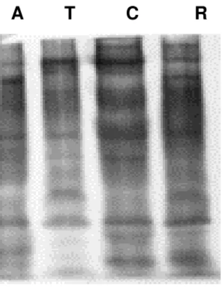
neutralization of all the separated protein bands (Figure 2).
There is difference between the geographical conditions of Ahmednagar and those of
Tamil Nadu, Ratnagiri, and Chiplun regions. The later are costal areas with an
average annual temperature of 29°C and the former presents 38°C average
temperature. There is also a difference in the average annual rainfall: around 700
mm in Ahmednagar, 2000 mm in Tamil Nadu, and 3500 mm in Ratnagiri and
Chiplun. The later regions have relatively high humidity [70-100] (15), which may
affect the scorpion food chain. These differences may lead to different critical lethal
dose (CLD) values of scorpion venoms. For red scorpion venoms from Ratnagiri,
S T C A R
{a}
S T C A R
{b}
Figure 1:Electrophoresis pattern of red scorpion (Mesobuthus tamulus) venoms.
{a} Electrophoresis using Coomassie blue staining {b} Electrophoresis using silver staining.
T - Tamil Nadu; C - Chiplun; A - Ahmednagar; R - Ratnagiri; S - Standard markers.
A T C R
Figure 2: Western blots of separated protein bands of all four scorpion venom samples.
A - Ahmednagar; T - Tamil Nadu; C - Chiplun; R - Ratnagiri.
Table 1: Molecular weights of resolved bands of scorpion venom samples, calculated
through comparison with the standard molecular weight markers, and their known
probable pharmacological activities.
Venom sample from Protein
band
No
Molecular
weight
(kDa)
Chiplun Ratnagiri Ahmednagar Tamil
Nadu
Probable
Pharmacological
actions
1 170 + + + +
2 115 + - - - Neurotransmitter
3 79.6 + + + + Hyluronidase
4 60 + + + + Hyluronidase
5 57 + + + + NMDR receptor
activator
6 51.5 + - - - Not known
7 43 + + + + Hyluronidase
8 38 + + + + Blocks chloride ion
9 29 + + + + Phospholipase A2
10 26 + + + - Hemorrhagic toxin
DISCUSSION
Ten bands of molecular weight ranging from 170 to 26 kDa were obtained for the red
scorpion venom of Chiplun region. As bands of more than 100 kDa (i.e. 115 kDa and
components. However, they were reported to have neurotransmitter releasing
property (6). The 51.5 kDa band has not been studied and its activity is unknown.
Bands of 79.6 kDa and 60 kDa molecular weights, which were present in all four
scorpion strains (from Ratnagiri, Ahmednagar, Tamil Nadu and Chiplun regions),
characterize proteins with hyluronidase activity(5, 7). The 57 kDa protein was cited to
be a NMDA (N-methyl-D-aspartic acid)-receptor activator (1). The 43 kDa protein has
hyluronidase activity (13), and that of 38 kDa was reported to have chloride-ion
blocking (4)and histamine releasing activities (3). Band of 29 kDa was characterized
as protein with phospholipase A2 activity(14). The last protein band of 26 kDa, which
is not present in Tamil Nadu scorpion venom, was cited to have hemorrhagic toxic
and phosphodiesterase activities (11). Silver staining of bands revealed the same
data.
Haffkine Biopharmaceutical Corporation Ltd., India, uses a mixture of venoms from
different regions for immunization, and the antisera produced was capable of
overcoming the effects of scorpion venoms from the Indian subcontinent. Western
blot of the venom against the anti-scorpion venom serum corroborated these data
(Figure 2).
Environmental conditions significantly affected venom lethality. According to the
present test, it is clear that the venom sample of red scorpion from Tamil Nadu is
more lethal at low concentrations. Out of the venom samples obtained from
scorpions of Maharashtra region, that of Chiplun was more lethal (8).
Differences in the band patterns of separated proteins in all venom samples clearly
suggest the existence of genetic variation among the scorpion strains of different
regions in western and southern India.
REFERENCES
1 BANKS BEC., SHIPOLINI AA. Chemistry and pharmacology of venom. In:
VENOMS OF HYMENOPTERA, Piek T (Ed.). Academic Press, 1986,
329-416.
3 CHATWAL GS., HABERMANN E. Neurotoxins, protease inhibitors and histamine
releasers in the venom of Indian red scorpion (Buthus tamulus); isolation and
partial characterization. Toxicon, 1981, 19, 807-23.
4 DHAWN R., JOSEPH S., LALA AK. Purification and characterization of a short
insect toxin from venom of scorpion Buthos tamulus. FEBS letters, 2002, 528,
261-6.
5 DIMITROV SD., NATCHAEV LA. Fractions of some bee venom components on a
new type of modified cellulose. Toxicon, 1977, 15, 447-8.
6 EGYPT. Govt. of Egypt. A report on venoms collected from different insects and
animals in Egypt. Egypt, 11/23/2000.
7 GAWADE SP. Excitatory effect of Buthus C-56 toxin on Drosophila larval
neuromuscular junction. J. Venom. Anim. Toxins, 2003, 9, 57-9.
8 KANKONKAR RC., KULKARNI DG., HULIKAVI CB. Preparation of a potent
anti-scorpion-venom-serum against the venom of red scorpion (Buthus tamulus), J.
Postgrad Med., 1998, 4, 85-91.
9 LAMMELI UK. Cleavage of structural proteins during the assembly of the head of
bacteriophage T4. Nature, 1970, 227-80.
10 LUDWING JA., RAYOLDS JF. Statistical ecology. A primer on methods and
computing, chapter 8, Diversity indices, John Willy and Sons, New York, 1988,
85-103.
11 MASTER RWP., RAO SS., SOMAN PD. Electrophoretic separation of biologically
active constituents of scorpion venom. Biochem. Biophys. Acta., 1963, 71,
422-8.
12 MEIR J., WHITE J. Handbook of clinical toxicology of animal venom and poisons.
C .R. Press, New York, 1995.
13 SCHMIDT JO. Biochemistry of insect venom. Ann. Rev. Ent., 1982, 27, 339-68.
14 SOBOTKA AK., FRANKLIN RM., ADKINSON NF., VALENTINE MD., BEAR H.,
LICHTENSTEIN LM. Allergy to insect sting. II: The major allergen in venom. J.
Allergy Clin. Immunol., 1976, 57, 29-40.
15 VIDYAPEETH BSKK. A report on “Weather conditions in Maharashtra”. Dapoli,