Themed Section: Pharmacology of the Gasotransmitters
REVIEW
S-nitrosation and neuronal
plasticity
A I Santos
1,2,3, A Martínez-Ruiz
4and I M Araújo
1,21Department of Biomedical Sciences and Medicine, University of Algarve, Faro, Portugal,2IBB – Institute for Biotechnology and Bioengineering, Centre for Molecular and Structural Biomedicine, University of Algarve, Faro, Portugal,3Centre for Neuroscience and Cell Biology, University of Coimbra, Coimbra, Portugal, and4Servicio de Inmunología, Hospital Universitario de La Princesa, Instituto de Investigación Sanitaria Princesa (IP), Madrid, Spain
Correspondence
Inês M Araújo, Department of Biomedical Sciences and Medicine, University of Algarve, Faro, Portugal. E-mail:
[email protected] ---Received 30 January 2014 Revised 8 May 2014 Accepted 9 June 2014
Nitric oxide (NO) has long been recognized as a multifaceted participant in brain physiology. Despite the knowledge that was gathered over many years regarding the contribution of NO to neuronal plasticity, for example the ability of the brain to change in response to new stimuli, only in recent years have we begun to understand how NO acts on the molecular and cellular level to orchestrate such important phenomena as synaptic plasticity (modification of the strength of existing
synapses) or the formation of new synapses (synaptogenesis) and new neurons (neurogenesis). Post-translational modification of proteins by NO derivatives or reactive nitrogen species is a non-classical mechanism for signalling by NO. S-nitrosation is a reversible post-translational modification of thiol groups (mainly on cysteines) that may result in a change of function of the modified protein. S-nitrosation of key target proteins has emerged as a main regulatory mechanism by which NO can influence several levels of brain plasticity, which are reviewed in this work. Understanding how S-nitrosation contributes to neural plasticity can help us to better understand the physiology of these processes, and to better address pathological changes in plasticity that are involved in the pathophysiology of several neurological diseases.
LINKED ARTICLES
This article is part of a themed section on Pharmacology of the Gasotransmitters. To view the other articles in this section visit http://dx.doi.org/10.1111/bph.2015.172.issue-6
Abbreviations
BDNF, brain-derived neurotrophic factor; CaMKII, calcium/calmodulin-dependent protein kinase II; Cdk5, cyclin-dependent kinase 5; cGMP, cyclic GMP; CREB, cAMP response element-binding protein; DRG, dorsal root ganglion; Drp1, dynamin-related protein 1; eNOS, endothelial NOS; EPSP, excitatory postsynaptic potential; HDAC2, histone deacetylase 2; iNOS, inducible NOS; LTD, long-term depression; LTP, long-term potentiation; nNOS, neuronal NOS; NSC, neural stem cells; NSF, N-ethyl-maleimide sensitive factor; PSD-95, postsynaptic density protein 95; PTM, post-translational modifications; RNS, reactive nitrogen species; RyR, ryanodine receptor; sGC, soluble guanylate cyclase
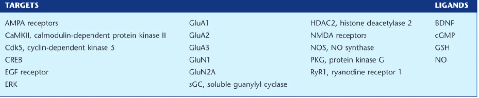
Table of Links
TARGETS LIGANDS
AMPA receptors GluA1 HDAC2, histone deacetylase 2 BDNF CaMKII, calmodulin-dependent protein kinase II GluA2 NMDA receptors cGMP Cdk5, cyclin-dependent kinase 5 GluA3 NOS, NO synthase GSH
CREB GluN1 PKG, protein kinase G NO
EGF receptor GluN2A RyR1, ryanodine receptor 1 ERK sGC, soluble guanylyl cyclase
This Table lists key protein targets and ligands in this article which are hyperlinked to corresponding entries in http:// www.guidetopharmacology.org, the common portal for data from the IUPHAR/BPS Guide to PHARMACOLOGY (Pawson et al., 2014) and are permanently archived in the Concise Guide to PHARMACOLOGY 2013/14 (Alexander et al., 2013a, b, c d).
Introduction
Brain plasticity is regarded as the ability of the brain to change its structure and function as a result of experience and changes in environment. Plasticity occurs mostly at three different levels: modification of the strength of existing syn-apses (synaptic plasticity), formation of new synsyn-apses (syn-aptogenesis) and formation of new neurons (neurogenesis). Many factors, including nitric oxide (NO), regulate and fine-tune these mechanisms.
Synaptic plasticity is described as the change in strength of a synapse in response to changes in synaptic activity. Short-term plasticity occurs on a scale from milliseconds to minutes, and is often associated with changes in the pres-ynaptic release of neurotransmitters. Longer forms of plas-ticity occur in the postsynaptic terminal, such as long-term potentiation (LTP) or depression (LTD); initial changes (in the first 1 or 2 h) in LTP or LTD are associated with altera-tions in the trafficking of glutamate receptors, while main-tenance of LTP and LTD for longer periods requires changes in gene expression. The increase in postsynaptic strength is triggered by activation of NMDA receptors, which causes calcium influx into the postsynaptic cell and activates signal transduction pathways that result in insertion in the mem-brane of a different type of glutamate receptors, AMPA receptors. The regulation of membrane insertion of AMPA receptors is key for efficient synaptic plasticity, and a large body of evidence can be found regarding the control of AMPA receptor trafficking (see Ho et al., 2011b), including by NO.
Synaptogenesis refers not only to formation of new syn-apses, but also to the remodelling of existing ones. Synapse formation occurs during development and in the adult brain, and may be changed by experience or disease (see Pérez-Domper et al., 2013; Robichaux and Cowan, 2014). NO has been consistently involved in such a rearrangement of existing synapses and formation of new ones (García-López et al., 2010; Shelly et al., 2010; Sunico et al., 2010).
Neurogenesis is not restricted to embryonic development, but occurs throughout adult life. New neurons and glial cells are continuously formed in the brain, from neural stem cells present in niches such as the subventricular zone and the dentate gyrus in the hippocampus (see Gage and Temple,
2013). Neurogenesis is a tightly regulated process, and NO is one of the many factors that influence the formation of new brain cells in physiological conditions and in disease states (see Carreira et al., 2012; Estrada and Murillo-Carretero, 2005).
In the brain, the free radical gaseous messenger NO is associated with the regulation of neuronal plasticity and cog-nitive functions, and also regulates biological functions such as the sleep-wake cycle, body temperature, appetite and hormone release, and modulates neuronal development, nociception and apoptosis (see Calabrese et al., 2007; Prast and Philippu, 2001). NO is mainly produced by nitric oxide synthases (NOS), which can be found in the CNS in three different isoforms: (i) neuronal NOS (nNOS or NOS1), which is constitutively expressed in neurons; (ii) endothelial NOS (eNOS or NOS3), constitutively expressed in endothelial cells; and (iii) inducible NOS (iNOS or NOS2), which is mainly expressed by activated microglial cells. While nNOS and eNOS are constitutively expressed and are transiently acti-vated by calcium/calmodulin and other regulators, iNOS is transcriptionally regulated by inflammation or toxic stimuli and its activity is independent of calcium regulation (Bredt, 1999; Guix et al., 2005).
The potential of NO as a modulator of synaptic transmis-sion was first proposed in 1988 (Garthwaite et al., 1988), and its presence was documented in several brain regions such as the hippocampus, striatum, hypothalamus and locus coer-uleus (Prast and Philippu, 2001). The small size of NO, its highly diffusible nature and its link to glutamate receptors make NO a special neurotransmitter. NO may be considered an atypical neurotransmitter, as it does not require vesicle storage or exocytotic release, being produced generally after receptor activation and readily diffused throughout cells and tissues. NO also modulates the release of several neurotrans-mitters such as acetylcholine, catecholamines, glutamate and GABA (see Prast and Philippu, 2001). NO is able to mediate both postsynaptic intracellular signalling and retrograde intercellular communication between different types of cells and tissues. However, the short half-life of NO and its diffu-sion coefficient (2.2× 10⁻⁵cm2s−1obtained in vivo) limit the range of action of NO in brain tissue, thus providing short-range responses in individual synapses (Santos et al., 2011; Martínez-Ruiz et al., 2013).
Classical NO signalling proceeds through its potent binding to soluble guanylate cyclase (sGC), which activates this enzyme that produces cyclic GMP (cGMP). cGMP in turn reacts with a family of cGMP-dependent protein kinases (also called PKG), triggering several signalling pathways (see Hofmann, 2005; Francis et al., 2010). A less classical mode of action of NO is through interaction with mitochondrial pro-teins, especially inhibition of cytochrome c oxidase. Non-classical NO signalling includes both the production of NO from alternative sources (mainly nitrite) and the formation of covalent post-translational modifications (PTM) in proteins (see Martínez-Ruiz et al., 2011). These PTM are not formed by NO itself, but by the production of the so-called reactive nitrogen species (RNS), small molecules produced from NO reactions. Although there are several RNS-induced PTM, three of them have been more intensively studied. Tyrosine nitra-tion, the incorporation of a nitro group (−NO2) to a tyrosine residue, is considered an almost irreversible modification, so it has been related to protein damage, although it is also implied in signalling pathways (Souza et al., 2008; Ischiropoulos, 2009). Cysteine S-glutathionylation, the for-mation of a mixed disulfide between a protein cysteine thiol and glutathione, is a reversible modification whose formation and removal may be mediated by enzymes such as glutar-edoxins. It has been involved in cell signalling and in oxida-tive stress, it can be formed from either reacoxida-tive oxygen species or RNS, and it is related to S-nitrosation in several ways, including the fact that S-nitrosation of a cysteine residue or of glutathione can lead to S-glutathionylation (see Martínez-Ruiz and Lamas, 2007; Mieyal et al., 2008; Sabens Liedhegner et al., 2012).
Protein S-nitrosation is the formation of a nitrosothiol (R-S-NO) in cysteine residues (denoted PSNO). It is also called S-nitrosylation (the terminology has been discussed in Martínez-Ruiz and Lamas, 2004; Heinrich et al., 2013), and both names could be used for the overall description of the PTM, as their difference relies mainly in the chemical mecha-nisms for their formation, which is not always known or considered (Heinrich et al., 2013). We will follow the recent recommendation in this Journal to use the term S-nitrosation as it includes the most part of the cases and can induce less confusion (Heinrich et al., 2013). Nitrosothiols are not formed from the direct reaction of NO with the cysteine residue, unless it is in the thiyl radical form (P-S•).
P-S• + •NO→P-S-NO
There are several chemical mechanisms that can lead to protein S-nitrosation, including formation of N2O3,
2NO O+ 2→2NO2
NO2+NO→N O2 3
a species that is a very effective nitrosating agent.
N O2 3+PSH→PSNO H+ ++NO2−
Nitrosothiols are not very stable in the cells compared with other PTM because of the lability of the bond, the presence of denitrosylases and other reductants (e.g. ascorbate), and the easy reaction with other thiols to transfer the nitroso group to either low molecular mass thiols or proteins (transnitrosation)
PSNO RSH+ →PSH RSNO+
PSNO P SH+ ′ →PSH P SNO+ ′
or to produce a mixed disulfide, leading to S-thiolation or S-glutathionylation.
PSNO RSH PSSR HNO S-thiolation RSH is a low
molecular mass
+ → + ( :
tthiol)
PSNO GSH PSSG HNO S-glutathionylation GSH is
glutathione
+ → + ( :
).
Enzymic catalysis is not strictly necessary for its formation and degradation in cell environment, so its specificity and involvement in cell signalling bears some properties that make it different from other signalling PTM such as phospho-rylation (see Martínez-Ruiz and Lamas, 2004; Martínez-Ruiz et al., 2011). Because of some of these properties, we have proposed that it is mainly used by cells as a short-range NO-derived signalling mechanism that can be favoured in proteins located at a short distance from the sources of NO, namely NOS, and in proteins interacting with them (Martínez-Ruiz et al., 2013).
In this work, we review the role of S-nitrosation and other NO signalling pathways in neuronal plasticity. The S-nitrosated proteins discussed in this review are summarized in Table 1.
S-nitrosation in synaptic plasticity
Synaptic plasticity refers essentially to the modification of the synaptic response upon intense or decreased synaptic input at a given synapse, resulting in long-lasting potentiation or depression of the synaptic function (see Ho et al., 2011b; Choquet and Triller, 2013). Both presynaptic and postsynap-tic changes in synappostsynap-tic function can occur, but for the purpose of this review, we will focus on postsynaptic altera-tions of synaptic strength. The presynaptic modulation of synaptic plasticity by NO has been the focus of a recent comprehensive review (Hardingham et al., 2013) and will not be discussed here. The changes in synaptic strength can be achieved by different mechanisms in different synapses throughout the brain. In the hippocampus, for instance, glu-tamate release and gluglu-tamate receptors are generally associ-ated with LTP, and LTP is achieved by altering the number of AMPA receptors on the postsynaptic membrane. More recep-tors activated each time glutamate is released means a sus-tained larger excitatory postsynaptic potential (EPSP), while the opposite (fewer AMPA receptors on the post synaptic membrane) holds true for LTD. LTP is usually expressed by a larger amplitude in EPSP following a high-frequency stimu-lation, while LTD is characterized by a decrease in the ampli-tude of EPSP after low-frequency stimulation. LTP can be dependent on NMDA receptors, which are fundamental for modifying synaptic strength in the initial stages of LTP, as they trigger the signalling pathways that result in the mobi-lization of more AMPA receptors towards the postsynaptic membrane (Bliss and Collingridge, 2013). NMDA-independent mechanisms for LTP are also characterized (Oren et al., 2009), but will not be discussed in this work. The mechanism for LTD induction may involve also NMDA
receptors in some brain regions, such as the hippocampus, while metabotropic glutamate receptors are involved in LTD induction in the cerebellum, for instance. Thus, the mecha-nism for LTP or LTD is region- and synapse-specific. For the purpose of this review, we will focus on NMDA receptor-dependent potentiation of synaptic activity.
Physiologically, the activation of NMDA receptors results in calcium influx through the receptor channel. To induce LTP, calcium will activate PKs, such as calcium/calmodulin-dependent PK II (CaMKII), which will phosphorylate AMPA receptors present in postsynaptic pools of vesicles, making
them insert into the postsynaptic membrane (see Ho et al., 2011b). Phosphorylation of AMPA receptors by CaMKII and other kinases will also increase channel conductance and opening probability (Lee et al., 2000; 2010). The calcium influx via NMDA receptors that triggers LTP is also coupled to the postsynaptic synthesis of NO, given the close proximity of nNOS with NMDA receptors because of the interaction with the scaffolding postsynaptic density protein 95 (PSD-95) (Sattler et al., 1999). The small distance between nNOS and the NMDA receptor allows the production of NO when calcium enters the postsynaptic neuron directly through the
Table 1
Proteins modified by S-nitrosation that contribute to brain plasticity
Protein
S-nitrosated
Cys Functional significance Experimental context References
NSF Cys91 Increase of membrane insertion of AMPA
receptors GluA2 subunits
NO donors increase NSF-GluA2 association; impairment of GluA2 surface expression in nNOS-deficient mice
Huang et al., 2005
Stargazin Cys3 Up-regulation of AMPA receptors in the
nucleus accumbens
NO donors elicit S-nitrosation of stargazin Selvakumar et al., 2009 GluA1 Cys875 Enhancement of Ser831 phosphorylation, facilitation of the associated increase in AMPA receptor conductance and endocytosis
GluA1 S-nitrosation in cells
overexpressing WT GluA1, but not GluA1-C875S, following exposure to NO donors or in HEK-nNOS cells
Selvakumar
et al., 2013
GluN2A Cys399 Down-regulation of NMDA receptor
activity and decrease of calcium influx
Exogenous and endogenous NO-related species elicit S-nitrosation of NR2A
in vitro
Choi et al., 2000; Kim
et al., 1999
GluN1 Cys744/Cys798 Regulation of calcium influx through the
NMDA receptor
S-nitrosation of GluN1 in cerebrocortical cultures and nNOS-HEK cells under hypoxic conditions
Takahashi et al., 2007
PSD-95 Cys3/Cys5 Reduction of synaptic clustering in granule
cells of the cerebellum
S-nitrosation following exposure to exogenous or endogenous NO, and in WT mice, but not in nNOS−/−mice
Ho et al., 2011a
Serine racemase
Cys113 Inhibition of enzyme activity and feedback
mechanism to decrease the presynaptic formation of D-serine
S-nitrosation and enzyme inhibition using NO donors in vitro
Mustafa et al., 2007
RyR1 Cys3635
Increase of calcium release from the endoplasmic reticulum
S-nitrosation of RyR1 following exposure to a NO donor
Kakizawa et al., 2012 HDAC2 Cys262/Cys274 Chromatin remodelling, dendritic growth
and branching, and the activation of genes associated with cortical
development, regulating radial migration of cortical neurons
Endogenous NO production by NOS, but not in nNOS−/−animals, and NO donors induces S-nitrosation of HDAC2
Nott et al., 2013; Nott
et al., 2008
Cdk5 Cys83/Cys157 Contribution to NMDA-induced dendritic
spine loss and neuronal apoptosis caused by amyloid-β damage
NO donors elicit Cdk5 activation; S-nitrosation of Cdk5 in human brain with Alzheimer’s disease
Qu et al., 2011
Drp1 Cys644
Dendritic spine loss and neurodegeneration in Alzheimer’s disease
NO donors and endogenous nNOS-derived NO induces Drp1 S-nitrosation
Cho et al., 2009
EGF receptor
Cys166/Cys305 Negative regulation of NSC proliferation in
the SVZ by down-regulation of the ERK/MAPK pathway
EGF receptor is S-nitrosated in vitro in the presence of NO donors
Murillo-Carretero
et al., 2009
H-Ras Cys118 Alteration of the active centre
conformation, allowing a faster transition from GDP- to GTP-binding state
NO donors activate WT H-Ras, but not H-Ras NO-insensitive mutant
Lander et al., 1997; Lander
NMDA receptor, while calcium entering the postsynaptic neuron by other channels does not necessarily increase NO production (Sattler et al., 1999). The importance of NO for LTP triggered by activation of NMDA receptors was demon-strated by several laboratories in the early 1990s, showing that LTP was abolished when neurons were treated with NOS inhibitors (Böhme et al., 1991; 1993; O’Dell et al., 1991; Mizutani et al., 1993; Zhuo et al., 1993). The membrane-impermeable scavenger of NO, haemoglobin, also blocks LTP when applied extracellularly, suggesting that NO must be released extracellularly to the synaptic cleft for the occur-rence of LTP (O’Dell et al., 1991). Furthermore, mice lacking only nNOS (O’Dell et al., 1994) or eNOS (Dinerman et al., 1994) are capable of normal LTP, but, in the presence of NOS inhibitors, LTP is reduced, suggesting that NO is important for LTP in these knockout mice. Moreover, an impairment of hippocampal LTP was observed in double-knockout mice for nNOS and eNOS, and no further reduction of LTP was found following the use of NOS inhibitors, demonstrating that NO derived from both NOS is involved in LTP (Son et al., 1996). However, in mature neurons of the neocortex of adult animals, synaptic potentiation was reduced (over 42%) in mice lacking nNOS (Dachtler et al., 2011), and LTP was com-pletely abolished in double-knockout mice for nNOS and the glutamate receptor subunit GluA1, but not in eNOS/GluA1 double-knockout mice. These findings suggest that nNOS-derived NO and GluA1 subunit of the AMPA receptor are required for experience-dependent plasticity in the neocortex (Dachtler et al., 2011), without any contribution by eNOS. The contribution of NO to LTP may also be through cGMP production and PKG activation (Zhuo et al., 1994; Gallo and Iadecola, 2011), but recent studies have shown that several intracellular proteins involved in LTP and in AMPA receptor trafficking can be S-nitrosated (see Tegeder et al., 2011; Martínez-Ruiz et al., 2013), including NMDA receptor subu-nits and proteins associated with AMPA receptor membrane insertion and internalization.
AMPA receptor trafficking to the postsynaptic membrane is fine-tuned by a variety of regulator and adaptor proteins. Synaptic plasticity is mostly driven by GluA1/GluA2-containing AMPA receptors being inserted into the mem-brane after high-frequency stimulation, while after synaptic strengthening a new equilibrium is achieved by the inser-tion of GluA2/GluA3 receptors (Shi et al., 2001). Membrane insertion of GluA1/GluA2 subunits is controlled by associa-tion with N-ethyl-maleimide sensitive factor (NSF), and S-nitrosation of Cys91 on the NSF protein was shown to drive the surface expression of AMPA receptors containing GluA2 subunits (Huang et al., 2005), by allowing the disso-ciation of protein interacting with Cα-kinase 1 from AMPA receptors and favouring membrane expression (Sossa et al., 2007). The surface expression of GluA1-containing AMPA receptors is regulated by stargazin, which is physiologically S-nitrosated on Cys3 (Selvakumar et al., 2009). Recently, S-nitrosation of stargazin was also shown to mediate the up-regulation of AMPA receptors in the nucleus accumbens shell of cocaine-sensitized rats during withdrawal (Selvakumar et al., 2014), which implicates S-nitrosation of stargazin in the pathophysiology of addiction mechanisms that involve up-regulation of membrane expression of AMPA receptors.
The GluA1 subunit itself is also a target for S-nitrosation. Cys875may be modified following NO production triggered by NMDA receptor activation by glutamate and thus enhance phosphorylation of Ser831, resulting in an increase in channel conductance (Selvakumar et al., 2013). In the same study, nitrosation of Cys875was also shown to facilitate binding of AP2 protein and promote endocytosis of AMPA receptors (Selvakumar et al., 2013).
S-nitrosation was also observed in NMDA receptor GluN2A and GluN1 subunits, with implications for the activ-ity and permeabilactiv-ity of NMDA receptors and, thus, on post-synaptic calcium influx. NMDA receptor GluN2 subunits are coupled to nNOS by the PSD-95/Dlg/ZO-1 (PDZ) domains of PSD-95, at the postsynaptic density (Christopherson et al., 1999) and nitrosation of GluN2A Cys399 down-regulates NMDA receptor activity and decreases calcium influx (Kim et al., 1999; Choi et al., 2000). Moreover, following hypoxia or stroke, GluN1 subunits appear nitrosated at the Cys744/ Cys798 pair, regulating calcium influx through the NMDA receptor, decreasing neurotoxicity (Takahashi et al., 2007). PSD-95 may also be regulated by S-nitrosation on Cys3/Cys5 (leading to reduced synaptic clustering); S-palmitoylation of the same residues favours synaptic localization of PSD-95 (Ho et al., 2011a). Furthermore, serine racemase, which converts L-serine to D-serine, was shown to be influenced by NO. D-Serine works as a co-agonist of NMDA receptors. Thus, in the presence of NO, serine racemase is S-nitrosated at Cys113 and its enzymic activity is inhibited, creating a feedback mechanism to decrease the presynaptic formation of D-serine (Mustafa et al., 2007) after activation of glutamate receptors, resulting in decreased signalling by the NMDA receptor.
In the cerebellum, burst stimulation of the parallel fibres triggers NO-dependent LTP in the parallel fibre-Purkinje cell synapse (Namiki et al., 2005). NO was shown to increase release of calcium from the endoplasmic reticulum via S-nitrosation of Cys3635in type 1 ryanodine receptors (RyR1), thus triggering the calcium increase necessary for increasing surface expression of AMPA receptors on the postsynaptic membrane in Purkinje cells (Kakizawa et al., 2012).
In dorsal root ganglion (DRG) neurons, which are impor-tant in nociception, NO inhibited the different types of sodium channels via S-nitrosation of C-type DRG neurons and consequently abolished sodium currents (Renganathan et al., 2002). The blockage of sodium channels conductance by a cGMP-independent mechanism could modulate the excitability of these neurons and contribute to impaired impulse in pathological conditions (Renganathan et al., 2002). Moreover, NO was also shown to activate ATP-sensitive potassium channels in large DRG neurons via S-nitrosation of cysteine residues in the SUR1 subunit (Kawano et al., 2009). Thus, by increasing the levels of NO, the currents of the ATP-sensitive potassium channels could be selectively enhanced even following a decrease in current caused by painful-like nerve injury (Kawano et al., 2009).
Overall, S-nitrosation of proteins involved in synaptic plasticity contributes to physiological receptor trafficking and synapse activity, and also affords neuroprotection in injury conditions. Recent evidence also indicates that S-nitrosation may be involved in sensitization to some drugs of abuse such as cocaine.
S-nitrosation and formation of
new synapses
Synaptogenesis is the process that involves the modification and elimination of existing synapses and the formation of new synapses, resulting in the enhancement of learning and memory formation, and can be modulated by NO. Despite the relatively little information regarding its role in synaptic rearrangement in adulthood, NO is mainly described as a positive modulator of developmental synaptogenesis (Roskams et al., 1994; Sunico et al., 2005). NO is required for the refinement of retinal axonal connections (Wu et al., 1994) and for the formation and maintenance of neuronal projec-tions in the developing and regenerating olfactory system (Roskams et al., 1994). Moreover, an increase in NOS expres-sion seems to follow the axonal regenerative phase (Roskams et al., 1994). In the hippocampus, the postsynaptic scaffold-ing protein PSD-95 interacts with NOS and induces NO sig-nalling, promoting synapse formation with nearby axons during development (Nikonenko et al., 2008). Another study with the NOS inhibitor Nω-nitro-L-arginine methyl ester dem-onstrated the relevance of NO to the formation of synaptic connections, as NOS inhibition decreased the immunoreac-tivity of synapsin I and NADPH-diaphorase acimmunoreac-tivity in the prefrontal cortex, hippocampus and dorsal thalamus (Sánchez-Islas and León-Olea, 2004). The close association between nNOS, the nNOS adapter protein, C-terminal PDZ domain ligand of nNOS, and synapsins (proteins associated with presynaptic membrane vesicles and important for the regulation of several functions including synaptic plasticity, neurotransmitter release, synaptogenesis and neuronal pro-liferation) seems to be responsible for this effect, and NO deficiency affects learning and memory as a result of impair-ment in synaptic connections (Sánchez-Islas and León-Olea, 2004).
More recently, some studies report the effect of NO on synaptogenesis during adulthood. An increase in the immu-noreactivity of synaptophysin was observed in the hip-pocampal dentate gyrus, 72 h following epileptic seizures, suggesting increased levels of synaptogenesis in this region. Treatment with the selective nNOS inhibitor Nω -propyl-L-arginine suppresses the increase in synaptogenesis, suggest-ing that nNOS plays an important role in seizure generation and in injury-induced synaptogenesis (Beamer et al., 2012). Indeed, studies with in vivo models of epileptic seizures showed a high rate of post-injury synaptic recovery in the hippocampus (Nadler et al., 1980; Phelps et al., 1991). More-over, in hippocampal slices, following anoxia/hypoglycaemia stimuli, the postsynaptic NMDA receptors are activated, thus increasing the calcium-induced release of NO, which stimu-lates growth of presynaptic filopodia-like protrusions and remodelling of presynaptic varicosities, contributing to syn-aptogenesis (Nikonenko et al., 2003). On the other hand, there are several reports showing a negative effect of NO on motoneuron synaptogenesis (Sunico et al., 2005; 2010; Moreno-López et al., 2011). During adulthood, NO produced following injury to motoneuron axons is anti-synaptotrophic as it leads to synapse detachment by cGMP-dependent mechanisms, and is also anti-synaptogenic because of the inhibition of synapse formation through a
S-nitrosation-mediated mechanism (Sunico et al., 2005). Indeed, a recovery of synaptic coverage was observed following nNOS inhibi-tion, suggesting that neuronal NO blocks the CNS restorative events by a cGMP-dependent mechanism (Sunico et al., 2005). Moreover, NO from nNOS inhibits the formation of synapses necessary to recover normal functionality after injury through an increase in S-nitrosation of proteins in the synaptic bouton-like structures. However, the protein targets of S-nitrosation remain to be identified (Sunico et al., 2005).
Neurotrophins such as brain-derived neurotrophic factor (BDNF) have a pivotal role in the differentiation, cell survival and synapse formation in the CNS. Increasing BDNF levels enhances axon arborization and synapse formation during development, thus reversing the effect of NO, and suggesting that the anti-synaptotrophic and anti-synaptogenic func-tions of NO manifested following an injury might be due to abnormal levels of neurotrophic factors (Alsina et al., 2001). In neurons, BDNF triggers the synthesis of NO and subse-quent S-nitrosation of two cysteine residues (Cys262 and Cys274) of histone deacetylase 2 (HDAC2) in embryonic cor-tical neurons. In turn, S-nitrosated HDAC2 promotes chro-matin remodelling, dendritic growth and branching, and the activation of genes associated with development (Nott et al., 2008).
Regarding the involvement of S-nitrosation in synaptic remodelling in neurodegenerative disorders, evidence of cyclin-dependent kinase 5 (Cdk5) being a target for the effect of NO in spine remodelling was found. S-nitrosation of Cys83 and Cys157promote the activation of Cdk5, and this event was further demonstrated to contribute to dendritic spine loss caused by amyloid-β damage (Qu et al., 2011). Dynamin-related protein 1 (Drp1) can also be S-nitrosated in Cys644and this modification has been linked to dendritic spine loss and neurodegeneration in Alzheimer’s disease (Cho et al., 2009), although phosphorylation of Drp1 in Ser616 has been described in another study as a more likely modification that promotes mitochondrial dysfunction and synaptic loss (Bossy et al., 2010). Other proteins have been described to be involved in neurodegenerative processes and this topic has been recently reviewed by others (García-García et al., 2012; Halloran et al., 2013; Nakamura et al., 2013).
More information is needed concerning the proteins involved in synapse formation that are S-nitrosated, but the relevance of this PTM is already becoming clear. The identification of more proteins susceptible of being S-nitrosated during the synaptogenic event as well as identifying the cysteines modified is critical. As several neurodegenerative diseases also affect the de novo formation of synapses, targeting the critical residues could be of therapeutic interest in limiting the progression and promot-ing the recovery of neurons and neuronal function in these pathologies.
S-nitrosation and neurogenesis
Neurogenesis is the process that involves the formation of new neural cells from neural stem cells (NSC). Neurogenesis occurs primarily during embryogenesis, when self-renewing NSC are multipotent and give rise to the different
neuroec-todermal lineages of the CNS (Gage et al., 1995). Nowadays, it is commonly accepted that neurogenesis also occurs throughout adult life. In the adult brain, there are two main neurogenic niches where NSC reside and can be recruited to give rise to new neurons: the subventricular zone in the wall of the lateral ventricles and the subgranular zone in the dentate gyrus of the hippocampus (see Gage and Temple, 2013). During adulthood, constitutive neurogenesis is involved in neural turnover in the olfactory bulb, and in learning and memory in the hippocampus respectively. The neurogenic process comprises several steps including prolif-eration, migration, survival and integration of the newly formed neurons into the neuronal networks of the CNS. Moreover, following certain conditions, such as neurodegen-erative diseases and acute brain injuries, neurogenesis is impaired and modulation of endogenous neurogenesis could be a strategy to overcome these pathologies (Whitney et al., 2009).
NO has been reported as a regulator of neurogenesis (see Carreira et al., 2012). The role of NO in neurogenesis has been described as neurogenic or antineurogenic in several studies. Although some studies using NO donors and/or transgenic animals describe NO as an antineurogenic molecule (Packer et al., 2003; Moreno-López et al., 2004; Zhu et al., 2006; Torroglosa et al., 2007), others show the potential of NO in increasing different steps of neurogenesis, including prolif-eration of NSC, migration and differentiation into new neu-ronal cells (Cheng et al., 2003; Lu et al., 2003; Zhu et al., 2003; Contestabile and Ciani, 2004; Covacu et al., 2006; Luo et al., 2007; Carreira et al., 2010; 2013; Tegenge et al., 2010). Fur-thermore, the amount and the source of NO are determining factors for this dual role of NO. While NO derived from nNOS has an inhibitory effect (Park et al., 2002; Sun et al., 2005; Fritzen et al., 2007), NO produced by iNOS in response to an injury, such as repeated seizures, ischemia or traumatic brain injury, increases the proliferation rate of NSC and the migration and differentiation into new neuronal cells (Reif et al., 2004; Luo et al., 2007; Carreira et al., 2010).
Although most of the effects of NO in NSC proliferation are mediated by the activation of ERK/MAPK and sGC/cGMP/ PKG pathways (Carreira et al., 2013), protein S-nitrosation appears to be important for the activation and/or inhibition of several proteins involved in the neurogenic process. Several proteins susceptible to S-nitrosation are involved in the control of cell proliferation. In the subventricular zone, the EGF receptor is S-nitrosated following treatment with NO donors and, thus, negatively regulates proliferation of NSC by down-regulation of the ERK/MAPK pathway. Moreover, a study with NO-insensitive mutants identified Cys166 and Cys305
of the EGF receptor as the targets of S-nitrosation (Murillo-Carretero et al., 2009). A more recent study reported that NO was able to stimulate NSC proliferation by the acti-vation of ERK/MAPK pathway, an effect that bypasses the EGF receptor, suggesting that NO acts downstream of this receptor in the modulation of cell proliferation by this pathway; moreover, Ras, an element downstream of the EGF receptor, appears likely to be the first target of NO (Carreira et al., 2010). It has been described that NO triggers the activation of H-Ras through S-nitrosation of Cys118, positioned near the active centre; this modification alters the active centre con-formation, allowing a faster transition from GDP- to
GTP-binding state (Lander et al., 1995; 1997). It remains to be established whether S-nitrosation of Ras is directly correlated to the up-regulation of neurogenesis by NO.
Some studies have attributed the down-regulation of cAMP response element-binding protein (CREB) transcrip-tional activity to the action of NO produced by nNOS, which has an antiproliferative effect, both in the hippocampal dentate gyrus and in the subventricular zone (Packer et al., 2003; Moreno-López et al., 2004). CREB is activated by phos-phorylation following an extracellular signal such as neuro-trophins or neurotransmitters. CREB activity (but not CREB itself) seems to be modulated by a NO-dependent signalling pathway through S-nitrosation, as CREB activation is induced by BDNF and is dependent on S-nitrosation of components in the CREB DNA-binding complex. Thus, BDNF induces S-nitrosation of proteins associated with the c-fos, nNOS and VGF promoters and S-nitrosation of histone H3 (Riccio et al., 2006), thus regulating CREB activity through S-nitrosation of some of its binding partners. However, the molecular pathway linking NO to down-regulation of CREB activity in neurogenesis remains to be characterized.
HDAC2 is involved in neurogenesis, and the catalytic function of HDAC2 was thought to be required only for adult neurogenesis, but not for embryonic neurogenesis (Jawerka et al., 2010). However, during development of the cortex, S-nitrosation of HDAC2 at Cys262/Cys274was observed follow-ing the increase of NO production induced by BDNF, result-ing in the regulation of the radial migration of neurons during cortical development (Nott et al., 2013). S-nitrosation of regulators of neuronal development is part of the physi-ological signalling of embryogenesis.
As there is evidence showing that S-nitrosation is able to modify a number of transcription factors and regulate gene expression (see Sha and Marshall, 2012), it would be interest-ing to investigate if their modification could affect neurogen-esis and brain development. The identification of the cysteine residues of proteins involved in the neurogenic process is essential for the better understanding of the functional role of the modifications.
Conclusion
NO has been widely recognized for its physiological functions at the CNS level, which can be both beneficial and detrimen-tal. Pathophysiological levels of NO, similar to those found in neurodegenerative diseases, can be responsible for the pro-gression of the disease by the S-nitrosation and other PTM of several proteins (see Nakamura et al., 2013). Additionally, the identification of the proteins that are abnormally S-nitrosated in a particular neurodegenerative disorder will help to better target those proteins and, thus, address their function or signalling pathway in order to develop possible therapeutic approaches. In this regard, the constant development of more precise thiol redox proteomic techniques (Chouchani et al., 2011; Izquierdo-Álvarez and Martínez-Ruiz, 2011; Martínez-Acedo et al., 2012; López-Sánchez et al., 2014) and their application to the field may be a valuable help in this task.
In physiological conditions, S-nitrosation is involved in the regulation of brain plasticity, as described in this review,
including synaptic plasticity and formation of new neurons and of new synapses. Here, we reviewed several proteins modified by S-nitrosation of specific Cys residues that con-tribute, directly or indirectly, to brain plasticity. Although some of these proteins may be possible therapeutic targets for increasing brain plasticity and, thus, ameliorate brain recov-ery following a lesion of a chronic neurodegenerative state, more studies are needed in order to better understand the implications of S-nitrosation in the pathogenesis and future therapeutic possibilities of neurodegenerative disorders.
Acknowledgements
The work in our laboratories is supported by FEDER funds via Programa Operacional Factores de Competitividade (COMPETE), by the COST action BM1005 (ENOG: European Network on Gasotransmitters), by the Foundation for Science and Technology (FCT, Portugal; grants PTDC/SAU-OSD/0473/ 2012, PEst-C/SAU/LA0001/2013–2014, and PEst-OE/EQB/ LA0023/2013–2014), by the Spanish Government (grants PS09/00101 and PI12/00875) and by the Spanish-Portuguese Integrated Action grant PRI-AIBPT-2011-1015/E-10/12. A. I. S. was supported by FCT (fellowship SFRH/BD/77903/2011) and is a PhD student of the PhD programme in Biomedical Sci-ences of the University of Algarve. A. M-R. is supported by the I3SNS programme (ISCIII, Spanish Government).
Author contributions
A. I. S., A. M-R. and I. M. A. wrote, reviewed and edited the manuscript.
Conflict of interest
The authors of this paper declare no conflict of interests.
References
Alexander SPH, Benson HE, Faccenda E, Pawson AJ, Sharman JL, Catterall WA et al (2013a). The Concise Guide to PHARMACOLOGY 2013/14: Ligand-Gated Ion Channels. Br J Pharmacol 170:
1582–1606.
Alexander SPH, Benson HE, Faccenda E, Pawson AJ, Sharman JL, Catterall WA et al (2013b). The Concise Guide to PHARMACOLOGY 2013/14: Ion Channels. Br J Pharmacol 170: 1607–1651.
Alexander SPH, Benson HE, Faccenda E, Pawson AJ, Sharman JL Spedding M et al (2013c). The Concise Guide to PHARMACOLOGY 2013/14: Catalytic Receptors. British Journal of Pharmacology, 170: 1676–1705.
Alexander SPH, Benson HE, Faccenda E, Pawson AJ, Sharman JL Spedding M et al (2013d). The Concise Guide to PHARMACOLOGY 2013/14: Enzymes. British Journal of Pharmacology, 170:
1797–1867.
Alsina B, Vu T, Cohen-Cory S (2001). Visualizing synapse formation in arborizing optic axons in vivo: dynamics and modulation by BDNF. Nat Neurosci 4: 1093–1101.
Beamer E, Otahal J, Sills GJ, Thippeswamy T (2012). N
(w)-propyl-L-arginine (L-NPA) reduces status epilepticus and early epileptogenic events in a mouse model of epilepsy: behavioural, EEG and immunohistochemical analyses. Eur J Neurosci 36: 3194–3203.
Bliss TV, Collingridge GL (2013). Expression of NMDA
receptor-dependent LTP in the hippocampus: bridging the divide. Mol Brain 6: 5.
Böhme GA, Bon C, Stutzmann JM, Doble A, Blanchard JC (1991). Possible involvement of nitric oxide in long-term potentiation. Eur J Pharmacol 199: 379–381.
Böhme GA, Bon C, Lemaire M, Reibaud M, Piot O, Stutzmann JM
et al. (1993). Altered synaptic plasticity and memory formation in
nitric oxide synthase inhibitor-treated rats. Proc Natl Acad Sci U S A 90: 9191–9194.
Bossy B, Petrilli A, Klinglmayr E, Chen J, Lutz-Meindl U, Knott AB
et al. (2010). S-Nitrosylation of DRP1 does not affect enzymatic
activity and is not specific to Alzheimer’s disease. J Alzheimers Dis 20 (Suppl. 2): S513–S526.
Bredt DS (1999). Endogenous nitric oxide synthesis: biological functions and pathophysiology. Free Radic Res 31: 577–596. Calabrese V, Mancuso C, Calvani M, Rizzarelli E, Butterfield DA, Stella AM (2007). Nitric oxide in the central nervous system: neuroprotection versus neurotoxicity. Nat Rev Neurosci 8: 766–775. Carreira BP, Morte MI, Inácio A, Costa G, Rosmaninho-Salgado J, Agasse F et al. (2010). Nitric oxide stimulates the proliferation of neural stem cells bypassing the epidermal growth factor receptor. Stem Cells 28: 1219–1230.
Carreira BP, Carvalho CM, Araújo IM (2012). Regulation of injury-induced neurogenesis by nitric oxide. Stem Cells Int 2012: 895659.
Carreira BP, Morte MI, Lourenço AS, Santos AI, Inácio A, Ambrósio AF et al. (2013). Differential contribution of the guanylyl
cyclase-cyclic GMP-protein kinase G pathway to the proliferation of neural stem cells stimulated by nitric oxide. Neurosignals 21: 1–13. Cheng A, Wang S, Cai J, Rao MS, Mattson MP (2003). Nitric oxide acts in a positive feedback loop with BDNF to regulate neural progenitor cell proliferation and differentiation in the mammalian brain. Dev Biol 258: 319–333.
Cho DH, Nakamura T, Fang J, Cieplak P, Godzik A, Gu Z et al. (2009). S-nitrosylation of Drp1 mediates beta-amyloid-related mitochondrial fission and neuronal injury. Science 324: 102–105. Choi YB, Tenneti L, Le DA, Ortiz J, Bai G, Chen HS et al. (2000). Molecular basis of NMDA receptor-coupled ion channel modulation by S-nitrosylation. Nat Neurosci 3: 15–21.
Choquet D, Triller A (2013). The dynamic synapse. Neuron 80: 691–703.
Chouchani ET, James AM, Fearnley IM, Lilley KS, Murphy MP (2011). Proteomic approaches to the characterization of protein thiol modification. Curr Opin Chem Biol 15: 120–128. Christopherson KS, Hillier BJ, Lim WA, Bredt DS (1999). PSD-95 assembles a ternary complex with the N-methyl-D-aspartic acid receptor and a bivalent neuronal NO synthase PDZ domain. J Biol Chem 274: 27467–27473.
Contestabile A, Ciani E (2004). Role of nitric oxide in the regulation of neuronal proliferation, survival and differentiation. Neurochem Int 45: 903–914.
Covacu R, Danilov AI, Rasmussen BS, Hallen K, Moe MC, Lobell A
et al. (2006). Nitric oxide exposure diverts neural stem cell fate from
neurogenesis towards astrogliogenesis. Stem Cells 24: 2792–2800. Dachtler J, Hardingham NR, Glazewski S, Wright NF, Blain EJ, Fox K (2011). Experience-dependent plasticity acts via GluR1 and a novel neuronal nitric oxide synthase-dependent synaptic mechanism in adult cortex. J Neurosci 31: 11220–11230. Dinerman JL, Dawson TM, Schell MJ, Snowman A, Snyder SH (1994). Endothelial nitric oxide synthase localized to hippocampal pyramidal cells: implications for synaptic plasticity. Proc Natl Acad Sci U S A 91: 4214–4218.
Estrada C, Murillo-Carretero M (2005). Nitric oxide and adult neurogenesis in health and disease. Neuroscientist 11: 294–307. Francis SH, Busch JL, Corbin JD, Sibley D (2010). cGMP-dependent protein kinases and cGMP phosphodiesterases in nitric oxide and cGMP action. Pharmacol Rev 62: 525–563.
Fritzen S, Schmitt A, Koth K, Sommer C, Lesch KP, Reif A (2007). Neuronal nitric oxide synthase (NOS-I) knockout increases the survival rate of neural cells in the hippocampus independently of BDNF. Mol Cell Neurosci 35: 261–271.
Gage FH, Temple S (2013). Neural stem cells: generating and regenerating the brain. Neuron 80: 588–601.
Gage FH, Ray J, Fisher LJ (1995). Isolation, characterization, and use of stem cells from the CNS. Annu Rev Neurosci 18: 159–192. Gallo EF, Iadecola C (2011). Neuronal nitric oxide contributes to neuroplasticity-associated protein expression through cGMP, protein kinase G, and extracellular signal-regulated kinase. J Neurosci 31: 6947–6955.
García-García A, Zavala-Flores L, Rodríguez-Rocha H, Franco R (2012). Thiol-redox signaling, dopaminergic cell death, and Parkinson’s disease. Antioxid Redox Signal 17: 1764–1784. García-López P, García-Marín V, Freire M (2010). Dendritic spines and development: towards a unifying model of spinogenesis – a present day review of Cajal’s histological slides and drawings. Neural Plast 2010: 769207.
Garthwaite J, Charles SL, Chess-Williams R (1988).
Endothelium-derived relaxing factor release on activation of NMDA receptors suggests role as intercellular messenger in the brain. Nature 336: 385–388.
Guix FX, Uribesalgo I, Coma M, Muñoz FJ (2005). The physiology and pathophysiology of nitric oxide in the brain. Prog Neurobiol 76: 126–152.
Halloran M, Parakh S, Atkin JD (2013). The role of s-nitrosylation and s-glutathionylation of protein disulphide isomerase in protein misfolding and neurodegeneration. Int J Cell Biol 2013: 797914. Hardingham N, Dachtler J, Fox K (2013). The role of nitric oxide in pre-synaptic plasticity and homeostasis. Front Cell Neurosci 7: 190. Heinrich TA, da Silva RS, Miranda KM, Switzer CH, Wink DA, Fukuto JM (2013). Biological nitric oxide signalling: chemistry and terminology. Br J Pharmacol 169: 1417–1429.
Ho GP, Selvakumar B, Mukai J, Hester LD, Wang Y, Gogos JA et al. (2011a). S-nitrosylation and S-palmitoylation reciprocally regulate synaptic targeting of PSD-95. Neuron 71: 131–141.
Ho VM, Lee JA, Martin KC (2011b). The cell biology of synaptic plasticity. Science 334: 623–628.
Hofmann F (2005). The biology of cyclic GMP-dependent protein kinases. J Biol Chem 280: 1–4.
Huang Y, Man HY, Sekine-Aizawa Y, Han Y, Juluri K, Luo H et al. (2005). S-nitrosylation of N-ethylmaleimide sensitive factor mediates surface expression of AMPA receptors. Neuron 46: 533–540.
Ischiropoulos H (2009). Protein tyrosine nitration–an update. Arch Biochem Biophys 484: 117–121.
Izquierdo-Álvarez A, Martínez-Ruiz A (2011). Thiol redox proteomics seen with fluorescent eyes: the detection of cysteine oxidative modifications by fluorescence derivatization and 2-DE. J Proteomics 75: 329–338.
Jawerka M, Colak D, Dimou L, Spiller C, Lagger S, Montgomery RL
et al. (2010). The specific role of histone deacetylase 2 in adult
neurogenesis. Neuron Glia Biol 6: 93–107.
Kakizawa S, Yamazawa T, Chen Y, Ito A, Murayama T, Oyamada H
et al. (2012). Nitric oxide-induced calcium release via ryanodine
receptors regulates neuronal function. EMBO J 31: 417–428. Kawano T, Zoga V, Kimura M, Liang MY, Wu HE, Gemes G et al. (2009). Nitric oxide activates ATP-sensitive potassium channels in mammalian sensory neurons: action by direct S-nitrosylation. Mol Pain 5: 12.
Kim WK, Choi YB, Rayudu PV, Das P, Asaad W, Arnelle DR et al. (1999). Attenuation of NMDA receptor activity and neurotoxicity by nitroxyl anion, NO-. Neuron 24: 461–469.
Lander HM, Ogiste JS, Pearce SF, Levi R, Novogrodsky A (1995). Nitric oxide-stimulated guanine nucleotide exchange on p21ras. J Biol Chem 270: 7017–7020.
Lander HM, Hajjar DP, Hempstead BL, Mirza UA, Chait BT, Campbell S et al. (1997). A molecular redox switch on p21(ras). Structural basis for the nitric oxide-p21(ras) interaction. J Biol Chem 272: 4323–4326.
Lee HK, Barbarosie M, Kameyama K, Bear MF, Huganir RL (2000). Regulation of distinct AMPA receptor phosphorylation sites during bidirectional synaptic plasticity. Nature 405: 955–959.
Lee HK, Takamiya K, He K, Song L, Huganir RL (2010). Specific roles of AMPA receptor subunit GluR1 (GluA1) phosphorylation sites in regulating synaptic plasticity in the CA1 region of hippocampus. J Neurophysiol 103: 479–489.
López-Sánchez LM, López-Pedrera C, Rodríguez-Ariza A (2014). Proteomic approaches to evaluate protein S-nitrosylation in disease. Mass Spectrom Rev 33: 7–20.
Lu D, Mahmood A, Zhang R, Copp M (2003). Upregulation of neurogenesis and reduction in functional deficits following administration of DEtA/NONOate, a nitric oxide donor, after traumatic brain injury in rats. J Neurosurg 99: 351–361.
Luo CX, Zhu XJ, Zhou QG, Wang B, Wang W, Cai HH et al. (2007). Reduced neuronal nitric oxide synthase is involved in
ischemia-induced hippocampal neurogenesis by up-regulating inducible nitric oxide synthase expression. J Neurochem 103: 1872–1882.
Martínez-Ruiz A, Lamas S (2004). S-nitrosylation: a potential new paradigm in signal transduction. Cardiovasc Res 62: 43–52. Martínez-Ruiz A, Lamas S (2007). Signalling by NO-induced protein S-nitrosylation and S-glutathionylation: convergences and
divergences. Cardiovasc Res 75: 220–228.
Martínez-Ruiz A, Cadenas S, Lamas S (2011). Nitric oxide signaling: classical, less classical, and nonclassical mechanisms. Free Radic Biol Med 51: 17–29.
Martínez-Ruiz A, Araújo IM, Izquierdo-Álvarez A,
S-nitrosylation: a short-range mechanism for NO signaling? Antioxid Redox Signal 19: 1220–1235.
Martínez-Acedo P, Núñez E, Gómez FJ, Moreno M, Ramos E, Izquierdo-Álvarez A et al. (2012). A novel strategy for global analysis of the dynamic thiol redox proteome. Mol Cell Proteomics 11: 800–813.
Mieyal JJ, Gallogly MM, Qanungo S, Sabens EA, Shelton MD (2008). Molecular mechanisms and clinical implications of reversible protein S-glutathionylation. Antioxid Redox Signal 10: 1941–1988. Mizutani A, Saito H, Abe K (1993). Involvement of nitric oxide in long-term potentiation in the dentate gyrus in vivo. Brain Res 605: 309–311.
Moreno-López B, Romero-Grimaldi C, Noval JA, Murillo-Carretero M, Matarredona ER, Estrada C (2004). Nitric oxide is a physiological inhibitor of neurogenesis in the adult mouse subventricular zone and olfactory bulb. J Neurosci 24: 85–95.
Moreno-López B, Sunico CR, González-Forero D (2011). NO orchestrates the loss of synaptic boutons from adult ‘sick’ motoneurons: modeling a molecular mechanism. Mol Neurobiol 43: 41–66.
Murillo-Carretero M, Torroglosa A, Castro C, Villalobo A, Estrada C (2009). S-Nitrosylation of the epidermal growth factor receptor: a regulatory mechanism of receptor tyrosine kinase activity. Free Radic Biol Med 46: 471–479.
Mustafa AK, Kumar M, Selvakumar B, Ho GP, Ehmsen JT, Barrow RK et al. (2007). Nitric oxide S-nitrosylates serine racemase, mediating feedback inhibition of D-serine formation. Proc Natl Acad Sci U S A 104: 2950–2955.
Nadler JV, Perry BW, Cotman CW (1980). Selective reinnervation of hippocampal area CA1 and the fascia dentata after destruction of CA3-CA4 afferents with kainic acid. Brain Res 182: 1–9.
Nakamura T, Tu S, Akhtar MW, Sunico CR, Okamoto S, Lipton SA (2013). Aberrant protein s-nitrosylation in neurodegenerative diseases. Neuron 78: 596–614.
Namiki S, Kakizawa S, Hirose K, Iino M (2005). NO signalling decodes frequency of neuronal activity and generates
synapse-specific plasticity in mouse cerebellum. J Physiol 566 (Pt 3): 849–863.
Nikonenko I, Jourdain P, Muller D (2003). Presynaptic remodeling contributes to activity-dependent synaptogenesis. J Neurosci 23: 8498–8505.
Nikonenko I, Boda B, Steen S, Knott G, Welker E, Muller D (2008). PSD-95 promotes synaptogenesis and multiinnervated spine formation through nitric oxide signaling. J Cell Biol 183: 1115–1127.
Nott A, Watson PM, Robinson JD, Crepaldi L, Riccio A (2008). S-Nitrosylation of histone deacetylase 2 induces chromatin remodelling in neurons. Nature 455: 411–415.
Nott A, Nitarska J, Veenvliet JV, Schacke S, Derijck AA, Sirko P et al. (2013). S-nitrosylation of HDAC2 regulates the expression of the chromatin-remodeling factor Brm during radial neuron migration. Proc Natl Acad Sci U S A 110: 3113–3118.
O’Dell TJ, Hawkins RD, Kandel ER, Arancio O (1991). Tests of the roles of two diffusible substances in long-term potentiation: evidence for nitric oxide as a possible early retrograde messenger. Proc Natl Acad Sci U S A 88: 11285–11289.
O’Dell TJ, Huang PL, Dawson TM, Dinerman JL, Snyder SH, Kandel ER et al. (1994). Endothelial NOS and the blockade of LTP by NOS inhibitors in mice lacking neuronal NOS. Science 265: 542–546.
Oren I, Nissen W, Kullmann DM, Somogyi P, Lamsa KP (2009). Role of ionotropic glutamate receptors in long-term potentiation in rat hippocampal CA1 oriens-lacunosum moleculare interneurons. J Neurosci 29: 939–950.
Packer MA, Stasiv Y, Benraiss A, Chmielnicki E, Grinberg A, Westphal H et al. (2003). Nitric oxide negatively regulates mammalian adult neurogenesis. Proc Natl Acad Sci U S A 100: 9566–9571.
Park C, Kang M, Kim-Kwon Y, Kim J, Ahn H, Huh Y (2002). Inhibition of neuronal nitric oxide synthase increases
adrenalectomy-induced granule cell death in the rat dentate gyrus. Brain Res 933: 81–84.
Pérez-Domper P, Gradari S, Trejo JL (2013). The growth factors cascade and the dendrito-/synapto-genesis versus cell survival in adult hippocampal neurogenesis: the chicken or the egg. Ageing Res Rev 12: 777–785.
Phelps S, Mitchell J, Wheal HV (1991). Changes to synaptic ultrastructure in field CA1 of the rat hippocampus following intracerebroventricular injection of kainic acid. Neuroscience 40: 687–699.
Prast H, Philippu A (2001). Nitric oxide as modulator of neuronal function. Prog Neurobiol 64: 51–68.
Pawson AJ, Sharman JL, Benson HE, Faccenda E, Alexander SP, Buneman OP, Davenport AP, McGrath JC, Peters JA, Southan C, Spedding M, Yu W, Harmar AJ; NC-IUPHAR. (2014). The IUPHAR/BPS Guide to PHARMACOLOGY: an expert-driven knowledge base of drug targets and their ligands. Nucl. Acids Res. 42 (Database Issue): D1098–1106.
Qu J, Nakamura T, Cao G, Holland EA, McKercher SR, Lipton SA (2011). S-Nitrosylation activates Cdk5 and contributes to synaptic spine loss induced by beta-amyloid peptide. Proc Natl Acad Sci U S A 108: 14330–14335.
Reif A, Schmitt A, Fritzen S, Chourbaji S, Bartsch C, Urani A et al. (2004). Differential effect of endothelial nitric oxide synthase (NOS-III) on the regulation of adult neurogenesis and behaviour. Eur J Neurosci 20: 885–895.
Renganathan M, Cummins TR, Waxman SG (2002). Nitric oxide blocks fast, slow, and persistent Na+ channels in C-type DRG neurons by S-nitrosylation. J Neurophysiol 87: 761–775.
Riccio A, Alvania RS, Lonze BE, Ramanan N, Kim T, Huang Y et al. (2006). A nitric oxide signaling pathway controls CREB-mediated gene expression in neurons. Mol Cell 21: 283–294.
Robichaux MA, Cowan CW (2014). Signaling mechanisms of axon guidance and early synaptogenesis. Curr Top Behav Neurosci 16: 19–48.
Roskams AJ, Bredt DS, Dawson TM, Ronnett GV (1994). Nitric oxide mediates the formation of synaptic connections in developing and regenerating olfactory receptor neurons. Neuron 13: 289–299.
Sabens Liedhegner EA, Gao XH, Mieyal JJ (2012). Mechanisms of altered redox regulation in neurodegenerative diseases – focus on S – glutathionylation. Antioxid Redox Signal 16: 543–566.
Sánchez-Islas E, León-Olea M (2004). Nitric oxide synthase inhibition during synaptic maturation decreases synapsin I immunoreactivity in rat brain. Nitric Oxide 10: 141–149. Santos RM, Lourenço CF, Gerhardt GA, Cadenas E, Laranjinha J, Barbosa RM (2011). Evidence for a pathway that facilitates nitric oxide diffusion in the brain. Neurochem Int 59: 90–96.
Sattler R, Xiong Z, Lu WY, Hafner M, MacDonald JF, Tymianski M (1999). Specific coupling of NMDA receptor activation to nitric oxide neurotoxicity by PSD-95 protein. Science 284: 1845–1848.
Selvakumar B, Huganir RL, Snyder SH (2009). S-nitrosylation of stargazin regulates surface expression of AMPA-glutamate neurotransmitter receptors. Proc Natl Acad Sci U S A 106: 16440–16445.
Selvakumar B, Jenkins MA, Hussain NK, Huganir RL, Traynelis SF, Snyder SH (2013). S-nitrosylation of AMPA receptor GluA1 regulates phosphorylation, single-channel conductance, and endocytosis. Proc Natl Acad Sci U S A 110: 1077–1082.
Selvakumar B, Campbell PW, Milovanovic M, Park DJ, West AR, Snyder SH et al. (2014). AMPA receptor upregulation in the nucleus accumbens shell of cocaine-sensitized rats depends upon
S-nitrosylation of stargazin. Neuropharmacology 77: 28–38. Sha Y, Marshall HE (2012). S-nitrosylation in the regulation of gene transcription. Biochim Biophys Acta 1820: 701–711.
Shelly M, Lim BK, Cancedda L, Heilshorn SC, Gao H, Poo MM (2010). Local and long-range reciprocal regulation of cAMP and cGMP in axon/dendrite formation. Science 327: 547–552. Shi S, Hayashi Y, Esteban JA, Malinow R (2001). Subunit-specific rules governing AMPA receptor trafficking to synapses in hippocampal pyramidal neurons. Cell 105: 331–343.
Son H, Hawkins RD, Martin K, Kiebler M, Huang PL, Fishman MC
et al. (1996). Long-term potentiation is reduced in mice that are
doubly mutant in endothelial and neuronal nitric oxide synthase. Cell 87: 1015–1023.
Sossa KG, Beattie JB, Carroll RC (2007). AMPAR exocytosis through NO modulation of PICK1. Neuropharmacology 53: 92–100. Souza JM, Peluffo G, Radi R (2008). Protein tyrosine nitration – functional alteration or just a biomarker? Free Radic Biol Med 45: 357–366.
Sun Y, Jin K, Childs JT, Xie L, Mao XO, Greenberg DA (2005). Neuronal nitric oxide synthase and ischemia-induced neurogenesis. J Cereb Blood Flow Metab 25: 485–492.
Sunico CR, Portillo F, González-Forero D, Moreno-López B (2005). Nitric-oxide-directed synaptic remodeling in the adult mammal CNS. J Neurosci 25: 1448–1458.
Sunico CR, González-Forero D, Domínguez G, García-Verdugo JM, Moreno-López B (2010). Nitric oxide induces pathological synapse
loss by a protein kinase G-, Rho kinase-dependent mechanism preceded by myosin light chain phosphorylation. J Neurosci 30: 973–984.
Takahashi H, Shin Y, Cho SJ, Zago WM, Nakamura T, Gu Z et al. (2007). Hypoxia enhances S-nitrosylation-mediated NMDA receptor inhibition via a thiol oxygen sensor motif. Neuron 53: 53–64. Tegeder I, Scheving R, Wittig I, Geisslinger G (2011). SNO-ing at the nociceptive synapse? Pharmacol Rev 63: 366–389.
Tegenge MA, Rockel TD, Fritsche E, Bicker G (2010). Nitric oxide stimulates human neural progenitor cell migration via
cGMP-mediated signal transduction. Cell Mol Life Sci 68: 2089–2099.
Torroglosa A, Murillo-Carretero M, Romero-Grimaldi C, Matarredona ER, Campos-Caro A, Estrada C (2007). Nitric oxide decreases subventricular zone stem cell proliferation by inhibition of epidermal growth factor receptor and
phosphoinositide-3-kinase/akt pathway. Stem Cells 25: 88–97. Whitney NP, Eidem TM, Peng H, Huang Y, Zheng JC (2009). Inflammation mediates varying effects in neurogenesis: relevance to the pathogenesis of brain injury and neurodegenerative disorders. J Neurochem 108: 1343–1359.
Wu HH, Williams CV, McLoon SC (1994). Involvement of nitric oxide in the elimination of a transient retinotectal projection in development. Science 265: 1593–1596.
Zhu DY, Liu SH, Sun HS, Lu YM (2003). Expression of inducible nitric oxide synthase after focal cerebral ischemia stimulates neurogenesis in the adult rodent dentate gyrus. J Neurosci 23: 223–229.
Zhu XJ, Hua Y, Jiang J, Zhou QG, Luo CX, Han X et al. (2006). Neuronal nitric oxide synthase-derived nitric oxide inhibits neurogenesis in the adult dentate gyrus by down-regulating cyclic AMP response element binding protein phosphorylation. Neuroscience 141: 827–836.
Zhuo M, Small SA, Kandel ER, Hawkins RD (1993). Nitric oxide and carbon monoxide produce activity-dependent long-term synaptic enhancement in hippocampus. Science 260: 1946–1950. Zhuo M, Hu Y, Schultz C, Kandel ER, Hawkins RD (1994). Role of guanylyl cyclase and cGMP-dependent protein kinase in long-term potentiation. Nature 368: 635–639.