Arq. Bras. Med. Vet. Zootec., v.69, n.6, p.1513-1520, 2017
Measurements of femoral angles, femur length, and hip width in cat radiographs
[Medidas dos ângulos femorais, comprimento do fêmur e largura do quadril em radiografias de gatos]
R.L. Fonseca1, A.R. Lobo-Jr2, M.I.S. Santana1
1
Faculdade de Agronomia e Medicina Veterinária ˗UnB ˗ Brasília, DF
2Instituto de Ciências Agrárias ˗ UFVJM ˗ Unaí, MG
ABSTRACT
Femoral angle, femur length, and hip width were measured in radiographs of 92 intact domestic cats, males and females of mixed breed from the Center for Zoonosis Control of the Federal District. The animals showed no trauma, orthopedic diseases or angular deformities and had closed physeal lines. Accordingly, we measured aLPFA (anatomical lateral proximal femoral angle, aLDFA (anatomical lateral distal femoral angle), mLPFA (mechanical lateral proximal femoral angle), mLDFA (mechanical lateral distal femoral angle), IA (femoral inclination angle), FL (femur length) and HW (hip width) using ventrodorsal radiographs, with both hindlimbs in a single exposure to an X-ray beam centered on the hip. The mean values of the variables were: mLPFA: 82.5±3.62°; aLPFA: 80.1±4.29°; mLDFA: 96.1±3.51° (males) and 97.3±2.05° (females); aLDFA: 94,3±3.43°; IA: 136.6±3.86°; FL: 12.9±0.55cm (males) and 13.4±0.66cm (females); and HW: 3.1cm±0.23 (males) and 3.5±0.26cm (females). These values will serve as a reference for the diagnosis of angular deformities and as support for planning corrective osteotomies in domestic cats.
Keywords:mixed-breed cats, angles, femur, radiography, corrective osteotomy, angular deformity
RESUMO
Foram realizadas mensurações radiográficas dos ângulos femorais e medidas lineares do quadril e do fêmur em 92 gatos domésticos, machos e fêmeas, SRD, oriundos do Centro de Controle de Zoonoses do Distrito Federal, livres de traumas, doenças ortopédicas e deformidades angulares em membros pélvicos, portadores de linhas fiseais fechadas, não castrados. Para tanto, foram obtidos os ângulos aLPFA (ângulo anatômico lateral proximal do fêmur), aLDFA (ângulo anatômico lateral distal do fêmur), mLPFA (ângulo mecânico lateral proximal do fêmur), mLDFA (ângulo mecânico lateral distal do fêmur), AI (ângulo de inclinação do fêmur), CF (comprimento femoral) e LQ (largura de quadril), empregando radiografias em projeções ventrodorsais, com ambos os membros em uma única exposição ao feixe de raios-X, centrada no quadril. Após a apuração dos resultados, os valores médios obtidos foram: mLPFA: 82,5°±3,62, aLPFA: 80,1°±4,29; mLDFA: 96,1°±3.51 (machos) e 97,3°±2,05 (fêmeas), aLDFA: 94,3,6°±3,43; AI: 136,6°±3,86; CF: 136,6°±3,86; 12,9cm±0,55 (machos) e 13,4cm±0,66 (fêmeas) e LQ: 3,1cm±0,23 (machos) e 3,5cm±0,26 (fêmeas), respectivamente. Esses valores servem como referenciais para diagnósticos de deformidades angulares e apoio para planejamento de osteotomias corretivas em felinos domésticos.
Palavras-chave: gatos sem raça definida, ângulos, fêmur, radiografia, osteotomia corretiva, deformidade angular
INTRODUCTION
The use of ventrodorsal radiographs makes it possible to assess the mechanical and anatomical axes of long bones and their articular orientation
Recebido em 12 de novembro de 2016 Aceito em 16 de abril de 2017 E-mail: [email protected]
Corrective osteotomies are usually performed to treat misalignment of the pelvic limb, and therefore, it is crucial to perform preoperative planning when treatment success is desired (Tomlinson et al., 2007).
Angular deviations have been recorded in British Shorthaired, Devon Rex and Siamese cats and in a variety of mixed breeds, and can mainly cause patellar dislocations (Denny; Butterworth, 2006). Recently, some angular measurements using magnetic resonance imaging have been reported, but there is no current standardization of measurements that can be widely used for the diagnosis of angular deformities in cats (Kawakami et al., 2002).
Considering the growth of the population of cats and how much knowledge can still be gained to improve the quality of life and longevity of this species, the aim of this work was to determine a normal reference range for the variables aLPFA (anatomical lateral proximal femoral angle, aLDFA (anatomical lateral distal femoral angle), mLPFA (mechanical lateral proximal femoral angle), mLDFA (mechanical lateral distal femoral angle), IA (femoral inclination angle), FL (femur length) and HW (hip width) in cats. These values can be of help for the diagnosis and planning of corrective surgeries.
MATERIALS AND METHODS
This study was previously submitted to the Ethics Committee in the Use of Animals (CEUA), under the protocol UnBDoc n°111871/2014, and approved.
The cadavers of 92 mixed-breed cats were used, including 45 males and 47 females, with body weight ranging between 3.2 and 5.1kg and age between one and three years (estimated by dental observation); they were from the Zoonosis Control Center of the Federal District (CCZ-DF) and died for reasons not related to this study. The animals were examined and the exclusion criteria were: traumatic involvement, orthopedic diseases, angular deformities, open bone fractures and presence of excessive rotation of the tibia, identified by the deviation of the crest of the medial border of the calcaneus by more than 50% of the width of the cochlea of the tibia, with the patella centered, as suggested by Dismukes et al., (2007).
The reproductive status (neutered or not) of the animals was obtained through registration information from CCZ-DF. The following clinical examinations were performed: drawer test, Ortolani, joint laxity test, joint crepitation evaluation, joint flexion and extension of the pelvic limbs; we also checked for radiographic abnormalities such as bony calluses, coxofemoral dysplasia, and patellar dislocation. All cadavers with at least one of the above-mentioned observations were excluded from the study. Thus, to reach a sample of 92 animals, 141 cats were previously radiographed during the study period, resulting in a 35% alteration rate.
The radiographic examination was performed with model FNX 200 radiology equipment, at 200mA, with digital processing, and evaluated by a single observer. For this, the animals were placed in a trough in dorsal decubitus, with pelvic limbs extended and attached with adhesive tape, avoiding angular tension or twisting of the limbs, as recommended by (Swanson et al., 2012).
A pilot study with 10 cadavers was previously performed to define the best positioning of the animal for the radiographic examination, besides the collimation to be applied in the examination. The best results were obtained when the center of the X-ray was centered on the hip, between the two wings of the ilium, at the same height, to the proximal aspect of the fibula, in a ventrodorsal position, on a foam trough.
Figure 1. Bilateral radiographic image of the hip and femur of one of the animals of the sample, in ventrodorsal position, showing the correct radiographic position: patella placed symmetrically between the lateral and medial sesamoid bones of the gastrocnemius muscle, on the center of the distal femur, considering the minor trochanter visible in the proximal femur; uniformity between the distances of the ischium to the femur, on each side; visualization of the proximal fibula bilaterally; and alignment and parallel positioning of the femurs. A and A': lateral and medial sesamoid, respectively, of the gastrocnemius muscle; B: patella; C: minor trochanter; D: right antimere; E: left antimere.
The angles (1) aLPFA, (2) mLPFA, (3) aLDFA, (4) mLDFA and (5) IA already defined were measured by the method described by Paley & Herzenbergddfsf (2002). The proximal articular reference lines were determined: EF, starting from a point centered on the femoral head to the most dorsal and distal point of the greater trochanter, and XY, represented by the points that touch the most distal aspect of the medial and lateral femoral condyles (Figure 2). The anatomical axis was then established by a bisecting line of the proximal third of the femoral diaphysis (AB). Two points, 33% (one third) and 50% (middle) from the proximal aspect of the femoral neck, were selected on this axis and served as reference points for the femoral diaphysis (Figure 2). The mechanical axis was determined by a line from the center of the femoral head to the center of the intercondylar fossa of the femur (CD) (Figure 2).
As the angle formed between the anatomical axis (AB) and the proximal articular reference line (EF) (Figure 2) aLPFA was measured; while aLDFA was measured using the anatomical axis (AB) and the distal articular line (XY) (Figure 2). The intersection of the mechanical axis (CD) with the proximal femoral joint reference line (EF) (Figure 2) determined mLPFA, while mLDFA was determined by the intersection of the mechanical axis (CD) with the distal articular reference line (XY) (Figure 2). The inclination angle of the femoral neck (IA) was determined by the point of intersection of the anatomical axis and a line originating in the center of the femoral head bisecting the femoral neck (TU) (Figure 3). Femur length (FL) was measured from the midpoint of the femoral neck to the intercondylar fossa, distally (Figure 3). Hip width (HW) was determined by measuring the distance between the ischial spines (GH) (Figure 3).
The images were analyzed by the investigator and measured using the program Agfa HealthCare NX 8700 SU1.
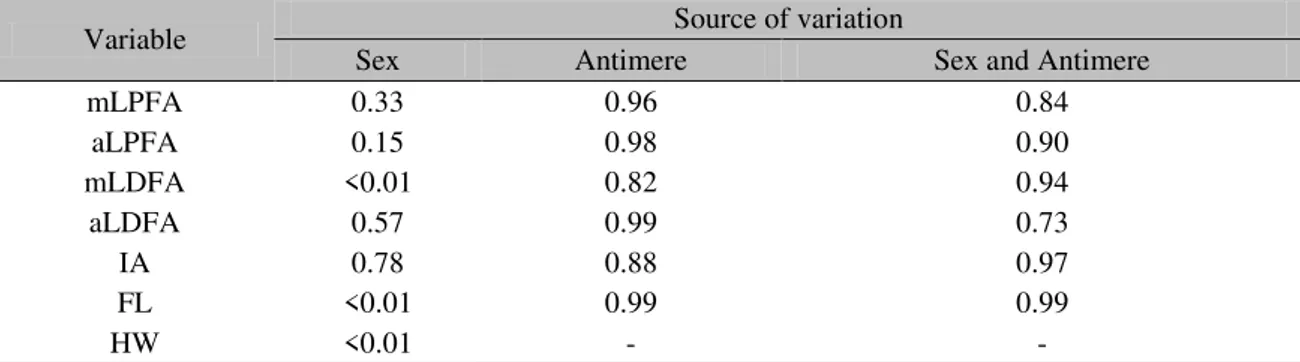
Analysis of variance (ANOVA) was performed for variables mLPFA, aLPFA, mLDFA, aLDFA, IA and FL according to a completely randomized design (CRD), in a 2 (sex: male and female) 2 (antimere: right and left) factorial arrangement using 92 cats (45 males and 47 females). Accordingly, a linear model considering the fixed effects of sex, antimere and their interactions were considered in the model. For the variable HW, ANOVA was also performed according to a CRD, but considering in the model only the fixed effect of sex. To describe the behavior of the data, a descriptive analysis was also performed according to the result obtained in ANOVA.
The variance and descriptive analysis of the data were respectively conducted using the MIXED and MEANS procedures of the Statistical Analysis System software (SAS, 2008). A probability of 5% was adopted for the F test to consider a significant effect in ANOVA.
Figure 3. Bilateral ventrodorsal radiogra
Figure 3. Bilateral ventrodorsal radiograph of hip and femur. Left: indicating the location of the IA (femoral inclination angle), formed by the intersection of the anatomical axis (AB) and a line originating in the center of the femoral head bisecting the femoral neck (TU). Right: indicating the measurement of the femoral length (FL), proximally from the midpoint of the femoral neck to the femoral intercondylar fossa, distally; (HW), between the dorsal margins of the acetabulum (points G and H).
RESULTS
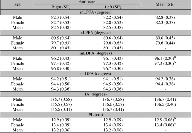
Regarding sex, statistical differences were detected for mLDFA, FL and HW measurements (F test <0.01) (Table 2 and 3), and no significant difference was observed when correlating sex and antimere (Table 3). Also, no differences were found in relation to antimere for any other variable (Table 2 and 3). However, mLDFA was
significantly higher in females (97.3±0.30º) than in males (96.1±0.30º) (Table 2). Male cats had a lower mean FL (12.9±0.06cm) compared to females (13.4±0.06cm) (Table 2), while HW in females (3.5±0.04cm) was greater than in males (3.1±0.04cm).
Table 1. Mean, median, standard deviation (SD), coefficient of variation (CV), and minimum and maximum values of the variables mLPFA, aLPFA, aLDFA and IA in the hip of cats, independent of sex and antimere
Variable N Mean
(degrees) Median (degrees) SD CV (%) Minimum Maximum mLPFA 184 82.5 82.0 3.62 4.4 74.0 93.0
aLPFA 184 80.1 81.0 4.29 5.4 69.0 99.0 aLDFA 184 94.3 95.0 3.43 3.6 83.0 101.0
IA 184 136.6 137.0 3.86 2.8 129.0 147.0 mLPFA = mechanical lateral proximal femoral angle; aLPFA = anatomical lateral proximal femoral angle; aLDFA = anatomical lateral distal femoral angle; mLDFA = mechanical lateral distal femoral angle; IA = femoral inclination angle; N = number of limbs evaluated.
Table 2. Influence of sex on the variables mLPFA, aLPFA, mLDFA, aLPFA, IA and FL in cats
Sex Antimere Mean (SE)
Right (SE) Left (SE) mLPFA (degrees)
Male 82.3 (0.54) 82.2 (0.54) 82.8 (0.37) Female 82.7 (0.53) 82.8 (0.53) 82.3 (0.38)
Mean 82.5 (0.38) 82.5 (0.38) -
aLPFA (degrees)
Male 80.5 (0.64) 80.6 (0.64) 80.6 (0.45) Female 79.7 (0.63) 79.6 (0.63) 79.6 (0.44)
Mean 80.1 (0.45) 80.1 (0.45) -
mLDFA (degrees)
Male 96.2 (0.43) 96.1 (0.43) 96.1 (0.30)B Female 97.4 (0.42) 97.3 (0.42) 97.3 (0.30)A
Mean 96.8 (0.30) 96.7 (0.30) -
aLDFA (degrees)
Male 94.2 (0.51) 94.1 (0.51) 94.2 (0.36) Female 94.4 (0.50) 94.5 (0.50) 94.4 (0.36)
Mean 94.3 (0.36) 94.3 (0.36) -
IA (degrees)
Male 136.7 (0.58) 136.7 (0.58) 136.7 (0.41) Female 136.5 (0.57) 136.6 (0.57) 136.5 (0.40)
Mean 136.6 (0.41) 136.7 (0.41) -
FL (cm)
Male 12.9 (0.09) 12.9 (0.09) 12.9 (0.06)B Female 13.4 (0.09) 13.4 (0.09) 13.4 (0.06)A
Mean 13.2 (0.06) 13.2 (0.06) -
Table 3. Probability values for F test of analysis of variance applied to the variables mLPFA, aLPFA, mLDFA, aLPFA, IA, FL and HW in cats
Variable Source of variation
Sex Antimere Sex and Antimere
mLPFA 0.33 0.96 0.84
aLPFA 0.15 0.98 0.90
mLDFA <0.01 0.82 0.94
aLDFA 0.57 0.99 0.73
IA 0.78 0.88 0.97
FL <0.01 0.99 0.99
HW <0.01 - -
mLPFA = mechanical lateral proximal femoral angle; aLPFA = anatomical lateral proximal femoral angle; mLDFA = mechanical lateral distal femoral angle; aLDFA = anatomical lateral distal femoral angle; IS = femoral inclination angle; FL = femur length; HW = hip width.
Table 4. Mean, median, standard deviation (SD), coefficient of variation (CV), and minimum and maximum values of the variables mLDFA, FL and HW, according to sex of cats, independent of antimere studied
Sex N Mean Median SD CV (%) Minimum Maximum mLDFA (degrees)
Male 90 96.1 96.0 3.51 3.7 79.0 102.0 Female 94 97.3 97.0 2.05 2.1 92.0 102.0
CF (cm)
Male 90 12.9 13.0 0.55 4.2 11.8 14.1 Female 94 13.4 13.6 0.66 4.9 12.0 14.7
LQ (cm)
Male 45 3.1 3.1 0.23 7.4 2.4 3.9
Female 47 3.5 3.5 0.26 7.4 2.9 3.9
mLDFA = mechanical lateral distal femoral angle; N = number of limbs evaluated; FL = femur length; HW = hip width.
DISCUSSION
The increasing population of domestic cats continue to generate the need for new knowledge about the species and the diseases that can affect them. In this sense, obtaining angle reference values directly contributes to the improvement of the diagnosis of osteoarticular diseases and the planning of orthopedic corrections, as determined for dogs (Soparat et al., 2012). These values provide information about the alignment of the pelvic limbs of this species, allowing better decision making for treatment, mainly because they were obtained in a population of clinically and radiographically normal cats.
A pilot study was carried out with 10 cadavers to determine the positioning of the body and the radiographic collimation to be applied in the research. Tomlinson et al. (2007) recommend
that collimation for radiological studies of the hip and femur in dogs be performed with the focus of the X-ray beam on the femur, and Swanson et al. (2012) reproduced the method in cats. However, the conclusion of the pilot showed that the small femoral muscular thickness in cats, compared to the hip, makes it impossible to obtain a radiographic image of acceptable quality when performed in this way. Radiographs were then performed with collimation of the focus of the beam on the hip between the two wings of the ilium at the same height up to the proximal aspect of the fibulae.
that reported by Swanson et al. (2012), and one explanation for this is the possible cadaveric rigidity in the animals used by the authors, as opposed to the greater muscular flexibility observed in the animals studied here, since they were radiographed immediately after euthanasia. Cadaveric rigidity causes muscle retraction, and it is not possible to impart any passive movement to the joints, after an initial period of relaxation and flaccidity of the entire musculature following death (Stevens, 2002). In this way, the active contraction of the musculature before rigor mortis can modify the radiographic positioning (Corley, 1992). As this type of alteration was not the focus of this work it opens a possibility for the future investigation on the differences in radiological study between cadavers versus living or recently dead animals.
The females in this study showed a higher mean for FL, since they were larger than the males in the sample analyzed. Despite this, no positive correlation was found between FL and other variables, suggesting that femur size does not alter the femoral reference angles. However, the angle of femoral anteversion should be studied, since Santanu et. al. (2014) found a moderate correlation between the size of humans and the angle of femoral anteversion. This possible relation in cats could explain the presence of degenerative arthropathies such as hip dysplasia in large cats, for example, deserving research attention.
At the birth of the animal, the hip is undifferentiated with respect to sex, but as it develops, hormonal interactions linked to 17β-estradiol and progesterone, influence the growth of bones, ligaments, joint capsule and muscle mass, producing forces that result in the ability to move the femoral head, moving it on its dorsal and lateral axis, out of the acetabular cavity (Corley, 1992). This is due to the fact that in this study, since X-rayed females in the study were not spayed, the mean HW was higher than that of males, probably due to these physiological specificities (Morgan et al., 2000).
In the present study, no significant difference was observed in relation to the antimeres, for none of the variables examined. Therefore, the contralateral antimere can be used for surgical planning of corrections of angular deformities or
fractures involving femoral alignment (Swanson et al., 2012).
The results of this work showed that the mean IA for the cats studied was 136.7±0.41º (males) and 136.5±0.40º (females), therefore far from the means found in reports of this angle for dogs, respectively 140º and 150º. This angle represents a fundamental component in the result of postoperative forces and in the kinematics of total replacement of the coxofemoral joint (Schulz; Dejardin, 2003).
The coxofemoral prostheses available commercially for use in veterinary medicine, especially in animals of less than 10kg body weight and therefore in cats, are microprostheses that have a total length of around 5cm (Liska et al., 2009). These implants are made with an inclination angle between 137º and 156º, depending on the brand chosen by the surgeon, complying with the femoral anatomy of the dog, according to Hauptman et al., (1985). However, the mean IA in cats was 136.7±0.41º (males) and 136.5±0.40º (females), therefore with a difference range of 0.4º to 19.4º, which may lead to implant failure. Liska et al., (2009) describes that failure of implants in total hip replacement surgeries is up to 20% in dogs, apparently at odds with this new information. Thus, additional postoperative studies are required in cats to determine the real possibility of such occurrence.
The aLDFA values found here (94.3±0.36º) are very similar to the values reported by Soparat et al. (2012) (95.21±3.48º), working with Pomeranians. On the other hand, the feline mLDFA values (96.7±0.30º) were significantly lower than in these dogs (99.46±4º). It is possible to infer, therefore, that there is a similarity between the small breed of dogs and the cats studied here; however, the difference in mLDFA between the species explains the lower prevalence of congenital medial dislocation in cats (Johnson, 1986).
mechanical axis may favor medial dislocation in female cats compared to males, as seen in dogs.
This work extends our understanding of the anatomy of femoral angles in the domestic cat and opens points for discussion on the impact of this knowledge on orthopedic afflictions in this species related to the hip and femur. In addition, it allows the planning of corrections of femoral angle deviations and the understanding of the occurrence of patellar dislocations. It was also possible to point out new research directions regarding the form of coxofemoral prostheses used in cats, relationships between patellar dislocation and reference angles in cats, and animal size and the presence of degenerative arthropathies.
CONCLUSION
The angles aLPFA, aLDFA, mLPFA, mLDFA and IA, HW and FL are measurements in ventrodorsal X-rays of cats, and the values obtained in this study provide reference values for application in surgeries and radiographic examinations.
REFERENCES
CORLEY, E.A. Role of the orthopaedic foundation for animals in the control of canine hip dysplasia. Vet. Clin. N. Am. Small Anim. Pract., v.22, p.579-593, 1992.
DENNY, H.R.; BUTTERWORTH, S.J. Cirurgia ortopédica em cães e gatos. 3.ed. São Paulo: Roca, 2006, 498p.
DISMUKES, D.I.; TOMLINSON, J.L.; FOX, D.B. et al. Radiographic measurement of the proximal and distal mechanical joint angles in the canine tibia. Vet. Surg., v.36, p.699-704, 2007.
FOX, D.B.; TOMLINSON, J.L.; BRESHEARS, L.B. Principles of uniapical and biapical radial deformity correction using dome osteotomies and the center of rotation of angulation methodology in dogs. Vet. Surg., v.35, p.67-77, 2006.
HARANSEN, G. Patellar luxation. Can. Vet. J. v.47, p.46-77, 2006.
HAUPTMAN, J., CARDINET, G.H., MORGAN, J.P. Angles of inclination and anteversion in hip dysplasia in the dog. Am. J. Vet. Res., v.46, p.2033-2036, 1985.
JOHNSON, M.E. Feline patellar luxation: a retrospective case study. J. Am. Anim. Hosp. Assoc., v.22, p.835-839, 1986.
KAWAKAMI, H.; NOBUHIKO, S.; TAKASHI, N. et al. 3D Analysis of the alignment of the lower extremity in high tibial osteotomy. Lect. Notes Comput. Sci., v.2489, p.261-267, 2002.
LISKA, W.D.; DOYLE, N.; MARCELLIN-LITTLE, D.J. et al. Total hip replacement in three cats: surgical technique, short-term outcome and comparison to femoral head ostectomy. Vet. Comp. Orthop. Traumatol., v.6, p.89-95, 2009.
MORGAN, J.P.; WIND, A.; DAVIDSON, A.P. Hereditary bone and joint disease in the dog. Hannover: Schlütersche, 2000. p.109-202.
PALEY, D., HERZENBERG, J.E. Principles of deformity correction. Heidelberg: Springer-Verlag, 2002. p.84-97.
SANTANU, B., PITBARAN, C., ANYNDYA, M. Correlation between neck shaft angle of femur with age and anthropometry: a radiographic study. Indian J. Basic Appl. Med. Res., v.3, p.100-107, 2014.
SAS. Statistical Analysis System. User’s guide: Statistics. Version 9.2 Edition. SAS Institute Inc., Cary, NC, 2008.
SCHULZ, K.S., DEJARDIN, L.M. Surgical treatment of canine hip dysplasia. In: SLATTER, D. Textbook of small animal surgery, 3.ed. Philadelphia: Elsevier, 2003. p.2029-2059.
SOPARAT, C.; WANGDEE, C.; CHUTHATEP, S. et al. Radiographic measurement for femoral varus in Pomeranian dogs with and without medial patellar luxation. Vet. Comp. Orthop. Traumatol., v.3, p.168-170, 2012.
STEVENS, L.J. Patologia. 2.ed. Manole: São Paulo, 2002. p.128-132.
SWANSON, E.A.; TOMLINSON, J.L.; DISMUKES, D.I. et al. Measurement of femoral and tibial joint reference angles and pelvic limb alignment in cats. Vet. Surg., v.1, p.1-9, 2012.