(Annals of the Brazilian Academy of Sciences)
Printed version ISSN 0001-3765 / Online version ISSN 1678-2690
www.scielo.br/aabc
http://dx.doi.org/10.1590/0001-3765201420140067
Attenuation of oxidative stress, inflammation and apoptosis by ethanolic and aqueous extracts of Crocus sativus L. stigma after chronic constriction injury of rats
BAHAREH AMIN1, KHALIL ABNOUS2, VAHIDEH MOTAMEDSHARIATY3 and HOSSEIN HOSSEINZADEH4
1Department of Pharmacology and Physiology, Cellular and Molecular Research Center, School of Medicine, Sabzevar University of Medical Sciences, 5714445994 Sabzevar, Iran
2
Department of Biotechnology, School of Pharmacy, Mashhad University of Medical Sciences, 1365-91775 Mashhad, Iran. 3
Pharmaceutical Research Center, Mashhad University of Medical Sciences, 1365-91775 Mashhad, Iran 4Pharmaceutical Research Center, Department of Pharmacodynamics and Toxicology, School of Pharmacy, Mashhad University of Medical Sciences, 1365-91775 Mashhad, Iran
Manuscript received on February 12, 2014; accepted for publication on June 30, 2014
ABSTRACT
In our previous study, the ethanolic and aqueous extracts of Crocus sativus elicited antinociceptive effects in the chronic constriction injury (CCI) model of neuropathic pain. In this study, we explored
anti-inflammatory, anti-oxidant and anti-apoptotic effects of such extracts in CCI animals. A total of 72
animals were divided as vehicle-treated CCI rats, sham group, CCI animals treated with the effective dose
of aqueous and ethanolic extracts (200 mg/kg, i.p.). The lumbar spinal cord levels of proinflammatory cytokines including tumor necrosis factor α (TNF-α), interleukin-1β (IL-1β) and interleukin 6 (IL-6), were
evaluated at days 3 and 7 after CCI (n=3, for each group). The apoptotic protein changes were evaluated
at days 3 and 7 by western blotting. Oxidative stress markers including malondialdehyde (MDA) and glutathione reduced (GSH), were measured on day 7 after CCI. Inflammatory cytokines levels increased in
CCI animals on days 3 and 7, which were suppressed by both extracts. The ratio of Bax/ Bcl2 was elevated on day 3 but not on day 7, in CCI animals as compared to sham operated animals and decreased following
treatment with both extracts at this time. Both extracts attenuated MDA and increased GSH levels in CCI
animals. It may be concluded that saffron alleviates neuropathic pain, at least in part, through attenuation
of proinflammatory cytokines, antioxidant activity and apoptotic pathways.
Key words: Crocus sativus, chronic constriction injury, inflammatory cytokines, oxidative stress, apoptosis.
Correspondence to: Hossein Hosseinzadeh E-mail: [email protected]
INTRODUCTION
For years, medicinal plants have been identified as
an important source of new chemical substances with potential therapeutic effects. Saffron, the dried stigmas of Crocus sativus L., (Iridaceae family),
has been used for coloring and flavoring in food preparations (Abdullaev 1993). Traditionally, this
plant has been used as a color and seasoning in food. Moreover, in folklore medicine, saffron has been used in the treatment of numerous illnesses including cough, asthma, menstruation problems, insomnia, pain relief, colic, chronic uterine hemorrhage,
cardiovascular disorders and tumors (Abdullaev and Espinosa-Aguirre 2004, Hosseinzadeh and Nassiri-Asl 2013). Scientific studies have verified
extracts and its active constituents, safranal and
crocin, have shown anti-cancer (Abe and Saito 2000,
Rastgoo et al. 2013), anti-oxidant (Hosseinzadeh et al. 2009), anti-convulsant (Hosseinzadeh et al. 2008), anti-depressant (Hosseinzadeh et al. 2003),
anti-anxiety (Hosseinzadeh and Noraei 2009) and
memory improvement properties (Hosseinzadeh and Ziaei 2006), in addition to antinociceptive and
anti-inflammatory properties in different kinds
of in vitro and in vivo studies (Hosseinzadeh and Younesi 2002, Hosseinzadeh and Shariaty 2007). Pain initiated or caused by a primary lesion or
dysfunction in the nervous system is defined as being
a neuropathic pain and triggers a cascade of events in the central or peripheral nervous system, including
excitotoxicity, oxidative stress, inflammatory process
and apoptotic like cell death mechanisms (Maione et al. 2002). In our previous work, antiallodynic and antihyperalgesic effects of ethanolic and aqueous extracts of saffron were shown in rats that underwent chronic constriction injury (CCI) of the sciatic nerve
(Amin and Hosseinzadeh 2012). However, the exact
mechanisms which underlie the antinociceptive effect
of this plant is still unknown. Anti-inflammatory
and antioxidant effects of saffron and its main constituents have been demonstrated in many investigations. The serum levels of prostaglandin
F2α, a marker of lipid peroxidation was significantly
decreased by crocin and safranal following subacute intoxication by an organophosphorus, diazinone. In addition, crocin, reduced serum tumor necrosis
factor α (TNF-α) level (Hariri et al. 2010). LPS-stimulated release of TNF-α, interleukin-1β (IL-1β), and intracellular reactive oxygen species
(ROS) were attenuated by crocin and crocetin in rat
brain microglial cells (Nam et al. 2010). Safranal
attenuated oxidative damage induced by an ischemia reperfusion injury (IRI) model in hippocampus of rats (Hosseinzadeh and Sadeghnia 2005). Radical scavenging activity of plant extracts and their bioactive consti tuents, crocin and safranal, was shown in DPPH (1,1-diphenyl-2-dipicrylhydrazyl)
radical scavenging test (Assimopoulou et al. 2005)
as well as microsomal lipid peroxidation induced
by Fe2+/ascorbat (Hosseinzadeh et al. 2009). In
this study crocin exhi bited more antioxidant effect than safranal moiety. Saffron extract and safranal showed cardioprotective effect by decreasing
lactate dehydrogenase (LDH) and creatine kinase
(CK-MB) and myocardial lipid peroxidation in isoproterenol-induced myocardial infarction in Wistar rats (Mehdizadeh et al. 2013). Crocin also prevented cytotoxicity induced by a potent neurotoxin, acrylamide as well as diazinone-induced hepatotoxicity in Wistar rats via decreasing Bax/Bcl2 ratio, as well as ROS generation inhibition (Mehri
et al. 2012, Lari et al. in press). Consequently, this
investigation was aimed at evaluating neuroprotective effects of both ethanolic and aqueous extracts of C. sativus by assessing time course of changes in the
proinflammatory factors (TNF-α, IL-1β and IL-6)
and apoptotic changes with measuring Bax/Bcl2 ratio in the spinal cord lumbar enlargement of 3 and 7
days-CCI animals. At the end of the experiment, the
alteration of oxidative damage markers including glutathione reduced (GSH), the major sulfhydryl (-SH) antioxidant (Cooper and Kristal 1997) and
malondialdehyde (MDA), the last product of lipid
peroxidation (Cheesman 1993) were also evaluated in the spinal cord lumbar section of animals.
MATERIALS AND METHODS ANIMALS
All experiments were performed on adult male
Wistar rats, weighing 220-260 g, randomly obtained
from the animal room of the Faculty of Pharmacy,
Mashhad University of Medical Sciences, Iran.
Animals were kept in a 12 h light–dark cycle
environment. Tap water and standard food pellets were available ad libitum. All procedures were approved by Mashhad University of Medical
MATERIALS
Stigma of C. sativus L. was purchased from Novin Saffron Co. (Mashhad, Iran) collected from Ghaen,
Khorasan province, Northeast of Iran and analyzed in accordance to ISO/TS 67321–2. Aqueous and
ethanolic extracts were extracted from saffron’s stigma in our laboratory. The saffron extracts were dissolved in normal saline (0.9%) immediately before injection and administered intraperitoneally once a day, from the day of surgery, for 7 consecutive days. Ketamine and xylazine were purchased from
Alfasan Co. (Woerden, Holland).
PREPARATION OF EXTRACTS
Ethanolic extract: 10 g of saffron powder was mixed with 25ml ethanol 80% at 0 °C and shaked
by vortex for 2 min. After centrifugation at 4000 g for
10 min, the supernatant was separated. Extraction by ethanol from sediment was repeated 6 other
times. As a result, the total volume of solvent con-sumption for 10 g of saffron stigmas was 200 mL (8×25mL). The resulting solution was dried in a
rotary evaporator system in darkness at 35 °C.
Aqueous extract: Saffron powder was mixed
with distilled water (1/50 W/V) and left on a shaking incubator at 8 °C for 48 h. The solution was centrifuged at 4000 g for 10 min. The resulting supernatant was retained and sediment was suspended in the half amount of mentioned distilled water and placed on the shaking incubator for another 24 h. The centrifugation was repeated again and the yielded supernatant was separated
and stored at −20 °C in a freezer. Sublimation of
solvent was performed by freeze drying.
CCISURGERY OF SCIATIC NERVE
Mononeuropathy was induced by performing chronic constriction injury model on the left sciatic nerve of animals as previously described (Bennett
and Xie 1988). At first, rats were anaesthetized
under a cocktail of ketamine and xylazine (64/1.6
mg/kg, i.p.). After the incision of the skin, the sciatic
nerve was exposed and four ligatures of 4-0 gauge chromic catgut were tied loosely with an interval of 1mm, until a slight twitching was observed in the
expected hind paw. Finally, muscle and skin were
separately sutured with 4-0 silk catgut and animals were placed in a warm condition until recovery.
STUDY PROTOCOL
Based on our previous study, CCI led to the
significant development of mechanical allodynia
(4.3 ± 0.6 g vs 53 ± 6.7 g) and cold allodynia (73.3 ± 8.4 % vs 8 ± 4.9 %) on day 3 in comparison
to sham group, as revealed by von Frey hairs
and acetone drop, respectively. Pain behaviors increased progressively during the study on days 5 and 7. In that study, the mechanical allodynia and
cold allodynia were significantly attenuated by the
aqueous (16 ± 2.4 g and 32.5 ± 3.4% respectively) and ethanolic extracts of saffron (32.8 ± 8/4 g and 17.5 ± 5.7%, respectively) at the dose of
200 mg/kg on day 3 (Amin and Hosseinzadeh
2012). In the present study, to examine the time
course of changes in proinflammatory cytokines
and apoptosis-related proteins in the spinal cord of CCI rats, three animals from each group were harvested on postoperative day 3, or 7 after the behavioral tests. In order to measure spinal cord
MDA or GSH, three animals from each group were sacrificed on postoperative day 7.
Hence, 72 animals were randomly assigned into the following groups:
Groups 1, 2: The animals were subjected to
CCI surgery, treated with normal saline (NS) at a
dose of 1 ml/kg and killed on day 3 for evaluation
of proinflammatory cytokines (n=3) and apoptotic
factors (n=3).
Groups 3, 4: Sham group: The animals underwent a similar surgery with the exception that the sciatic nerves were not ligated and they were treated with the normal saline and killed on
day 3 for evaluation of proinflammatory cytokines
CCI animals were treated with ethanolic extract of saffron (200 mg/kg, administered at a dose of 1 ml/kg) for seven days and killed on day 3 for
evaluation of proinflammatory cytokines (n=3) and
apoptotic factors (n=3). Groups 7, 8: CCI animals were treated with aqueous extracts of saffron (200 mg/kg, administered at a dose of 1 ml/kg) for seven days and killed on day 3 for evaluation of
proinflammatory cytokines (n=3) and apoptotic
factors (n=3). Groups 9-12: The CCI animals were
treated with NS, and killed on day 7 for evaluation of proinflammatory cytokines (n=3), apoptotic factors (n=3), MDA (n=3) and GSH (n=3).
Groups 13-16: Sham operated animals were killed on day 7 for evaluation of proinflammatory
cytokines (n=3), apoptotic factors (n=3), MDA
(n=3) and GSH (n=3). Groups 17-20: CCI animals were treated with ethanolic extract of saffron (200 mg/kg) for seven days and killed on day 7 for evaluation of proinflammatory
cytokines (n=3), apoptotic factors (n=3), MDA
(n=3) and GSH (n=3). Groups 21-24: CCI animals were treated with aqueous extracts of saffron (200 mg/kg) for seven days and killed on day 7 for evaluation of proinflammatory
cytokines (n=3), apoptotic factors (n=3), MDA
(n=3) and GSH (n=3).
For the protein extraction, the lumbar spinal
cord was rapidly ejected from the vertebral column
using a saline-filled syringe, as quickly as possible
and then separated on dry ice. The choice of
examining the L4 and L5 segments was based on
the consideration that these lumbar segments are the major contributors to the sciatic nerve (Bennett and Xie 1988).
TISSUE PREPARATION
Tissue samples were placed in the individual tubes and quickly frozen in liquid nitrogen and then
stored at –80 °C until usage. Determination of
protein content was performed by Bradford assay kit (BioRad) and adjusted (Bradford 1976).
DETECTION OF TNF-Α , IL-1Β AND IL-6, IN THE LUMBAR
SPINAL CORD BY ELISA
At the time of experiment, samples were
immedia-tely homogenized in the extraction buffer (20 mM
Tris (pH 7.4, Sigma-Aldrich), containing 150 mM NaCl Aldrich), 1 mM EDTA (Sigma-Aldrich), 2 mM 2-N-morpholinoethanesulfonic acid (2 ME) (Sigma-Aldrich) and one complete protease
inhibitor tablet (Roche, Mannheim, Germany) (Reece et al. 2004). Cytokine levels in the lumbar spinal cord of rats were measured according to the
manufacturer's instructions, specific for TNF-α , IL-1β and IL-6 (Koma Biotech, Korea). The content of
each sample was obtained according to the standard curve at 450 nm.
WESTERN BLOTTING
Western blot analysis of Bax and Bcl2 proteins, in the lumbar spinal cord
Samples of tissues were homogenized in the lysis buffer containing 50 mM Tris-HCl (pH:
7.4), 2 mM EDTA, 2 mM EGTA, 10 mM NaF, 1 mM sodium orthovanadate (Na3VO4), 10 mM β-glycerophosphate, 0.2% W/V sodium deoxy-cholate, 1 mM phenylmethylsulfonyl fluoride (PMSF), and complete protease inhibitor cocktail
(Roche, Mannheim, Germany). Homogenate was sonicated on ice with three 10-sec bursts at high intensity with a 10-sec cooling period between each burst and then centrifuged at 10,000g for 10 min at
4 °C. After determining protein content by Bradford
protein assay kit (BioRad) and adjusting the protein concentration, each sample was mixed 1:1 v:v with
2x SDS blue buffer. After boiling for 5 min, samples were aliquoted and stored at –80 °C in a freezer.
At the time of experiment, samples containing 50 μg of protein were loaded on a 12% sodium
dodecyl polyacrylamide gel and electrotrensferred
to polyvinylidene fluoride (PVDF) membranes. After transfer, nonspecific binding sites were
mM Tris-HCl pH 7.6, 137mM NaCl (TBS-T)
at 4 °C overnight. The probed membranes were then incubated with primary antibodies including: rabbit polyclonal anti-B-cell lymphoma 2 (Bcl2), rabbit polyclonal anti-Bcl-2-associated
X protein (Bax) and rabbit polyclonal anti-β-actin antibodies (Cell Signaling, 1: 1000) for 1–2 hours at room temperature. After three washes
with TBST, blots were incubated with rabbit horseradish peroxidase-conjugate anti-rabbit IgG (Cell Signaling, 1:2000) as a secondary antibody for 1 hour. Enhanced chemiluminescence (Pierce)
and Alliance 4.7 Gel doc (UK) were used to
visualize the peroxidase-coated bands. Protein
bands were densitometrically quantified using
UVtec software (UK). To compare the differences between control and treatment groups, the relative
optical density of each specific band to sham
group was normalized against the density of the
corresponding internal loading band, β-actin as a
control protein.
MEASUREMENT OF MDALEVELS IN THE LUMBAR
SPINAL CORD
Estimation of lipid peroxidation was performed by
measuring the Malondialdehyde (MDA) using the
thiobarbituric acid assay (Uchiyama and Mihara
1978). At the time of experiment, each sample was
weighed and a homogenate (10%) was prepared in
1.15% potassium chloride solution. At first, 3 ml of phosphoric acid (1%) and 1 ml of TBA (0.6%)
were added to 0.5 ml of homogenate and the mixture was heated for 45 min in a boiling water
bath. After cooling, 4 ml of n-butanol was added to
the mixture and vortex-mixed for 1 min followed by centrifugation at 3000 rpm for 15 minutes. The organic layer was transferred to a fresh tube and its
absorbance was read at 532 nm. A calibration curve was constructed using MDA (Sigma-Aldrich)
as standard. Protein content was determined by Bradford protein assay kit (BioRad) and the results were expressed as nmol/mg protein.
MEASURMENT OF GSHLEVELS IN THE LUMBAR SPINAL CORD
Total SH groups belonging to GSH were measured
using DTNB (2, 2´- dinitro-5, 5´-dithiodibenzoic acid) as the reagent. A 10% tissue homogenate
in buffer phosphate 7.4 was mixed with an equal
volume of 10% trichloro acetic acid (TCA) and
vortexed. The contents were then centrifuged at 5000 rpm for 10 min. Each 3.5 ml reaction mixture contained 0.5 of supernatant, 2.5 ml 0.1M phosphate
buffer (pH 8.4) and 0.5 ml DTNB. The absorbance
was measured at 412 nm using a spectrophotometer within 15 min. GSH was determined from a standard curve constructed using commercially
available standard GSH (Sigma-Aldrich). Protein
content was determined by Bradford protein assay
kit (BioRad). Levels of GSH were expressed as
nmol/mg protein (Moron et al. 1979).
STATISTICAL METHODS
Data were expressed as means±SEM for 3 rats in each group and statistically analyzed by one-way
ANOVA followed by Tukey's post hoc tests, using SPSS version 13. A P value of ˂0.05 was considered to be significant.
RESULTS
THE EFFECTS OF Crocus sativusEXTRACTS ON THE
PROINFLAMMATORY CYTOKINES
CCI resulted in a significant elevation of spinal cord TNF-α especially on day 3 (P < 0.001) as well as on day 7 after CCI (P < 0.01) in comparison to the sham group (Fig. 1A). IL- 1β also significantly
increased on days 3 and 7 after CCI in comparison
to the sham group (P < 0.01, Fig. 1B). IL-6,
however, with a delay (no difference on day 3)
showed a significant increase only on the 7th day
post-CCI in comparison to the sham operated
rats (P < 0.001, Fig. 1C). As shown in fig. 1A, the contents of TNF-α was significantly lower on days 3 (P < 0.05) and 7 (P < 0.05) in animals
mg/kg of both extracts. The contents of IL-1β was significantly lower on days 3 (P < 0.05) and 7 (P < 0.05) in animals treated with intraperitoneal administration of ethanolic extract (Fig. 1B).
Co-treatment with aqueous extract (200 mg/kg) for
seven days attenuated the levels of IL-1β on day 3 (P < 0.01) and day 7 (P < 0.05) after CCI (Fig.
1B). Seven days treatment with extracts attenuated
spinal cord IL-6 in comparison to saline treated CCI animals (P < 0.05, Fig.1 C).
THE EFFECTS OF Crocus sativusEXTRACTS ON THE MDA AND
GSHLEVELS
MDA and GSH were measured one week after surgery. As shown in Table I, a significantly higher spinal cord MDA level was observed in the CCI
group treated by normal saline relative to sham
animals (P < 0.01). CCI caused a significant
decrease in the amount of GSH of spinal cord of
animals, in comparison to the sham group (P < 0.05) (Table I). MDA levels were significantly attenuated following the 7 days treatment of ethanolic (P < 0.01) and aqueous extracts of saffron (P < 0.05). As indicated in Table I, lumbar spinal cord contents
of GSH was restored by co-treatment of aqueous
and ethanolic extracts of saffron (P < 0.05).
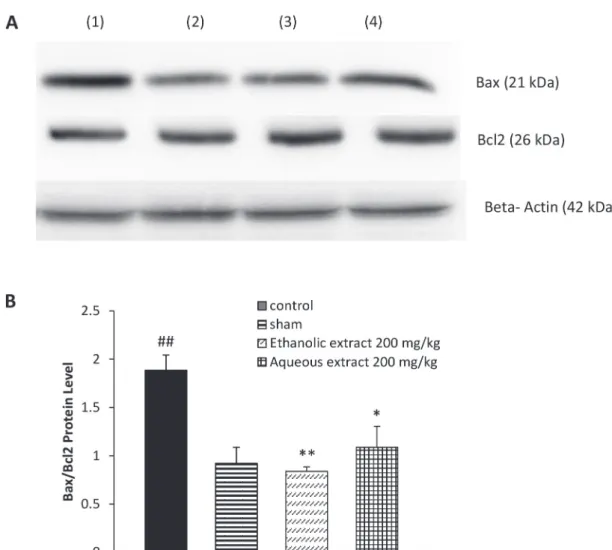
proapoptotic protein, Bax, increased within 3 days following sciatic nerve CCI, in comparison to the
sham group (P < 0.01). A slight and not significant
change was observed in the anti-apoptotic protein
Bcl-2 (Fig. 2A). As shown in Fig. 2B, a significant
raise was observed in the Bax/Bcl2 ratio of the CCI animals on day 3 post-CCI. CCI animals treated with 200 mg/kg of both ethanolic and aqueous
extracts of saffron, for 7 days, showed a significant
decrease in the Bax level on the day 3 post CCI,
with a slight but not significant increase in the anti-apoptotic protein Bcl-2, resulting in a significant
reduction in the Bax/Bcl2 ratio in comparison
to the control group (P < 0.01 and P < 0.05,
respectively). The levels of Bax protein decreased
in the spinal cord of CCI-NS animals on day 7 after
CCI, whereas the Bcl2 increased; consequently, no
significant differences were observed in the Bax/
Bcl2 ratio among control, sham group and animals treated with the extracts (data not shown).
DISCUSSION
Through our previous study, antiallodynia and antihyperalgesia effects of ethanolic and aqueous extracts of C. sativus (200 mg/kg, i.p.) for seven days were demonstrated in sciatic nerve chronic cons-triction injury of rats. In that study, threshold of pain in sham-operated animals was very similar to that
observed in naive group (Amin and Hosseinzadeh
2012) which is consistent with many studies (Decostered and Woolf 2000, Urban et al. 2013).
Today many investigations have focused on
the role of neuroinflammation in the pathogenesis
of neuropathic pain which could contribute to cell death (Kawasaki et al. 2008, Jancalek et al.
2010). Agents that suppress inflammatory cytokine elevation including TNF-α, IL-I β and IL-6, have
been advocated to be useful for the treatment of neuropathic pain (Kandhare et al. 2012). Therefore, in the present study, we evaluated potential
anti-apoptotic, anti-inflammatory and antioxidative
effects of C. sativus in the lumbar spinal cord of Sham
group
Control group (CCI rats with NS)
CCI- Ethanolic
extract
CCI- Aqueous
extract
MDA (nmol/g protein) 2.98 ± 0.1 10.72 ± 1.3## 4.84 ± 0.5** 4.99 ± 0.9*
GSH (nmol/g protein) 63.14± 4.14
36.6 ± 7.7#
60.2 ± 5.46*
62.08 ± 5.8*
TABLE I
The effect of aqueous extract (200 mg/kg) and ethanolic extract (200 mg/kg) of Crocus sativus
on the lumbar spinal cord levels of MDA and GSH levels in 7 days CCI rats.
Administration of extracts was started from the day of surgery and continued for seven consecutive days. CCI caused a significant increase in the spinal cord MDA contents, which was accompanied by a significant decrease in GSH level in comparison to sham operated animals. Data are presented as mean±SEM for n=3/group. One-way ANOVA followed by Tukey’s post-hoc test was used for multiple comparisons. *P < 0.05 and **P < 0.01 indicate a statistically significant difference when compared to CCI-NS treated rats; # P < 0.05, ## P < 0.01: Difference between CCI-NS and sham group.
THE EFFECTS OF Crocus sativusEXTRACTS ON THE BAX AND
BCL2PROTEIN LEVELS
CCI animals. CCI accompanied by a significant increase in the spinal cord levels of TNF-α and IL-1β, which were more prominent on day 3 but
remained high on day 7 post CCI. This result was consistent with the study of Kuang et al. 2012.
However, IL-6 (with a delay) peaked on day 7 post CCI. A slow release of IL-6 may be attributed to a more important role of this inflammatory
cytokine in the maintenance phase of neuropathic
pain or involvement of different mechanisms for producing proinflammatory cytokines. Induction
of IL-6 via TNF-α receptor 1 has been reported recently in a model of neuropathic pain (Lee et al. 2009). A week of daily administration of 200 mg/kg aqueous and ethanolic extracts significantly attenuated the contents of IL-1β, IL-6, and to a lesser extent, TNF-α, in the spinal cord of animals
subjected to nerve injury.
Figure 2 - The effect of aqueous extract (200 mg/kg) and ethanolic extract (200 mg/kg) of Crocus sativus on the spinal cord protein levels of Bax (21 kDa) and Bcl2 (26 kDa) following western blotting (A) and relative density of Bax/ Bcl2 (B), 3 days after CCI. Administration of extracts was started from the day of surgery and continued for seven
The balance between Bax, a pro-apoptotic protein, and Bcl-2, an antiapoptotic protein, stabi-lizing membrane permeability is very important in apoptosis induction (Oltvai et al. 1993). Our western blot data showed that Bax/Bcl-2 ratio was elevated following CCI, supporting evidence that in the CCI of sciatic nerve, the development of neuropathic pain may be associated with the activation of apoptosis (Siniscalco et al. 2007, Maione et al. 2002). Bax/Bcl2 ratio declined in the spinal cord of rats treated with saffron’s extracts on day 3.
However, depending on the time point of study, there were differences in the pattern of apoptosis occurrence. Bcl2 was gradually elevated
in CCI animals by day 7, as a result no significant
difference was observed among groups. It seems that as a modulatory mechanism, apoptotic process,
via mitochondria, is limited to the first few days
after nerve injury. In agreement with this, an early apoptosis (2-3 days post-CCI), occurred transiently by the increased ratio of bax/bcl-2 gene in a study
by de Novellis and co-workers (De Novellis et al. 2004). Authors in that study found an inversed
pattern of bcl-2 family genes expression at later
stages. Thus, there was a significant lowering in bax/bcl-2 and bcl-Xs/bcl-xL ratios over time
as a consequence of increased expression of
antiapoptotic bcl-2 and bcl-xL. The increased ratio
between pro- and antiapoptotic gene bax/bcl-2 expression in the spinal cord of neuropathic rats
was also limited to the first few days following
nerve injury (Costa et al. 2006). However, it might be possible that apoptosis process will be activated again, after the time course of our study. Hence taking more stage samples, for example days 10 or 14 could help better characterize time course of apoptotic factors activation.
CCI- induced neuropathy was associated with
the elevated levels of MDA and decreased levels
of GSH in the spinal cord of 7 days CCI rats which is in line with previous studies, suggesting the contributory role of oxidative stress in neuropathic
pain (Siniscalco et al. 2007). Seven days treatment of CCI animals with both ethanolic and aqueous
extracts exhibited decreased the levels of MDA and
restored the contents of GSH, as compared with vehicle treated control group.
Our results in this study confirm the complexity
of underlying mechanisms in the neuropathic pain
(Nickel et al. 2012). It seems that apoptosis is an
early rapid event following the induction of injury,
while contents of proinflammatory cytokines
and oxidative stress markers contribute to both induction and maintenance of neuropathic pain.
Beneficial neuroprotective effects of ethanolic
and aqueous extracts of saffron could be attributed to their active ingredients including polar carotenoids (crocins) that are glucosyl esters of crocetin (a polyene dicarboxylic acid), monoterpene aldehydes, like picocrocin (a glycosidic precursor of safranal) and safranal (Rezaee and Hosseinzadeh 2013,
Alavizadeh and Hosseinzadeh 2013). Flavonoids
such as rutin, quercetin and kaempferol, phenolic compounds such as gallic acid and pyrogallol, as well as other minor terpenoids, alkaloids and saponin contents are also present in stigma of saffron (Moraga et al. 2009, Hosseinzadeh and Younesi 2002, Karimi et al. 2010).
The antioxidant and anti-inflammatory activities
of these compounds have been demonstrated in various studies (Karimi et al. 2010, Ishige et al. 2001).
Although the comparison of the chemical constituents
of aqueous and ethanolic of C. sativus was not determined in this study, based on our data as well as
previous behavioral study (Amin and Hosseinzadeh 2012), it seems that no significant difference exists
between the effects of the ethanolic extract to those of the aqueous extract. It is plausible that constituents of aqueous extract are more water hydrophilic/soluble ingredients such as crocins, with less permeability
to CNS. While, ingredients of ethanolic extract are
more oil soluble/hydrophobic including crocetin and safranal (Rezaee and Hosseinzadeh 2013), with more
However, there is some evidence that crocins may be able to penetrate to the blood-brain barrier. Intraperitoneal administration of crocin to
rats improved spatial memory deficit induced by
chronic cerebral hypoperfusion (Hosseinzadeh et al. 2012). In another study, intraperitoneal injection of crocin enhanced functional recovery after sciatic nerve crush injury in rats (Tamaddonfard et al. 2013). In fact, based on our results, we cannot be sure about the central or peripheral and the real mechanisms of these combinations. However, involvement of both peripheral and central mechanisms is susceptible which is supported previous studies (Bridges et al. 2001).
In summary, data from current study suggest that ethanolic and aqueous extracts-mediated antinociceptive effects of saffron (stigma of
C. sativus L.) could be at least in part through
the modulation of oxidative stress, release of
proinflammatory cytokines, as well as interfering
with mitochondrial apoptosis pathways in CCI rats.
Although, further studies examining the exact
constituents of this valuable plant and underlying mechanisms of saffron’s antinociceptive effect are needed.
ACKNOWLEDGMENTS
We are thankful to the Pharmaceutical Research Center and the Vice Chancellor of Research, Mashhad University of Medical Sciences for
financial support.
RESUMO
Em nosso estudo anterior, os extratos etanólico e aquoso de Crocus sativus provocaram efeitos antinociceptivos no modelo de lesão por constrição crônica (CCI) da
dor neuropática. Neste estudo, exploramos os efeitos anti-inflamatório, anti-oxidante e anti-apoptótico de tais
extratos em animais CCI. Um total de 72 animais foi dividido em ratos CCI tratados com veículo, grupo de
simulação, animais CCI tratados com a dose eficaz de
extratos aquoso e etanólico (200 mg / kg, ip). Os níveis
de citocinas pró-inflamatórias da medula espinhal lombar, incluindo o fator de necrose tumoral α (TNF-α), a interleucina-1β (IL-1β) e a interleucina 6 (IL-6), foram
avaliados nos dias 3 e 7 após a CCI (n = 3, para cada
grupo). As alterações de proteínas apoptóticas foram
avaliadas nos dias 3 e 7 por western blotting. Marcadores
de estresse oxidativo, incluindo malondialdeído (MDA)
e glutationa reduzida (GSH), foram medidos no dia
7 após a CCI. Os níveis de citocinas inflamatórias
aumentaram em animais CCI nos dias 3 e 7, o que
foi suprimido por ambos os extratos. A proporção
de Bax / Bcl2 estava elevada no dia 3, mas não no 7° dia, nos animais CCI em comparação com os animais com operação simulada (sham), e diminuíram após o tratamento com ambos os extratos neste momento.
Ambos os extratos atenuaram o MDA e aumentaram os
níveis de GSH em animais CCI. Pode-se concluir que o açafrão alivia a dor neuropática, pelo menos em parte,
por meio de atenuação de citocinas pró-inflamatórias,
atividade antioxidante e vias apoptóticas.
Palavras-chave: Crocus sativus, lesão por constrição
crônica, citocinas inflamatórias, estresse oxidativo,
apoptose.
REFERENCES
ABDULLAEV F. 1993. Biological effects of saffron. BioFactors
4: 83-86.
ABDULLAEV F AND ESPINOSA-AGUIRRE J. 2004. Biomedical properties of saffron and its potential use in cancer therapy and chemoprevention trials. Cancer Detect Prevent 28: 426-432.
ABE K AND SAITO H. 2000. Effects of saffron extract and its constituent crocin on learning behaviour and long-term potentiation. Phytother Res 14: 149-152.
ALAVIZADEH SH AND HOSSEINZADEH H. 2013. Bioactivity assessment and toxicity of crocin: A comprehensive review. Food Chem Toxicol 64: 65-80.
AMIN B AND HOSSEINZADEH H. 2012. Evaluation of aqueous and ethanolic extracts of saffron, Crocus sativus L., and its constituents, safranal and crocin in allodynia and hyperalgesia induced by chronic constriction injury model of neuropathic pain in rats. Fitoterapia 83: 888-895. ASSIMOPOULOU A, SINAKOS Z AND PAPAGEORGIOU V. 2005.
Radical scavenging activity of Crocus sativus L. extract and its bioactive constituents. Phytother Res 19: 997-1000. BENNET GJ AND XIE YK. 1988. A peripheral mononeuropathy
BRADFORD MM. 1976. A rapid and sensitive method for the
quantitation of microgram quantities of protein utilizing the principle of protein-dye binding. Anal Biochem 72: 248-254.
BRIDGES D, THOMPSON SW AND RICE AS. 2001. Mechanisms of neuropathic pain. Br J Anaesth 87: 12-26.
CHEESMAN KH. 1993. Lipid peroxidation in biological
systems. In: HALLIWELL B AND ARUOMA OI (Eds), DNA and Free Radicals, London: Ellis Horwood, p. 12-17.
COOPER AJ AND KRISTAL BS. 1997. Multiple roles of
glutathione in the central nervous system. Biol Chem 378: 793-802.
COSTA B, SINISCALCO D, TROVATO AE, COMELLI F, SOTGIU
ML, COLLEONI M, MAIONE S, ROSSI F AND GIAGNONI
G. 2006. AM404, an inhibitor of anandamide uptake,
prevents pain behaviour and modulates cytokine and apoptotic pathways in a rat model of neuropathic pain. Br J Pharmacol 148: 1022-1032.
DECOSTERED I AND WOOLF CJ. 2000. Spared nerve injury: an animal model of persistent peripheral neuropathic pain. Pain 87: 149-158.
DE NOVELLIS V, SINISCALCO D, GALDERISI U, FUCCIO C,
NOLANO M, SANTORO L, CASCINO A, ROTH KA, ROSSI
F AND MAIONE S. 2004. Blockade of glutamate mGlu5 receptors in a rat model of neuropathic pain prevents early over-expression of pro-apoptotic genes and morphological changes in dorsal horn lamina II. Neuropharmacology 46: 468-479.
HARIRI AT, MOALLEM SA, MAHMOUDI M, MEMAR B AND
HOSSEINZADEH H. 2010. Subacute effects of diazinon on biochemical indices and specific biomarkers in rats: protective effects of crocin and safranal. Food Chem Toxicol 48: 2803-2808.
HOSSEINZADEH H, KARIMI G AND NIAPOOR M. 2003.
Antidepressant effect of Crocus sativus L. stigma extracts and their constituents, crocin and safranal, in mice. ISHS Acta Horticulturae 650: 435-445.
HOSSEINZADEH H AND NASSIRI-ASLM. 2013. Avicenna's (Ibn
Sina) the canon of medicine and saffron (Crocus sativus): a review. Phytother Res 27:475-483.
HOSSEINZADEH H AND NORAEI NB. 2009. Anxiolytic and
hypnotic effect of Crocus sativus aqueous extract and its constituents, crocin and safranal, in mice. Phytother Res 23: 768-774.
HOSSEINZADEH H AND SADEGHNIA HR. 2005. Safranal, a constituent of Crocus sativus (saffron), attenuated cerebral ischemia induced oxidative damage in rat hippocampus. J Pharm Pharm Sci 8: 394-399.
HOSSEINZADEH H, SADEGHNIA HR, GHAENI FA,
MOTAMEDSHARIATY VS AND MOHAJERI SA. 2012. Effects of saffron (Crocus sativus L.) and its active constituent, crocin, on recognition and spatial memory after chronic cerebral hypoperfusion in rats. Phytother Res 26: 381-386. HOSSEINZADEH H, SADEGHNIA HR AND RAHIMI A. 2008.
Effect of safranal on extracellular hippocampal levels of glutamate and aspartate during kainic acid treatment in anesthetized rats. Planta Med 74: 1441-1445.
HOSSEINZADEH H, SHAMSAIE F AND MEHRI S. 2009.
Anti-oxidant activity of aqueous and ethanolic extracts of Crocus sativus L. stigma and its bioactive constituents, crocin and safranal. Pharmacogn Mag 5: 419-424. HOSSEINZADEH H AND SHARIATY V. 2007. Anti-nociceptive
effect of safranal, a constituent of Crocus sativus (saffron), in mice. Pharmacologyonline 2: 498-503.
HOSSEINZADEH H AND YOUNESI HM. 2002. Antinociceptive
and anti-inflammatory effects of Crocus sativus L. stigma and petal extracts in mice. BMC Pharmacol 2: 7-12. HOSSEINZADEH H AND ZIAEI T. 2006. Effects of Crocus sativus
stigma extract and its constituents, crocin and safranal, on intact memory and scopolamine-induced learning deficits in rats performing the morris water maze task. J Med Plants 5: 40-50.
ISHIGE K, SCHUBERT D AND SAGARA Y. 2001. Flavonoids
protect neuronal cells from oxidative stress by three distinct mechanisms. Free Radic Biol Med 30: 433-446. JANCALEK R, DUBOVY P, SVIZENSKA I AND KLUSAKOVA I.
2010. Bilateral changes of TNF alpha and IL-10 protein
in the lumbar and cervical dorsal root ganglia following a unilateral chronic constriction injury of the sciatic nerve. J Neuroinflammation 7: 11.
KANDHARE AD, RAYGUDE KS, GHOSH P, GHULE AE AND
BODHANKAR SL. 2012. Neuroprotective effect of naringin
by modulation of endogenous biomarkers in streptozotocin induced painful diabetic neuropathy. Fitoterapia 83: 650-659. KARIMI E, OSKOUEIAN E, HENDRA R AND JAAFAR HZ. 2010.
Evaluation of Crocus sativus L. stigma phenolic and flavonoid compounds and its antioxidant activity. Molecules 15: 6244-6256.
KAWASAKI Y, ZHANG L, CHENG JK AND JI RR. 2008. Cytokine mechanisms of central sensitization: distinct and overlapping role of interleukin-1β, interleukin-6, and tumor necrosis factor-α in regulating synaptic and neuronal activity in the superficial spinal cord. J Neurosci 28:5189-5194.
KUANG X, HUANG Y, GU HF, ZU XY, ZOU WY, SONG ZB AND
GUO QL. 2012. Effects of intrathecal epigallocatechin gallate, an inhibitor of Toll-like receptor 4, on chronic neuropathic pain in rats. Eur J Pharmacol 676: 51-56. LARI P, ABNOUS K, IMENSHAHIDI M, RASHEDINIA M, RAZAVI M
AND HOSSEINZADEH H. IN PRESS. Evaluation of diazinon-induced hepatotoxicity and protective effects of crocin. Toxicol Ind Health.
LEE KM, JEON SM AND CHO HJ. 2009. Tumor necrosis factor receptor 1 induces interleukin-6 upregulation through NF-kappaB in a rat neuropathic pain model. Eur J Pain 13: 794-806.
MAIONE S, SINISCALCO D, GALDERISI U, DE NOVELLIS V,
ULIANO R, DI BERNARDO G, BERRINO L, CASCINO A AND ROSSI F. 2002. Apoptotic genes expression in the
lumbar dorsal horn in a model of neuropathic pain in rat. Neuroreport 13: 101-106.
MEHDIZADEH R, PARIZADEH MR, KHOOEI AR, MEHRI S AND
MEHRI S, ABNOUS K, MOUSAVI SH, SHARIATY VM AND
HOSSEINZADEH H. 2012. Neuroprotective effect of crocin
on acrylamide-induced cytotoxicity in PC12 cells. Cell Mol Neurobiol 32: 227-235.
MORAGA AR, RAMBLA JL, AHRAZEM O, GRANELL A AND
GOMEZ-GOMEZ L. 2009. Metabolite and target transcript analyses during Crocus sativus stigma development. Phytochemistry 70: 1009-1016.
MORON MS, DEPIERRE JW AND MANNERVIK B. 1979. Levels
of glutathione, glutathione reductase and glutathione S-transferase activities in rat lung and liver. Biochim Biophys Acta 582: 67-78.
NAM KN ET AL. 2010. Anti-inflammatory effects of crocin and
crocetin in rat brain microglial cells. Eur J Pharmacol 648: 110-116.
NICKEL FT, SEIFERT F, LANZ S AND MAIHOFNER C. 2012.
Mechanisms of neuropathic pain. Eur Neuropsychophar-macol 22: 81-91.
OLTVAI ZN, MILLIMAN CL AND KORSMEYER SJ. 1993. Bcl-2 heterodimerizes in vivo with a conserved homolog, Bax, that accelerates programmed cell death. Cell 74: 609-619. RASTGOO M, HOSSEINZADEH H, ALAVIZADEH H, ABBASI A,
AYATI Z AND JAAFARI MR. 2013. Antitumor activity of
PEGylated nanoliposomes containing crocin in mice bearing C26 colon carcinoma. Planta Med 79: 447-451. REECE TB, OKONKWO DO, ELLMAN PI, WARREN PS, SMITH
RL, HAWKINS AS, LINDEN J, KRON IL, TRIBBLE CG AND
KERN JA. 2004. The evolution of ischemic spinal cord injury in function, cytoarchitecture, and inflammation and the effects of adenosine A2A receptor activation. J Thorac Cardiovasc Surg 128: 925-932.
REZAEE R AND HOSSEINZADEH H. 2013. Safranal: from an aromatic natural product to a rewarding pharmacological agent. Iran J Basic Med Sci 16: 12-26.
SINISCALCO D, FUCCIO C, GIORDANO C, FERRARACCIO F,
PALAZZO E, LUONGO L, ROSSI F, ROTH KA, MAIONE S AND
DE NOVELLIS V. 2007. Role of reactive oxygen species and spinal cord apoptotic genes in the development of neuropathic pain. Pharmacol Res 55: 158-166.
TAMADDONFARD E, FARSHID AA, AHMADIAN E AND
HAMIDHOSEYNI A. 2013. Crocin enhanced functional recovery after sciatic nerve crush injury in rats. Iran J Basic Med Sci 16: 83-90.
UCHIYAMA M AND MIHARA M. 1978. Determination of malonaldehyde precursor in tissues by thiobarbituric acid test. Anal Biochem 86: 271-278.
URBAN R, SCHERRER G, GOULDING EH, TECOTT LH AND
BASBAU AI. 2011. Behavioral indices of ongoing pain are largely unchanged in male mice with tissue or nerve injury-induced mechanical hypersensitivity. Pain 152: 990-1000.