Steroidogenic enzymes mRNA expression profile and steroids production in bovine theca
cells cultured
in vitro
and stimulated with angiotensin II
Perfil de expressão de RNAm de enzimas esteroidogênicas e produção de esteroides a partir de células da teca bovina cultivadas in vitro e estimuladas por Angiotensina II
Melânia Lazzari RigoI Andressa Minussi Pereira DauI Werner Giehl GlanznerI Manoel MartinsII
Renato ZanellaII Tiele Medianeira RizzettiII Fabio Vasconcellos ComimI, III
Paulo Bayard Dias GonçalvesI ISSN 0103-8478
ABSTRACT
The main objective of this study was to detect the steroidogenic effects of Ang II in bovine theca cells in vitro. Bovine theca cells were obtained from follicles (larger than 10mm of diameter) collected from a local abattoir and submitted to different treatments in a sequence of experiments. In experiment 1,
CYP17A1 mRNA profile was evaluated in LH- (10ng ml-1) and Ang
II-treated (0.1µM) theca cells. In experiment 2, a dose-response effect of Ang II (0.001; 0.1 e 10µM) plus insulin (100ng ml-1) and
LH (100ng ml-1) was evaluated on steroidogenesis of bovine theca cells. Experiment 3 explored the effects of saralasin (an antagonist of Ang II receptors) on steroid production and steroidogenic
enzymes regulation in theca cells. After 24 hours, culture media from experiments 2 and 3 was collected to evaluate testosterone and androstenedione levels by High-Performance Liquid
Chromatography. In parallel, mRNA levels of key steroidogenic
enzymes (HSD3B2, CYP11A1, CYP17A1) and STAR were assessed by RT-PCR. There was no difference in testosterone and
androstenedione production between treated and controls groups, as well as in mRNA levels of the evaluated genes. In conclusion, the results suggest that Ang II does not regulate steroidogenesis in bovine theca cells.
Key words: RAS, LH, steroidogenesis, ovary.
RESUMO
O objetivo deste trabalho foi verificar o efeito da
Angiotensina II (Ang II) sobre a esteroidogenese nas células da teca bovina, cultivadas in vitro. Para isso, células da teca bovina foram obtidas de folículos maiores que 10 mm de diâmetro de ovários oriundos de abatedouro e submetidas a diferentes tratamentos
em uma sequência de experimentos. No experimento 1, o perfil de
expressão do RNAm de CYP17A1 foi avaliado nas células da teca
em resposta ao LH (10ng ml-1) e/ou Ang II (0,1µM) em diferentes
momentos de tratamento. No experimento 2, foi investigado o efeito dose-resposta de Ang II (0,001; 0,1 e 10µM), acrescido de
insulina (100ng ml-1) e LH (100ng ̸ml) sobre a esteroidogênese nas células da teca bovina. O experimento 3 explorou os possíveis efeitos da Ang II por meio do tratamento de células da teca com
saralasina (antagonista dos receptores da Ang II). Após 24 horas, nos experimentos 2 e 3, o meio de cultura foi coletado e avaliado
quanto aos níveis de testosterona e androstenediona pela técnica
de HPLC. Em paralelo, a expressão gênica de enzimas-chave da esteroidogênese (HSD3B2, CYP11A1, CYP17A1) e STAR foi avaliada por qRT-PCR. Não se observou diferença na produção de
testosterona e androstenediona entre controle e grupos tratados, bem como, na expressão do RNAm para os genes estudados. Em conclusão, nossos resultados não demonstraram um papel da Ang II sobre a esteroidogenese nas células da teca bovina.
Palavra-chave: RAS, LH, esteroidogênese, ovário.
INTRODUCTION
Synthesis of androgens occurs in response of a LH peak (YOUNG & MCNEILLY, 2010). This process is initiated by the Steroidogenic acute regulatory protein (STAR), which transports cholesterol into mitochondria of theca cells. Inside the mitochondria, cholesterol is converted to pregnolone by the action of P450scc enzyme (CYP11A1). Pregnolone is transported out of the cytosol and is converted to progesterone by the enzyme
3β-hidroxiesteroide desidrogenase (HDS3B2).
Progesterone is converted to 17α-hidroxiprogesterone
ILaboratório de Biotecnologia e Reprodução Animal (BioRep), Universidade Federal de Santa Maria (UFSM) Santa Maria, RS, Brasil.
IILaboratório de Analise de Resíduos de Pesticidas, UFSM, Santa Maria, RS, Brasil.
IIIDepartamento de Clínica Médica, Curso de Medicina, UFSM, 97105-900, Santa Maria, RS, Brasil. E-mail:fabio.comim@bol.com.br.
by the enzyme 17α-hidroxilase (CYP17A1). The main hormone produced from 17α-hidroxiprogesterone
is androstenedione, which can be converted to testosterone by the isoenzyme 17b-hidroxisteroide
deidrogenase (HSD17B) in theca cells or to estrone by
aromatase (CYP19A1) in granulosa cells. In general, theca cells express the crucial enzymes responsible for the conversion of cholesterol to androgens, otherwise, the conversion of androgens to estradiol happens in granulosa cells.
Although there is a complex network of genes involved in steroidogenesis, the majority of them are not clearly elucidated in this process. There is evidence relating the Renin Angiotensin System (RAS) with steroidogenesis since 1995, when it was
first demonstrated that angiotensin II (Ang II) could
act on steroid production in both theca and granulosa
cells of rabbits (FERAL et al., 1995). In bovine, it has
also been shown that Ang II can affect steroidogenesis, mainly in granulosa cells.Ang II participates in the synthesis of estrogen through CYP19A1 during
follicular deviation (FERREIRA et al., 2011b).
However, it did not stimulate progesterone production and LH-induced STAR expression (PORTELA et al., 2011). In theca cells, there was no difference in
mRNA expression for CYP17A1, HSD3B2 and STAR
between dominant follicles treated with saralasin or
vehicle during follicular deviation (FERREIRA et al., 2011a). During the preovulatory period, variations in
AGTR2 mRNA levels were shown in theca but not in granulosa cells after the administration of analogue GnRH (SIQUEIRA et al., 2012). Nevertheless, it is still not clear if Ang II stimulates steroidogenesis in bovine theca cells treated with LH.
To better understand the role of Ang II on the regulation of steroidogenesis in theca cells, this study evaluated the function of Ang II alone
or in association with LH on the mRNA profile of
steroidogenic enzymes and steroid production. To achieve this objective, a validated in vitro theca cells
cultures system was used (SCHREIBER et al., 2012).
MATERIAL AND METHODS
All reagents were purchased from Sigma-Aldrich unless otherwise mentioned.
Cell Isolation, culture and treatment
Theca cells were obtained from ovaries of non-pregnant adult cows collected in a local slaughterhouse. The ovaries were transported in thermal compartments (30oC) with saline solution (NaCl 0.9%) supplemented with penicillin (100UI mL-1) and streptomycin (50µl mL-1). Theca cells were
harvested no longer than 3 hours after the collection of the ovaries. Cells were isolated from follicles larger than 10mm that were apparently healthy, i.e. those showing good vascularization, translucid follicular
fluid and absence of an active (>1cm) corpus luteum,
thus avoiding the use of atretic follicles. The internal
walls of the follicles were rinsed in PBS to remove
granulosa cells. The collected theca tissues were digested in a collagenase solution (1mg mL-1) for cell separation and the obtained cells were seeded in 60mm culture plates. Cells were cultured for 48 hours
in a basic medium: (DMEM/F12 supplemented with
10% fetal bovine serum (FBS), 1µg mL-1 transferrin,
1ng/mL selenium, 100UI mL-1 penicillin, 100µg mL-1 streptomycin and 2.5µg mL-1 amphotericin), at 38.5oC and 5% of CO
2 as described previously (COMIM et al., 2013; SCHREIBER et al., 2012). After this period, the cells used in experiment 1 were trypsinized and seeded (2x10⁵ cells well-1) in 4-well
culture plates (Thermo Scientific). In experiments 2
and 3, cells were seeded in 96-well plates (Corning) with a concentration of 3x10⁴ cells well-1 and cultured under the same culture conditions for 24 hours. The
cells were then treated in a final volume of 400µl
(experiment 1) and 150µl (experiments 2 and 3)
using the same basic medium in the absence of FBS.
In experiment 1, bovine theca cells were cultured for 30min, 1, 3, 6, 12 or 24 hours in the presence of Ang
II and LH to evaluate the mRNA expression profile of
extraction. All the experiments were performed in triplicate. The absence of CYP19A1 mRNA was used to verify the possible contamination with granulosa cells, and positive samples were eliminated from the study.
RNA extraction and quantification and reverse
transcription
Total RNA was obtained using Trizol® according manufactures instructions.
The quantification was performed using a spectrophotometer NanoDrop (ND1000 - Thermo Scientific) with a wavelength of 260nm. RNA integrity
was assessed by electrophoresis in a 1.2% agarose gel stained with ethidium bromide. Purity was assessed
through absorption rate OD260/OD280 and samples
showing a value inferior to 1.8 were discarded.
Total RNA (1μg) was treated with DNase (DNAse
Amplification Grade I - Invitrogen) at 37oC during
5min to digest any contaminating DNA. The reverse transcription reaction to cDNA was performed using
the iScript cDNA Synthesis Kit® (BioRad)according
tomanufactures instructions.
qRT-PCR
The real time PCR to assess mRNA levels was performed in the Step One Plus termocycler
(Applied Byossistems) using SYBR Green (Power SYBR Green PCR Master Mix – Life Technologies). After amplification, the melting curves were analyzed to verify the amplification of only one product. The
relative mRNA expression was calculated based
on the amplification of the reference gene GAPDH according to PFAFFL et al. (2001). The primers
used for the amplification of CYP17A1 (sense:
CCATCAGAGAAGTGCTCCGAAT and
anti-sense: GCCAATGCTGGAGTCAATGA), CYP11A1
(sense: CTTGCACCTTTCTGGCTAGG and
anti-sense: AAGGGGAAGAGGTAGGGTGA), HSD3B2
(sense: GCCCAACTCCTACAGGGAGAT and
anti-sense: TTCAGAGCCCACCCATTAGCT) and STAR
(sense: CCCAGCAGAAGGGTGTCATC and
anti-sense: TGCGAGAGGACCTGGTTGAT) were based
on previous studies (GASPERIN et al., 2012) and were synthetized by Invitrogen.
Hormonal dosage through UHPLC-MS/MS
The identification and quantification of
androstenedione and testosterone were performed using Ultra High Performance Liquid
Cromatography-Tandem Mass Spectrometry (UHPLC-MS/MS). For
extract preparation, 200µL samples were diluted in acetonitrile until reaching 500µL. This mixture was
homogenized by vortexing and injected in the UHPLC-MS/MS system. After that, an analytical solution (internal pattern) for androstenedione, testosterone and triphenylphosphate was added to the mixture to
reach a final concentration of 100ng L-1. This new
mixture was once again injected in to the system. The results were calculated through the Pattern Addition and the triphenylphosphate pattern was used to verify during chromatography assay. The linearity and the detection limits of each analytic were verify through analytic solutions with concentrations ranging from 50 to 5000ng L-1 injected in the UHPLC-MS/MS. Using the data obtained with dilutions, the calibration curves were obtained. The spectrophotometer was operated in the Selected Reaction Monitoring (SRM) mode, with two transitions for each analytic, one for
quantification and the second for confirmation. The
higher intensity transition corresponds to analytic
quantification and the second higher transition corresponds to confirmation. The ionization mode
used was Electrospray Ionization (ESI) in positive mode for androstenedione and testosterone, with column oven temperature at 40oC, pressure of 15000psi, capillary of 2.8kV, desolvation temperature at 500oC, gas flow 800L h-1, collision gas flow (argon) of 0,15mL min-1 and source temperature about 150oC. The mobile phase used was compose of aqueous solution (solvent A), 0.05% of ammonium hydroxide
and methanol (solvent B). A boiling gradient was used
with 0.150mL min-1 flow and an injection volume of 10µL and total running time of 5min.
Statistical analysis
Gene expression and hormonal synthesis data were analyzed comparing between treatments. The differences were tested by ANOVA and the differences found, tested by LSMEANS Student’s t between different treatments. All data were tested for normality and when necessary normalized through
data ranking. Data analyses were performed using
the JPM® software (SAS Institute). The results are expressed as mean±standard mean error and all the values were obtained from three individual and distinct replicates.
RESULTS AND DISCUSSION
(1995) demonstrated an increase in androstenedione concentration after treatment with insulin, in a dose response manner (1, 10 e 100ng ml-1), associated with LH (100ng/ml) in bovine theca cells cultured in
vitro for 24 hours. Because of that, in experiments
2 and 3, theca cells were treated with LH (100ng mL-1) and insulin (100ng mL-1). This experiment was performed to verify a possible stimulatory effect on mRNA expression of steroidogenic enzymes, as well as, on androstenedione and testosterone production
according to the methodology proposed by COMIM
et al. (2013) and SCHREIBER et al. (2012).
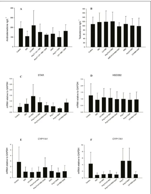
Angiotensin II, at the three different concentrations tested (10µM, 0.1µM e 0.01µM) in the presence of LH (100ng mL-1) and insulin (100ng
mL-1; Figure 2A-B), did not stimulate androstenedione
and testosterone production in bovine theca cells. Nevertheless, previous studies in rabbit have demonstrated that Ang II can stimulate testosterone production in theca cells cultured in vitro and treated
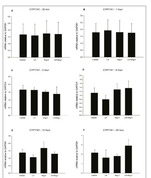
Figure 1 - Ang II effect on steroidogenesis in bovine theca cells cultured in vitro at different periods of treatment.
with hCG (FERAL et al., 1995). These differences are likely due to a specie-specific factors since the RAS
system is not uniform in mammals (GONÇALVES et al., 2012).In bovine, there was no effect of Ang II alone or in association with LH on androstenedione or testosterone synthesis in theca cells, neither on progesterone production induced by LH in granulosa
cells (PORTELA et al., 2011).Additionally, it was
observed a significant decrease in mRNA abundance
for the enzyme CYP19A1 in granulosa cells of dominant follicles after intrafollicular injection of
saralasin (FERREIRA et al., 2011a). Although it
seems that Ang II does not stimulate the synthesis of androstenedione and testosterone in theca cells, and
Figure 2 - Dose response effect of Ang II in steroidogenesis in bovine theca cells cultured in vitro, after 24
hours of treatment. Steroid measurements such as androstenedione (A) and testosterone (B) and mRNA expression for STAR (C), HSD3B2 (D), CYP11A1 (E) and CYP17A1 (F). The results are shown as
progesterone in granulosa cells, it might have a role in estrogen prodution in granulosa cells.
The theca cells analysis showed a
significant difference in CYP17A1 mRNA expression after 24 hours of treatment (Figure 2F). Similarly, mRNA levels for STAR, HSD3B2 and CYP11A1
tended to increase, but were not statistically different, in LH, insulin and Ang II treated groups in comparison
to control groups (Figure 2C-E). Even during
follicular deviation in the preovulatory period, Ang II seems not to affect mRNA expression ofCYP17A1
in bovine theca cells. Indeed, the intrafollicular injection of saralasin in the dominant follicle did not reduce CYP17A1 expression, which is in agreement
with findings in the present study showing that Ang II
do not increase CYP17A1 mRNA expression in LH-treated cells.
In the third experiment, the use of an antagonist of Ang II receptors in LH, insulin and Ang II-treated cells did not reduce androstenedione and testosterone production (Figure 3A-B). Similarly, there was no effect on mRNA expression for
Figure 3 - Effect of an Ang II receptor antagonist (Saralasin) over steroidogenesis in bovine theca cells
cultured in vitro, after 24 hours of treatment. Steroid measurements such as androstenedione
STAR (Figure 3C) and the steroidogenic enzymes
HDS3B2 (Figure 3D), CYP11A1 (Figure 3E) and
CYP17A1(Figure 3F) between different treatments. These results suggest that Ang II does not regulate steroidogenesis in bovine theca cells during the preovulatory period, such as in follicular deviation,
as first shown by FERREIRA et al. (2011a) in
bovine theca cells treat with saralasin. However, variances in the experimental conditions, such as the reproductive status and metabolism of the cows used for cell collection, might have accounted for the
non-homogenous findings.
In summary, findings from the
present study suggest that Ang II do not regulate steroidogenesis in theca cells, as evidenced by mRNA levels of genes encoding steroidogenic enzymes, as well as, steroid hormones production. Although a
significant decrease had been observed in CYP17A1
mRNA levels in treated cells compared to the control samples, there was no increase in androgen levels in response to Ang II or a decrease in response to
saralasin treatment. Besides of it well-characterized
roles and importance for oocyte maturation, ovulation
and follicular dynamics, Ang II seems not to influence
the production of androgens by in vitro theca cells.
CONCLUSION
Ang II alone or in association with LH do not stimulates steroidogenesis in bovine theca cells culture in vitro, at least under the conditions used in this study.
ACKNOWLEDGMENTS
The authors would like to thanks Frigorífico Silva
for the donation of bovine ovaries used in this research and to
Coordenação de Aperfeiçoamento de Pessoal de Nível Superior (CAPES), Conselho Nacional de Desenvolvimento Científico e Tecnológico (CNPq) and Fundação de Amparo à Pesquisa do Estado do Rio Grande do Sul (FAPERGS) for financial support.
REFERENCES
COMIM, F.V. et al. Adiponectin and its receptors in the
ovary: further evidence for a link between obesity and hyperandrogenism in polycystic ovary syndrome. PLoS One, v.8, n.11, p.e80416. 2013. Available from: <http://www.ncbi.nlm.
nih.gov/pubmed/24260388%3E>. Accessed: Jan. 15, 2014. doi:
10.1371/journal.pone.0080416.
FERAL, C. et al. Angiotensin II modulates steroidogenesis
in granulosa and theca in the rabbit ovary: its possible involvement in atresia. Eur J Endocrinol, v.133, n.6,
p.747-753, 1995. Available from: <http://www.ncbi.nlm.nih.gov/
pubmed/8548062%3E>. Accessed: Dec. 10, 2013.
FERREIRA, R. et al. Angiotensin II signaling promotes follicle
growth and dominance in cattle. Endocrinology, v.152, n.12, p.4957-4965, 2011a. Available from: <http://www.ncbi.nlm.nih.
gov/pubmed/22009728%3E>. Accessed: Nov. 13, 2013. doi:
10.1210/en.2011-1146.
FERREIRA, R. et al. Angiotensin II profile and mRNA encoding
RAS proteins during bovine follicular wave. J Renin Angiotensin
Aldosterone Syst, v.12, n.4, p.475-482, 2011b. Available
from: <http://www.ncbi.nlm.nih.gov/pubmed/21459786%3E>.
Accessed: Nov. 14, 2013. doi: 10.1177/1470320311403786.
GASPERIN, B.G. et al. FGF10 inhibits dominant follicle growth
and estradiol secretion in vivo in cattle. Reproduction, v.143, n.6, p.815-823, 2012. Available from: <http://www.ncbi.nlm. nih.gov/pubmed/22457435>. Accessed: Apr. 20, 2013. doi:
10.1530/REP-11-0483.
GONÇALVES, P.B. et al. Role of angiotensin in ovarian follicular
development and ovulation in mammals: a review of recent advances. Reproduction, v.143, n.1, p.11-20, 2012. Available from:
<http://www.ncbi.nlm.nih.gov/pubmed/22046052%3E>. Accessed:
Nov. 24, 2013. doi: 10.1530/REP-11-0192.
PFAFFL, M.W. A new mathematical model for relative
quantification in real-time RT-PCR. Nucleic Acids Res, v.29,
n.9, p.e45, 2001. Available from: <http://www.ncbi.nlm.nih.gov/ entrez/query.fcgi?cmd=Retrieve&db=PubMed&dopt=Citation&li
st_uids=11328886%3E>. Accessed: May 23, 2011.
PORTELA, V.M. et al. Role of angiotensin II in the periovulatory epidermal growth factor-like cascade in bovine granulosa cells in vitro. Biol Reprod, v.85, n.6, p.1167-1174, 2011. Available from:
<http://www.ncbi.nlm.nih.gov/pubmed/21849708%3E>. Accessed: Dec. 15, 2013. doi: 10.1095/biolreprod.111.094193.
SCHREIBER, N.B. et al. Expression and effect of fibroblast
growth factor 9 in bovine theca cells. J Endocrinol, v.215, n.1, p.167-175, 2012. Available from: <http://www.ncbi.nlm.nih.gov/
pubmed/22872763%3E>. Accessed: Nov. 10, 2013. doi: 10.1530/
JOE-12-0293.
SIQUEIRA, L.C. et al. Preovulatory changes in the angiotensin II system in bovine follicles. Reprod Fertil Dev, v.25, n.3, p.539-546, 2012. Available from: <http://www.ncbi.nlm.nih.gov/
pubmed/23464501>. Accessed: Oct. 28, 2013. doi: 10.1071/ RD11316 RD11316 [pii].
STEWART, R.E. et al. Effects of insulin-like growth factor I and
insulin on proliferation and on basal and luteinizing hormone-induced steroidogenesis of bovine thecal cells: involvement of glucose and receptors for insulin-like growth factor I and luteinizing hormone. J Anim Sci, v.73, n.12, p.3719-3731, 1995.
Available from: <http://www.ncbi.nlm.nih.gov/pubmed/8655449>.
Accessed: Oct. 28, 2010.
YOUNG, J.M.; MCNEILLY, A.S. Theca: the forgotten cell of the ovarian follicle. Reproduction, v.140, n.4, p.489-504, 2010. Available from: <http://www.ncbi.nlm.nih.gov/
pubmed/20628033>. Accessed: Oct. 28, 2010. doi: