Centro de Diagnóstico por Imagem – Hospital São Lucas da PUCRS Mailing address: Flavio José Petersen Velho – Av. Ipiranga, 6690/515 Cep 90610-000 – Porto Alegre, RS, Brazil - E-mail: [email protected] Received - 11/28/02
Accepted - 5/10/03
Arq Bras Cardiol, volume 82 (nº 1), 52-6, 2003
Flavio José Petersen Velho, Fernanda Dotta, Leonora Scherer, Eduardo Bartholomay, Daniela Augusta da Silva, Jefferson Gomes Fernandes, Marco Antonio Rodrigues Torres
Porto Alegre, RS - Brazil
Association Between the Effect of Spontaneous Contrast in the
Thoracic Aorta and Recent Ischemic Stroke Determined by
Transesophageal Echocardiography
Cerebrovascular diseases are important causes of mor-tality and disability. In Brazil, despite its medical-social im-portance as the first cause of death in the country, epide-miological studies are rare 1-3.
Cerebrovascular diseases may have many causes, and embolic events, presumably cardiac, are responsible for 20 to 40% of strokes 3.
The use of transesophageal echocardiography in the assessment of the heart as an embolic source has been wide-ly accepted because of its greater capacity for spatial reso-lution of basal cardiovascular structures, such as the left atrium, the left atrial apex, the interatrial septum, and the thoracic artery. Transesophageal echocardiography has enabled the diagnosis of other risk factors associated with an increased risk of stroke.
Thrombi, tumors, and intracavitary vegetations, rheu-matic valvular disease (especially mitral stenosis), left ven-tricle systolic dysfunction, left atrium spontaneous echo contrast, and the complex plaques in the proximal aorta are factors proved to be associated with an increased risk of stroke 4-8. Other factors, such as degenerative calcification
involving the mitral and the aortic valves; patent foramen ovale with right-left shunt; interatrial septal aneurysm; strands or Lambl’s excrescences in the aortic or mitral valve, or both valves; and, more recently, spontaneous echo con-trast in the aorta 6-9 have been described as potential risk
factors; however, some doubts still exist that will certainly be settled with future research in this area.
The meaning of the presence of spontaneous echo contrast in the aorta has been questioned, and recently it was linked to coronary artery disease and to ischemic events in general 10.
The objective of the study was to assess the indepen-dent association between the presence of echo contrast in the aorta and recent stroke events.
Methods
The study group of 224 individuals with a recent diag-nosis of stroke and the control group of 85 individuals un-Objective - To assess the independent association
between the presence of spontaneous echo contrast in the aorta and recent stroke events.
Methods - Two hundred and twenty-four individuals
with a diagnosis of recent stroke and 85 control indivi-duals who were examined due to various present/suspec-ted heart diseases were studied through transesophageal echocardiography. The effects of spontaneous contrast in the aorta and the presence of other potential sources of cardiac embolism associated with them were researched and a questionnaire was completed about patients’ clinical risk factors at the time of examination.
Results - The effects of contrast in the aorta was
asso-ciated with stroke (OR=2.83; CI = 95%, 1.65-4.46; P<0.001) in the bivariate analysis. In the multivariate ana-lysis, it remained associated with recent stroke (OR=2.05; CI = 90%, 1.10-3.85; P=0.06). Age ≥ 60 years, a history of systemic blood hypertension and smoking, and dyslipide-mia were risk factors independently associated with the ef-fects of contrast in the aorta. The presence of a spontaneous contrast effect in the left atrium and Lambl’s excrescences were echocardiographic factors independently associated with the effects of contrast in the aorta.
Conclusion - The effect of contrast in the aorta was
independently associated with recent stroke and with its clinical risk factors. These results reinforce the hypothesis that the phenomenon is a predictor of several risk factors.
5 3 5 3 5 3 5 3 5 3 derwent transesophageal echocardiography for several
present or suspected cardiac diseases.
The ethics committee of the institution approved the project, and all patients gave written informed consent.
The study group included individuals with a recent diagnosis of stroke (less than 30 days) treated from Novem-ber 1997 through NovemNovem-ber 1999 at the Serviço de Neurolo-gia do Hospital São Lucas da PUC (Neurology Service of the Hospital São Lucas from PUC), in the city of Porto Ale-gre. These patients underwent transesophageal echocar-diography to detect whether the source of emboli was the heart, the thoracic aorta, or both of these.
The control group comprised consecutive individuals studied through transesophageal echocardiography to diagnose various cardiac diseases.
Exclusion criteria for the study and control groups we-re a descending thoracic aorta diameter > 4 cm; documented thoracic aorta dissection; fractional shortening, obtained through M mode echocardiography, < 25% or, in cases of segmental systolic dysfunction, ejection fraction (obtained through bi-dimensional transthoracic echocardiography using Simpson’s method) of < 40%; definite disagreement on the diagnosis of the effects of contrast in the aorta bet-ween 2 echocardiographers; refusing to take part in the stu-dy; not signing written informed consent; and, only in the control group, a previous history of systemic or encephalic embolization.
Patients with complete ischemic stroke or transitory is-chemic attacks revealed with neurological evaluations were identified as ischemic stroke based on conventional clinical and tomographic criteria.
Diagnosis of the effects of contrast in the aorta was established when an echodense image was observed, moving with the cardiac cycle. When the echodense image observed did not move with the cardiac cycle as defined, we considered it as a technical artifact.
As a methodology, the patient or a relative answered a questionnaire about the patient’s current health status and about risk factors for ischemic stroke. All patients under-went transesophageal echocardiography performed by an experienced echocardiographer. Patients were sedated with midazolam before esophageal intubation, with doses ran-ging from 2 to 15 mg IV. Examinations were performed with XP10 from Acuson-USA, using a multiplanar esophageal transducer with a 5-MHz frequency. For diagnosis of patent foramen ovale, saline solution was injected into a peripheral vein in the study group patients. Examinations were recor-ded on VCR tapes and assessed by another echocardiogra-pher. The second observer was unaware of the diagnosis. In the cases where disagreement existed regarding the pre-sence or abpre-sence of contrast in the aorta, observers tried to reach a consensus, and when they could not, the patient was excluded from the study.
Initially, descriptive statistics were obtained for all study variables, including mean and standard deviation for the quan-titative and percentage values for the categorical variables.
To control the confusion effects, a multivariate analysis model was created using the logistic regression technique.
The evaluation of the association between ischemic stroke and the effects of contrast in the aorta was done using contingency tables, and relative risk estimations were obtai-ned using odds ratio (OR) with their respective confidence interval (CI), 95% for bivariate analysis and 90% for multiva-riate analysis. Statistical significance of the associations was determined using the chi-square test and Fisher’s exact test, when necessary. Similar techniques were used in the evalua-tion of the associaevalua-tion between the effects of contrast in the aorta and other factors considered in the study.
The most relevant variables, according to the literatu-re, were included in the mathematical model, regardless of the significance reached in the bivariate analysis.
We did not include all of the potential embolic sources detected through transesophageal echocardiography in the statistical analysis. Variables with an inexpressive pre-valence in the sample were not studied in the statistical ana-lysis. Patent foramen ovale was not included in the statisti-cal analysis because its diagnosis was performed, in the majority of cases, through isotonic saline containing micro-bubbles, which was not routinely performed in the patients from the control group. Mitral valve prolapse and mitral or aortic degeneration calcification were not included in the analysis, because these diagnoses are usually performed using transthoracic echocardiogram.
Statistical significance levels accepted were ∝ = 0.05, for bivariate analysis, and ∝ = 0.1, for multivariate analysis. Data were processed and analyzed with the SPSS software program (Statistical Package for the Social Sciences) version 6.0 for Windows.
Results
Of 355 individuals who underwent transesophageal echocardiography, 309 were included in the study, 224 in the study group, and 85 in the control group. Forty-six pa-tients were excluded from the study.
Age ranged from 15 to 85 years, with a mean of 62 ± 13 in the study group and 53.1 ± 18.1 in the control group. One hundred and seventy individuals were male.
Table I presents the occurrence of effects of contrast in the aorta and the frequency of clinical and echocardiogra-phic risk factors for ischemic stroke in both groups.
Contrast effects in the aorta were associated with ischemic stroke events (OR = 2.83) in the bivariate analysis (tab. II), which demonstrated an association between ische-mic strokes and systeische-mic blood hypertension (OR= 3.79), diabetes mellitus (OR = 2.96), dyslipidemia (OR = 2.77), and with age ≥ 60 years (OR = 2.54). Among the echocardiogra-phic findings, Lambl’s excrescences (OR = 6.50) and the pre-sence of complex plaques in the aorta (OR = 3.27) were asso-ciated with recent ischemic stroke events.
Multivariate analysis demonstrated that systemic blood hypertension was the only clinical risk factor (OR = 2.68) associated independently with ischemic stroke events, confirming the independent association between the complex plaques in the aorta and ischemic stroke events (OR = 2.92, CI 1.34-6.67, P=0.02). Lambl’s excrescence remained highly as-sociated with recent ischemic stroke events (OR = 6.93, CI = 90% 3.18-15.12, P<0.001) in the multivariate analysis.
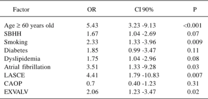
Regarding the multivariate analysis of the association between spontaneous echo contrast in the aorta and the presence of other risk factors for ischemic stroke, among the clinical variables, it was observed that age ≥ 60 years, syste-mic blood hypertension, smoking, and dyslipidemia were associated independently with echo contrast in the aorta. Among the echocardiographic variables, spontaneous echo contrast in the left atrium and Lambl’s excrescences re-mained associated independently with the effects of con-trast in the aorta (tab. IV).
Discussion
The spontaneous echo contrast in the left atrium in patients with atrial fibrillation is associated with greater risk of thromboembolism 5,11-13, and some evidence exists that
echo contrast in the aorta is associated with atherosclerotic coronary disease and with ischemic stroke events 10,14.
In our series, the echo contrast in the aorta had an in-creased prevalence, both in the study group and in the con-trol group (52.7% vs. 28.2%). In the bivariate analysis, an as-sociation between echo contrast in the aorta and recent is-chemic stroke events was demonstrated (OR = 2.83, CI =
95% 1.65 – 4.46, P < 0.001). Multivariate analysis confirms that this association is independent (OR = 2.05, CI = 90% 1.10 - 3.86, P = 0.06).
Although the presence of echo contrast in the aorta is attributed to the rouleau phenomenon observed in stagna-ting flows due to erythrocyte aggregation, other factors, such as anticardiolipin antibody, the increase in erythrocyte sedimentation rate, the increase in fibrinogen plasma levels, and plasma viscosity are also related to the phenomenon. These facts explain its presence even in the absence of stag-nating flows.
In our study, we excluded patients with diseases that certainly created stagnating flows in the thoracic artery, such as significant left ventricle systolic dysfunction, aortic dis-section, and aortic aneurysm. However, it may be possible that these factors involved in the erythrocyte aggregation (known or unknown) are mainly responsible for the appea-rance of contrast in the aorta in the study group. On the other hand, the increase in plasma fibrinogen, the increase in blood
Table II - Bivariate analysis
Study Control
Group Group
Factor f % f % RC CI 95% P
TASEC 118 52.7 24 28.2 2.83 1.65 - 4.46 <0.001 ≥ 60 years 138 63.9 34 41 2.54 1.52 - 4.28 <0.001 SBHH 151 67.4 30 35.3 3.79 2.24 - 6.41 <0.001 Smoking 65 29 17 20 1.63 0.98 - 2.99 0.11 DM 47 21 7 8.2 2.96 1.28 - 6.84 0.008 Dyslipidemia 70 31.3 12 14.1 2.77 1.41 - 5.41 0.002 Atrial 18 8 16 18.8 0.38 0.18 - 0.78 0.007 fibrillation
CAOP 68 30.4 10 11.8 3.27 1.59 - 6.71 <0.001 LASEC 28 12.5 13 15.3 0.79 0.38 - 1.16 0.52 EXVALV 74 33 6 7.1 6.50 2.71 -15.59 <0.001
TASCE- thoracic aorta spontaneous contrast effect; ≥ 60 years old - age equal to or greater than 60 years old; SBHH- systemic blood hypertension history; DM- diabetes mellitus history; CAOP- complex aortic plaque; LASCE- left atrium spontaneous contrast effect; EXVALV- Lambl’s excrescences in the mitral and/or aortic valve.
Table III - Multivariate analysis
Factor OR CI 90% P
TASEC 2.05 1.10 - 3.85 0.06
≥ 60 years 1.43 0.80 - 2.54 0.31
SBHH 2.68 1.61 - 4.44 0.001
Smoking 1.70 0.32 - 3.13 0.15
DM 1.52 0.67 - 3.45 0.40
Dyslipidemia 1.27 0.66 - 2.45 0.54 Atrial fibrillation 0.25 0.09 - 0.70 0.02
CAOP 2.92 1.34 - 6.67 0.02
LASCE 0.79 0.30 - 2.05 0.68
EXVALV 6.93 3.18 -15.12 <0.001
TASCE- thoracic aorta spontaneous contrast effect; ≥ 60 years old - age equal to or greater than 60 years old; SBHH- systemic blood hypertension history; DM- diabetes mellitus history; CAOP- complex aortic plaque; LASCE- left atrium spontaneous contrast effect; EXVALV- Lambl’s excrescences in the mitral and/or aortic valve.
Table I - Occurrence of spontaneous contrast in the aorta and of echocardiographic and clinical risk factors for ischemic
stroke events
Study Group Control Group
Factor f % f %
ASEC 118 52.7 24 28.2
Age ≥ 60 years 138 63.9 34 41
SBHH 151 67.4 30 35.3
Smoking 65 29 17 20
DM 47 21 7 8.2
Dyslipidemia 70 31.3 12 14.1
Atrial fibrillation 18 8 16 18.8
Vegetation 0 0 3 3.5
Myxoma 1 0.4 1 1.2
ANESIA 7 3.1 1 1.2
LA thrombi 11 4.9 1 1.2
LASCE 28 12.5 13 15.3
D P P 52 23.2 12 14.1
FOSH 56 25 5 5.9
CAOP 68 30.4 10 11.8
EXCVALV 74 33 6 7.1
5 5 5 5 5 5 5 5 5 5 viscosity, and the increase in erythrocyte sedimentation rate
are factors associated with heart diseases and vascular brain diseases 15-24. Their concomitant presence may explain the
as-sociation of echo contrast in the aorta with ischemic stroke events, even in the absence of stagnating flows.
Because the control group comprised nonvolunteer individuals with a suspicion of heart disease, some results of the study may have been influenced. The most frequently observed diseases in the control group were mitral stenosis, ischemic heart disease, atrial fibrillation, and infectious en-docarditis, which may be associated with stagnating flows and ischemic stroke events. The control group was formed because transesophageal echocardiography is an invasive investigation method that, although it has minimal risk, may cause discomfort, which prevented us from performing it in healthy individuals. It is very likely that, if the control group had comprised paired volunteers, the prevalence of echo contrast in the aorta would have been significantly lower and the association significantly more intense.
The results of the study demonstrate an intense and independent association between atrial fibrillation and echo contrast in the aorta. Atrial fibrillation is one of the main indications for transesophageal echocardiography,
both for risk stratification in chronic patients and as an addi-tional element after cardioversion, and it was present in 18.8% of the individuals in the control group, much more prevalent than the 8.0% of the study group. This fact was an important influence contrary to our hypothesis and was probably responsible for the numbers obtained in the biva-riate and multivabiva-riate analysis in which atrial fibrillation was a protective factor in ischemic stroke events. The results pertaining to atrial fibrillation were influenced by the forma-tion of the control group. The presence of atrial fibrillaforma-tion increases the risk of ischemic stroke 4 to 6 times 9,12,25-28. The
results regarding spontaneous echo contrast in the left atrium were also influenced by the composition of the con-trol group. The spontaneous echo contrast in the left atrium in this study appears to be a nonsignificant protective fac-tor for ischemic stroke events. This finding is in disagree-ment with that in the literature, and the reasons for that con-trast are the same found when we analyzed the association between atrial fibrillation and ischemic stroke events.
In our study, multivariate analysis did not confirm an independent association between the complex aortic pla-ques and the spontaneous echo contrast in the aorta. This finding, contrary to what was expected, was explained after the performance of several mathematical simulations, which verified that the complex aortic plaques were intensively and independently associated with the factor age ≥ 60 years (OR=8.4), and this factor was associated with the sponta-neous echo contrast in the aorta.
In conclusion, the echo contrast in the aorta is inde-pendently associated with recent ischemic stroke events. All clinical risk factors for ischemic stroke events evaluated were independently associated with the echo contrast in the aorta. These results together with the certainty of the in-dependent association between echo contrast in the aorta and ischemic stroke events, reinforce the hypothesis that the phenomenon is a marker of multiple risk factors. Among the echocardiographic variables, the presence of sponta-neous echo contrast in the left atrium and Lambl’s excres-cences were echocardiographic factors independently as-sociated with echo contrast in the aorta.
Table IV - Multivariate analysis of the association between aortic spontaneous contrast effects and the presence of other risk factors for
ischemic stroke events
Factor OR CI 90% P
Age ≥ 60 years old 5.43 3.23 -9.13 <0.001
SBHH 1.67 1.04 -2.69 0.07
Smoking 2.33 1.33 -3.96 0.009
Diabetes 1.85 0.99 -3.47 0.11
Dyslipidemia 1.75 1.04 -2.96 0.08
Atrial fibrillation 3.51 1.33 -9.28 0.03
LASCE 4.41 1.79 -10.83 0.007
CAOP 0.7 0.40 -1.23 0.31
EXVALV 2.06 1.23 -3.47 0.02
Age ≥ 60 years old – age equal to or superior than 60 years old; SBHH- systemic blood hypertension history; LASCE- left atrium spontaneous contrast effect; CAOP- complex aortic plaques; EXVALV- Lambl’s excrescence in the mitral and/or aortic valve.
1. Lessa I. Epidemiologia das doenças cerebrovascualres no Brasil. Rev Soc Cardiol Estado de São Paulo 1999; 9.
2. Fernandes J. Epidemiologia das doenças cerebrovasculares. Ass Med RGS 1989; 33:353-7.
3. Tatani S, Fukujima M, Lima J et al. Impacto clínico da ecocardiografia transesofá-gica em pacientes com acidente vascular cerebral sem evidência clínica de fonte emboligênica. Arq Bras Cardiol 2001;76:453-7.
4. Autuore C, Cartoni D, Piccininno M, et al. Multiplane tranesophageal echocar-diography and stroke. Am J Cardiol 1998;81(12A):79G-81G.
5. Zabalgoitia M, Hlaperin J, Pearce L, et al. Transesophageal echocardiographic correlates of clinical risk of thromboembolism in nonvalvular atrial fibrillation. J Am Coll Cardiol 1998;31:1622-6.
6. Khandheria B, Seward J, Tajik J, et al. Transesophageal echocardiography. Mayo Clin Proc 1994; 69: 856-63.
References
7. O’Brien, P. J. et al. Usefulness of transesophageal echocardiography in predic-ting mortality and morbidity in stroke patients without clinically known car-diac sources of embolus. Am J Cardiol 1998; 81:1144-51.
8. Cohen A, Tzourio C, Chauvel C, et al. Mitral valve strands and the risk of ische-mic stroke in eldery patients: the french study of aortic plaques in stroke. Stroke 1997; 28: 1575-8.
9. Sacco R. Patogênese, classificação e epidemiologia das doenças vasculares cere-brais.In Merrit Tratado de Neurologia. Rio de Janeiro: Guanabara, 1996 Koogan. p. 177-90.
10. Steinberg E, Madmon L, Wesolowsky H, et al. Prognostic of the spontaneous echo contrast in the thoracic aorta: relation with accelerated clinical progression of coronary artery disease. J Am Coll Cardiol 1997; 30: 71-5.
12. Stroke Prevention in Atrial Fibrillation Investigators. Predictors of thromboem-bolism in atrial fibrillation.II: Echocardiographic features in patients at risk. Ann Intern Med 1992;116:6-12.
13. The Stroke Prevention in Atrial Fibrillation investigators Commitee on Echo-cardiography. Transesophageal echocardiographic correlate of thromboembo-lism in high rick patients with nonvalvular atrial fibrillation. Ann Intern Med 1998;128: 639-47.
14. Finkelhor R, Lamont W, Ramanavarapu S, et al. Spontaneous echocardiographic contrast in the thoracic aorta: factors associated with its occurence and its asso-ciation with embolic events. Am Heart J 1995; 130: 1254-8.
15. Black I, Chersterman C, Hopkins A, et al. Hematologic correlates of left atrial spontaneous echo contrast and thromboembolism in nonvalvular atrial fibrilla-tion. JAm Coll Cardiol 1993; 21: 451-7.
16. Tanahashi N, Gotch F, Tomita M, et al. Enhanced erythrocyte aggregation in oc-clusive cerebrovascular disease. Stroke 1989; 20: 1202-7.
17. Lowe GD, Lee AJ, Rumley A, et al. Blood viscosity and risk of cardiovascular events: the Edinburgh Artery Study. Br J Haematol 1997; 96: 168-73. 18. Kannel WB. Influence of fibrinogen on cardiovascular disease. Drugs 1997; 54:
32-40.
19. Tribouilloy C, Peltier M, Colas L, et al. Fibrinogen is an independent marker for thoracic aortic atherosclerosis. Am J Cardiol 198l; 81: 321-6.
20. Kowal P. Quantitative study of fibrinogen molecules contribution to the
inter-red cells connections in selected clinical groups of stroke patients. Clin Hemor-heol Microcirc 1998; 18: 37-41.
21. de Maat MP; Kastelein JJ; Jukema JW, et al. A polymorphism of the beta-fibrinogen gene is associated with the progression of coronary atherosclerosis in sympto-matic men: proposed role for an acute-phase reaction pattern of fibrinogen. REGRESS group. Arterioscler Thromb Vasc Biol 1998; 18: 265-71. 22. De Bacquer D; De Backer G; Braeckman L, et al. Intra-individual variability of
fi-brinogen levels. J Clin Epidemiol 1997; 50: 393-9.
23. Cook PJ, Honeybourne D, Lip GY et al. Chlamydia pneumoniae antibody titers are significantly associated with acute stroke and transient cerebral ischemia: the West Birmingham Stroke Project. Stroke 1998; 29: 404-10.
24. Mattila KJ; Valtonen VV; Nieminen MS, et al. Role of infection as a risk factor for athe-rosclerosis, myocardial infarction, and stroke. Clin Infect Dis 1998; 26: 719-34. 25. Gorelick P, Sacco R, Smith D, Alberts M, et al. Prevention of a firt stroke: a review
of guideline and multidisciplinary consensus statemente from the national stro-ke association. JAMA 1999; 281: 1112-20.
26. Wolf P, Dagostino R, Belanger A,et al. Probability of the stroke: a risk profile from the Framingham Study. Stroke 1991; 22: 312-18.
27. Saccol L. Identifying patients populations at risk for stroke. Neurology 1998; 51: S27-30.