In vitro production of Ganoderma lucidum mycelium from
northeast Portugal: The antioxidant potential of tocopherols
extract in the preservation of natural yogurt
KHIRA AMARA
Dissertation Presented to the Polytechnic Institute of Bragança to obtain the Master
Degree in Biotechnological Engineering
Supervisors
Dr. Anabela Rodrigues Lourenço Martins
Dr. Isabel Cristina Fernandes Rodrigues Ferreira
Dr. Fethia Skhiri
Dissertation made under the agreement of
Double Diploma
between the Escola
Superior Agrária de Bragança|IPB and the High Institut of Biotechnology of
i
ACKNOWLEDGEMENT
In the name of ALLAH, the Most Gracious and the Most Merciful
“My Lord show me right from wrong
Give me light make me strong I know the road is long
Make me strong”
I know that the road of Science is long; I know that, sometimes, it gets too much; I know that, sometimes, I feel that I have lost touch, but I believe that I am in the road of right, the road of
light…
First of all, I praise GOD, the almighty, for showing me the right, giving me the light, making me strong and giving me this opportunity to undertake my master thesis successfully under the Erasmus mobility program.
At the very outset, I would like to express my pride to be supervised by widely known and highly experienced scientific researchers: Dr. Anabela Martins, Dr. Isabel C.F.R. Ferreira and Dr. Fethia Skhiri. I shall eternally be grateful to your understanding, patience, support and able guidance, engaging me in new ideas, and demanding a high quality of work in all my endeavors. I acknowledge the very interesting theme you proposed for my thesis and your high care for its fully successful accomplishment.
I owe my deepest sense of gratitude to Dr. Lillian Barros for her insightful decisions, her knack for solving practical difficulties with her constant warm smile. By side of my esteemed supervisors, she is an extremely reliable source of practical scientific knowledge. I, also, gratefully acknowledge Dr. João Barreira for supporting me in the Statistical analyzes.
ii I would like also to acknowledge the various members of the BioChemCore and Mountain Research Centre (CIMO), for their support and generosity.
I would like to offer my sincere thank to the laboratory of Biology and Biotechnology members,
for their infinite support and continuously help, namely “Donna Maria Isabel Afonso”, the
strong, hard- working and warm lady.
I extend my deep gratitude to my Tunisian and Portuguese professors for contributing to my training and giving me the knowledge basics that will allow me to pursue my research career. I gratefully acknowledge the founders and the funders of the international studentship Erasmus program for enabling me to have this amazing opportunity that deeply change in my personality and my vision for life.
I also sincerely thank all my friends and family members for their kindness and moral support during my study. A special thanks to my cousin Sabra for being for me more than a sister and for her infinite generosity.
A special mention for special and wonderful friends forever, Takwa, Maha, Amira, Chayma and Mohamed. Thank you for all the funny and great moments we had, for each smile and every tear, for every minute detail. Thank you for supporting me up in my difficult situations and for tolerating my idiosyncrasies and crazy habits. Thank you for your over spontaneity and sympathy. Love you all.
I would like also to express my special appreciation for the special girl Iness Jabeur for her valuable help and goodness and also for my lovable friend Cristina Cameirão for her generosity and help.
My beloved Mum Salma and Dad Ali, I have no words to acknowledge your invaluable patience
and sacrifice, your tears and pain…just I want to say a heartfelt thank you for believing in me
and encouraging me to follow my dreams, for giving me liberty to choose what I desired, for being by my side in every challenge in my life, for believing in me even when I kept blaming
you for not doing so…I know I never thank you enough and I will never, nevertheless Thank
you!
My beloved brothers Mohamed, Ilyess and Ayoub, you don’t know how much I am pride to be
your single sister, how much I love you. Thank you for your motivation, trust, love, and for feeling so close despite being so far.
iii
TABLE OF CONTENT
ACKNOWLEDGEMENT ... i
INDEX OF FIGURES ... v
INDEX OF TABLES ... vi
LIST OF ABREVIATIONS ... vii
ABSTRACT ... x
RESUMO ... xiii
INTRODUCTION ... 1
1. Food additives ... 1
1.1. Main classes and widespread using ... 1
1.2. A special emphasis in food antioxidants ... 4
1.3. Natural alternatives versus synthetic counterparts ... 5
2. Tocopherols as natural preservatives ... 9
2.1. Antioxidant properties of tocopherols ... 9
2.2. The use of tocopherols as food antioxidants ... 10
3. In vitro culture for the production of tocopherols ... 12
3.1. In vitro production of mushrooms mycelium ... 12
3.2. Biosynthetic pathway of tocopherols ... 14
4. Working plan ... 19
4.1. Mushroom species to be studied ... 19
4.2. Objectives ... 20
MATERIALS AND METHODS ... 22
1. Standards and reagents ... 22
iv
3. Tocopherols rich extract ... 25
3.1. Extraction procedure ... 25
3.2. Determination of tocopherols content ... 25
3.3. Evaluation of the antioxidant activity ... 26
4. Incorporation of the tocopherols rich extract in natural yogurt ... 28
4.1. Nutritional composition and evaluation of the antioxidant activity of the samples along the shelf-life period ... 29
5. Statistical analysis ... 32
RESULTS AND DISCUSSION... 33
1. Mycelium production ... 33
2. Incorporation of tocopherols in yogurt ... 36
2.1. Caracterization of different fortified yogurts ... 37
2.2. Linear Discriminant Analysis ... 43
CONCLUSIONS AND PERSPECTIVES ... 45
v
INDEX OF FIGURES
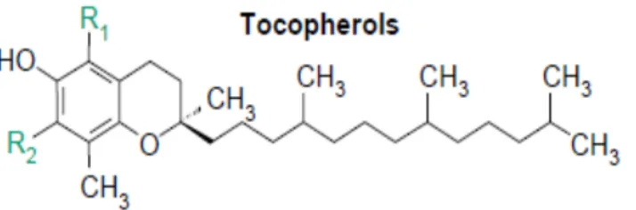
Fig 1: General structure of tocopherols (DellaPenna, 2005b). ... 14
Fig 2: Tocopherol biosynthesis in plants and cyanobacteria (DellaPenna, 2005b). ... 16
Fig3: Overview of tocopherols biosynthesis in plants showing the two pathways of PDP synthesis (Valentin, 2006). ... 17
Fig 4: Mycelium subculture under the laminar flow hood. ... 23
Fig 5: Radial growth measurements. ... 24
Fig 6: Mass production of Ganoderma lucidum mycelium in PDB liquid medium. ... 24
Fig 7: Mycelium recovery using sieves. ... 25
Fig 8: HPLC-UV equipment. ... 26
Fig 9: Microplates for the DPPH radical scavenging activity assay. ... 27
Fig 10: Microplates for the reducing power assay... 28
Fig 11: Preparation of the three groups of samples. ... 28
Fig 12: Antioxidant activity extracts preparation from modified yogurt ... 31
Fig 13:Ganoderma lucidum mycelium growth in solid and liquid media... 35
Fig 14: Means of radial growth of Ganoderma lucidum in PDA and MMN complete media during the growth period. ... 36
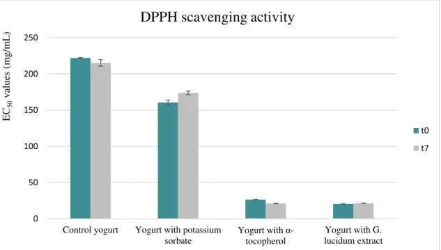
Figure 15. DPPH scavenging activity of different yogurt formulations assayed at preparation day and after 7 days of storage. ... 40
Figure 16. Reducing power of different yogurt formulations assayed at preparation day and after 7 days of storage ... 40
vi
INDEX OF TABLES
Table 1. Tocopherols structure/antioxidant activity relationship (DellaPenna, 2005a, 2005b). The
table indicates the number and position of the ring methyls in α-, β-, γ- and δ-isoforms. Relative
antioxidant activity refers to the vitamin E antioxidant activity of each tocopherol with α -tocopherol being 100%. ... 15
Table 2. Nutritional composition and energy values for different yogurt formulations (YF) and
storage times (ST). Results are presented as mean±standard deviation.1 ... 41
Table 3. Major (detected above 0.5%) fatty acids (relative percentage) of different yogurt
vii
LIST OF ABBREVIATIONS
AdeH Arogenate dehydrogenase ADI Acceptable Daily Intake
ADPPH Absorbance of the DPPH solution
AOAC procedures Official Methods of Analysis of the Association of Official Agricultural
Chemists
AS Absorbance of the Solution
At5g04490 (VTE5) Genes locus for the phytyl tail synthesis from chlorophyll-derived phytol
in Arabidopsis
BHA Butylated Hydroxyanisole BHT Butylated Hydroxytoluene
Cyclase Tocopherol cyclase
DMPBQ 2,3-Dimethyl-5-phytyl-1,4-benzoquinone
DPPH 2,2-Diphenyl-1-picrylhydrazyl
EC European council
EC50 Sample concentration providing 50% of antioxidant activity or 0.5 of
absorbance in the reducing power assay
EMM Estimated marginal means
EQ Ethoxyquin
EU European Union
EFSA European Food Safety Authority
FAME Fatty acids methyl ester
viii
FDA Food and Drug Administration FID Flame ionization detector γ–TMT γ-Tocopherol methyltransferase
GC Gas Chromatography
GGDP Geranylgeranyl diphosphate
GGR Geranylgeranyl Diphosphate Reductase GLM General linear model
HGA Homogentisic Acid
HPLC High-performance liquid chromatography
HPP p-Hydroxyphenylpyruvic acid
HPPD p-Hydroxyphenylpyruvic acid dioxygenase
HPT Homogentisate Prenyl Transferases IS Internal standard
LDA Linear discriminant analysis
LQBA Laboratory of Applied Chemistry and Biochemistry
MGGBQ 2-Methyl-6-geranylgeranylplastoquinol
MPBQ 2-Methyl-6-phytylplastoquinol or 2-methyl-6-phytyl-1,4- benzoquinone
MPBQ MT MPBQ methyltransferase
NaCl Sodium Chloride
PAT Prephenate amino transferase PDA Potato Dextrose Agar
ix
PG Propyl Gallate
PDS1 Gene locus encoding HPPD
PS Potassium sorbate
QS Quantum satis
RI Refraction index
ROS Reactive Oxygen Species RNS Reactive Nitrogen Species
RP Reducing power by Ferricyanide/Prussian blue assay SAM S-adenosyl methionine
SCF Scientific Committee on Food
SPSS Statistics Package for Social Sciences
ST Storage time
MMN Melin-Norkans medium
mMMN Modified Melin-Norkans medium
TAT Tyrosine amino transferase
TBHQ tert-Butylhydroquinone
Trolox 6-Hydroxy-2,5,7,8-tetramethylchroman-2-carboxylic acid Tyr transaminase Tyrosine transaminase
x
ABSTRACT
The increase in public awareness for the direct relationship between diet and health has led to concerns related to the use of commercial food additives, promoting the search for natural alternatives. Thus, natural antioxidants are among the most searched compounds nowadays, due to the issues related with the oxidative stress and its harmful effects on both food quality and human health. Tocopherols are among the most well-known and interesting natural antioxidants because its use as a food additive has multiple benefits. These are encompassed by the designation of vitamin E, which is an essential and extremely important micronutrient for human health that should be provided by the diet, being a strong wall against oxidation, especially in cooperation with vitamin C.
The use of natural tocopherols at an industrial level depends on the sustained production, hence the importance and choice of the in vitro culture as an alternative to their
production. The in vitro production of natural compounds allows a continuous year round availability of the demanded products in an independent way of environmental conditions and without ecological issues.
In recent years, mushrooms became more and more value and exploited due to their richness on valuable compounds with biotechnological interest and to their easier and faster
in vitro production compared to plants. Moreover, the in vitro production of mushrooms mycelium has been explored to improve the synthesis of antioxidants, namely tocopherols.
The present work aims to evaluate the tocopherols content of the well-known medicinal mushroom Ganoderma lucidum (Curtis) P. Karst from the Northeast of Portugal, produced by in vitro culture, and comparing with its fruiting body. The tocopherol levels were
xi additive was evaluated for the maintenance of the nutritional properties of the studied food matrix. These studies were carried out comparing the nutritional parameters of four yogurt formulations: i) control (yogurt without any type fortifying agent); ii) yogurt with potassium sorbate - E202; iii) yogurt with α-tocopherol - E307; and iv) yogurt with G. lucidum
mycelium tocopherol enriched extract.
Overall, with regard to the in vitro culture, G. lucidum mycelium did not show
significant differences in radial growth between the two used media.
The chemical analysis showed that the in vitro produced mycelium of G. lucidum was
particularly rich in -tocopherol (362±7 μg/g extract) and β-tocopherol (272±8 μg/g extract), among the total tocopherols content 717±12 μg tocopherols/g extract with relevant differences in comparison to the corresponding fruiting body. Besides, G. lucidum mycelium
showed good results in the DPPH radical scavenging activity assay (EC50 = 10.4±0.2
mg/mL), but especially good in the reducing power assay (EC50 = 0.32±0.01 mg/mL). While,
different results were registered in the tested yogurt formulations with the highest antioxidant activities observed in yogurts incorporated with α-tocopherol and G. lucidum extract.
Besides, no particularly significant differences were observed among 0 and 7 days, especially in yogurts incorporated with tocopherol enriched extract obtained from G. lucidum
mycelium.
xii According to the Linear Discriminant Analysis (LDA), it was possible to identify the most distinctive parameters of each YF, allowing to choose the functionalizing agent
(potassium sorbate, α-tocopherol or the enriched extract in tocopherols obtained from G.
lucidum mycelium), according to a specific objective (in this case preservative effect). Considering the values of the antioxidant activity, it can be concluded that the yogurt
incorporated with α-tocopherol or extract enriched in tocopherols obtained from the
mycelium of G. lucidum, presented a greater preservation potential.
Overall, the mycelium of G. lucidum might certainly be considered as a potential source
of tocopherols to be employed as food lipophilic antioxidants.
.
xiii
RESUMO
O aumento da consciencialização pública para a relação direta entre dieta e saúde levou a um aumento da preocupação relativamente ao uso de aditivos alimentares comerciais, promovendo a busca de fontes alternativas naturais. Assim, os antioxidantes naturais estão entre os compostos mais estudados hoje em dia, devido a questões relacionadas com o stresse oxidativo e os seus efeitos nocivos para a qualidade dos alimentos e saúde humana. Os tocoferóis estão entre os antioxidantes naturais mais conhecidos e interessantes devido aos seus múltiplos efeitos benéficos. Estes são abrangidos pela designação de vitamina E, que é um micronutriente essencial e extremamente importante para a saúde humana que deve ser fornecido pela dieta, na medida em que é uma barreira protetora contra a oxidação, especialmente quando atua em cooperação com a vitamina C.
O uso de tocoferóis naturais a nível industrial depende da sua produção sustentada, daí a importância e a escolha da cultura in vitro como alternativa à sua produção. A produção in vitro
de compostos naturais permite uma disponibilidade contínua durante todo o ano dos produtos desejados, de forma independente das condições ambientais e sem problemas ecológicos.
Nos últimos anos, os cogumelos tornaram-se uma mais-valia e são cada vez mais explorados devido à sua riqueza em compostos com interesse biotecnológico, e dado que a sua produção in vitro é mais fácil e rápida comparativamente com as plantas. Além disso, a produção in vitro de micélio de cogumelos já foi explorada para promover a síntese de compostos antioxidantes, nomeadamente tocoferóis.
O presente trabalho teve como objetivo avaliar o conteúdo em tocoferóis do cogumelo medicinal Ganoderma lucidum (Curtis) P. Karst do Nordeste de Portugal, produzido por cultura in vitro e comparando com o seu corpo frutífero. Os níveis de tocoferóis foram determinados por
xiv poder redutor. Avaliou-se também a eficácia do extrato enriquecido em tocoferóis como aditivo alimentar, na conservação das propriedades nutricionais da matriz alimentar estudada. Estes estudos foram realizados comparando os parâmetros nutricionais de quatro formulações de iogurtes: i) controle (iogurte sem qualquer tipo de agente fortificante); ii) iogurte com sorbato de potássio - E202; iii) iogurte com α-tocoferol - E307; e iv) iogurte com extrato enriquecido em tocoferóis obtido do micélio de G. lucidum.
Em geral, no que diz respeito à cultura in vitro, o micélio da espécie em estudo não mostrou
diferenças significativas no crescimento radial comparando os dois meios de cultura utilizados. A análise química mostrou que o micélio produzido in vitro é particularmente rico em β
-tocoferol (272 ± 8 μg/g de extrato) e δ-tocoferol (362 ± 7 μg/g de extrato), entre o total de tocoferóis 717 ± 12 μg de tocoferóis/g extrato e com diferenças relevantes em relação ao corpo de frutificação. O micélio apresentou capacidade de captação de radicais DPPH (EC50 = 10,4 ±
0,2 mg/mL), demonstrando ainda maior eficácia para o poder redutor (EC50 = 0,32 ± 0,01
mg/mL). As formulações de iogurte com maior atividade antioxidante pertenceram ao grupo incorporado com o aditivo α-tocoferol e com o extrato enriquecido em tocoferóis obtidos do micélio de G. lucidum. Contudo, não foram observadas diferenças significativas entre 0 e 7 dias, particularmente no iogurte incorporado com extrato de micélio de G. lucidum.
xv De acordo com a Análise Discriminante Linear (LDA), foi possível identificar os parâmetros
mais distintivos de cada YF, permitindo escolher o agente funcional (sorbato de potássio, α
-tocoferol ou extrato enriquecido em tocoferóis obtidos a partir do micélio de G. lucidum), de acordo com um objetivo específico, que neste caso era um efeito conservante. Atendendo aos valores da atividade antioxidante, pode concluir-se que os iogurtes incorporados com α-tocoferol ou extrato enriquecido em tocoferóis obtidos a partir do micélio de G. lucidum, apresentaram maior poder de conservação.
Em geral, o micélio de G. lucidum pode, de facto, ser considerado como uma potencial fonte
1
INTRODUCTION
1. Food additives
1.1. Main classes and widespread using
The use of food additives has a long tradition. They have been widely used for years and a continuous research has been carried out in order to meet consumer demand, who nowadays has a very hectic lifestyle, but is increasingly concerned with the acquisition of healthier habits, namely the consumption of beneficial compounds through the diet. Moreover, food preservation, allowing the increase and maintenance of products shelf-life, remains a focus of the researches performed today. Indeed, over the years man has been developing methods for food preservation, as well as flavour enhancers. These include salting, sunlight and oven drying, smoking, vinegar / pickling, fermentation, or addition of herbs. Moreover, additives to alter the colour of foods making them more attractive, as well as additives that confer biological properties to food have been also developed (Carocho et al., 2015; Gilsenan, 2011; Smithers, 2016).
Since pre-history, man has been developing techniques in order to feed himself. These techniques range from improvements in methods of hunting, domestication of animals and plants, development of food preservation methods, until the introduction of food additives (Carocho et al., 2014; Smithers, 2016).
2 In this way, the food industry was born, and its main objective is to transform huge agricultural and aquaculture products into different forms of food, processed in different ways, to meet the increasing demand for processed food and ensure a safe distribution, at a national and international level. Furthermore, the food industry also aims to offer a variety of food products to meet the market challenges (Fellows, 2000). In order to achieve these objectives, the food industry needs to use a variety of additives. Given the need to increase the production levels, the costs also increased and the use of the natural additives used initially becomes unviable. Thus, there were also advances in the chemistry area that seek to fill these problems, and offer as an alternative a wide variety of chemical additives providing a wide range of colours, flavours, aromas and textures to food (Moldes, Vecino, & Cruz, 2016). Actually, the incorporation of additives has been widely disseminated, leading to an excessive use, without taking into account possible impacts on consumer health. The development of sophisticated analytical methods in 1920, together with regulatory pressure, reduces significantly these problems, ensuring the development of safer products (Carocho et al., 2014).
In the XX century, there was a breakthrough in food science and technology (Smithers, 2016), and progress was made in the area of food additives (Tomaska & Brooke-Taylor, 2014). On the current century, the world population is estimated to reach 8 billion people by 2025 and the civilization became very dependent on processed and ready-to-eat food, so food additives remain the basis of food processing (Carocho et al., 2014, 2015; Floros et al., 2010).
The definition of food additive has been updated for many times since the first proposal in 1995 by the joint panel comprised by the Food and Agriculture Organization (FAO) and the
World Health Organization (WHO). Today, food additive is defined as “any substance not
normally consumed as a food by itself and not normally used as a typical ingredient of the food, whether or not it has nutritive value, the intentional addition of which to food for a technological (including organoleptic) purpose in the manufacture, processing, preparation, treatment, packing, packaging, transport or holding of such food results, or may be reasonably expected to result (directly or indirectly), in it or its by-products becoming a component of or otherwise affecting the characteristics of such foods. The term does not include contaminants or substances added to
3 Nowadays, additives are added to foods with diverse purposes, namely preserving their nutritional quality, regulating acidity, preventing food from adhering surfaces, reducing foaming,
improving texture, improving food’s baking quality or colour, improving the organoleptic
properties, or assisting in the manufacture, processing, storage and transportation (Gilsenan, 2011; Tomaska & Brooke-Taylor, 2014).
4
1.2. A special emphasis in food antioxidants
As previously referred, the use of additives for food preservation is required to maintain the quality, extend the shelf-life period, maintain the nutritional value and ensure the safety of the food products (Wedzicha, 2003). Advances in Food Science and Technology, Chemistry and Microbiology have contributed to the development of more effective preservatives and preservation technologies (Floros et al., 2010). The effectiveness of chemical preservatives depends on their concentration, food composition and on the type of microorganism or process to be inhibited (Angiolillo, Conte, & Nobile, 2014).
Food spoilage is a serious issue in food storage. Lipid peroxidation and rancidification are the prevalent type of oxidation occurred during food processing and storage. This oxidation causes undesirable off-flavours and odours, origins detrimental compounds, as well as alterations in the chemical composition and loss of the nutritional value of foodstuffs.
Oxidative reactions may also affect the food quality by changing the native colour to brown, the so called browning, in which polyphenol oxidase catalyses the conversion of polyphenols to quinones, occurring further the breakdown of these compounds and causing the darkness colour of food (Carocho et al., 2014). The browning process can also be non-enzymatic. In this type of browning occurs the production of melanoidin pigments due to various reactions such as the Maillard reaction, caramelization, chemical oxidation of phenols, and maderisation (the oxidation of a brand of wine called Madeira resulting in a darker colour and altered taste) (Carocho et al., 2014; Manzocco et al., 2001).
5 Antioxidants are divided in five groups: radical scavengers or chain-breaking antioxidants; chelators, which bind to metals and prevent them from initiating radical formation; quenchers, which deactivate high-energy oxidant species; oxygen scavengers, that remove oxygen from systems, avoiding their destabilization; and antioxidant regenerators that regenerate other antioxidants when these become radicalized (Carocho et al., 2015). Although antioxidants can be naturally present in the raw provisions, these are not considered as preservatives of the final processed food, since they can be destroyed during the food processing. Therefore, antioxidant additives are required. Several antioxidants are available for food preservation. However, their selection for food application is a serious concern, as the use of antioxidants in food is strictly controlled by legislation (Logan et al., 2013). Antioxidant additives are mainly used in meats, oils, fried foods, dressings, dairy products, baked goods and extruded snacks (Carocho et al., 2015).
The most common chemical antioxidants used are butylated hydroxyanisole (BHA, E320; Acceptable Daily Intake (ADI) 0.5 mg/kg bw), butylated hydroxytoluene (BHT, E321; ADI 0.05 mg/kg bw), propyl gallate, (PG, E310; ADI 1.4 mg/kg bw), ethoxyquin (EQ, E324; ADI 0.005 mg/kg bw), and tert-butylhydroquinone (TBHQ, E319; ADI 0.7 mg/kg bw).
Natural compounds are also used as alternatives, namely tocopherols, vitamin C, carotenoids and phenolic compounds (Carocho et al., 2014; Logan et al., 2015).
Overall, synthetic and natural antioxidants are used in food preservation. They must be effective at low doses, non-toxic and have no impact on flavour. For these reasons, and given the global trend for natural products, natural antioxidants are preferred for food applications (Logan et al., 2013).
1.3. Natural alternatives versus synthetic counterparts
6 For the most common chemical antioxidants widely used (BHA, BHT, PG, EQ, and TBHQ) some contradictory effects have been published. Some studies attributed to these compounds a potential toxicity and carcinogenic effects, while others considered them tumour suppressors (Bauer et al., 2001; Botterweck et al., 2000; Carocho & Ferreira, 2013; Vandghanooni et al., 2013). Indeed, some authors reported that many synthetic additives are toxic after long-term consumption (Zhang et al., 2016).
Many food preservation technologies have been developed and improved in the last years, such as mild-heat processing, modified atmosphere packaging, vacuum packaging, and refrigeration. In addition, new others like ultraviolet radiation, ionizing radiation, pulsed-light, high pressure, among other are being studied in order to restrict the use of preservatives in food (Carocho et al., 2014). However, currently, it is impossible to think about food free of additives / preservatives. Indeed, most of these technologies are not adapted yet to be widely used at the industrial level. They are not completely effective by themselves and they are also quite expensive, being imperative the use of additives in order to complement the preservative effect. Perhaps these limitations can be solved in a few years because of the fast development of technology and continuous innovation, demonstrating whether they actually constitute alternatives to food additives or not.
7 2015). Given this controversy, the food industry and scientific research have accepted the challenge of seeking to dispel doubts about food safety and quality in terms of food additives.
Due to the doubts that have arisen regarding the safety of the use of chemical additives, the search for natural additives has become a hot topic. This search has included natural antimicrobials and antioxidants. In contrast to synthetic preservatives, which are either neutral or harmful to human health, some natural preservatives show additional benefits towards human health namely in the prevention and resistance of several diseases (Ćilerdžiće et al., 2014). Some preservatives, namely antioxidants, in addition to their effect on the extent of shelf-life of food, have even been investigated for the prevention and/or treatment of some diseases such as cancer, cardiovascular and brain diseases and immunological conditions (Carocho et al., 2014). In addition to plants, mushrooms and algae have also become interesting sources of food antioxidants.
As mentioned above, the well-known and the most used molecules as preservatives are vitamins, polyphenols, and carotenoids (Carocho et al., 2014).
Polyphenols are excellent antioxidants that can act as scavengers, chelators, quenchers, and as ion or hydrogen donors, being effective in food preservation. They may also assure the food safety at the microbial level. Their preservative effectiveness can be achieved when incorporated as plant extracts, where synergistic effects may exist between the compounds present in the extract, or acting as single molecules (Carocho et al., 2014, 2015; Lucera et al., 2012). They have also beneficial effects on human health against cancer, osteoporosis, cataracts, cardiovascular dysfunctions, brain diseases, inflammation and immunological problems.
Carotenoids prevent also the oxidation of food but they are limited in use due to their susceptibility to oxidation by light (Carocho et al., 2015).
Vitamins are essential elements for human body that should be provided in the diet. Their
use as “green preservatives” enhances the nutritional value of food, helps to prevent food
8 gamma and delta). This vitamin constitutes a strong wall against food oxidation, especially when acting in cooperation with vitamin C. Indeed, vitamin C recycles vitamin E by reducing the generated radicals re-establishing its antioxidant activity (Carocho et al., 2014, 2015).
9
2. Tocopherols as natural preservatives
2.1. Antioxidant properties of tocopherols
The previously mentioned green preservative, vitamin E, is extremely important for human health as an essential micronutrient that should be provided by diet (Rizvi et al., 2014). It is widely known especially by its antioxidant activity preventing many diseases linked to the oxidative stress (Barros et al., 2008).
Vitamin E is the term used to refer to a group of eight compounds, which include both tocopherols and tocotrienols. Tocopherols are among the most well-known antioxidants (Carocho et al., 2015) that halt the propagation of the oxidation process through the stabilization of peroxyl radicals by donating their phenolic hydrogen. The oxidation of tocopherols produces powerless tocopheroxyl radicals which can be recycled to their native form (tocopherols) essentially by ascorbic acid, or undergo further reactions with others peroxyl or tocopheroxyl radicals forming more stable radicals (Barros et al., 2008; Carocho et al., 2014; Reis et al., 2010; Tomassi & Silano, 1986). Furthermore, tocopherols are quite effective in the direct neutralization of alkyl radicals when oxygen is present in low quantities and hydroperoxides are present in trace amounts. Besides, they can also react with alkoxy radicals formed in the propagation step (Reis et al., 2010).
Alpha-tocopherol is the most well-known isoform in terms of biological activity. This isoform is the one preferentially metabolized and absorbed in the human body, and due to its antioxidant potential, constitutes the first wall against cells lipid peroxidation (Barros et al., 2008; Lobo et al., 2010; Rizvi et al., 2014). Although α-tocopherol is the most recognized vitamin E isoform due to its antioxidant potential in vivo, γ-tocopherol have shown high
effectiveness. Indeed, several studies reported that γ- tocopherol is more effective protecting
against RNS comparing to α-tocopherol (Reis et al., 2010), and this isoform has been used for
10
2.2. The use of tocopherols as food antioxidants
It is known that the natural presence of vitamin E in vegetable oil and animal fats stabilizes them when stored under minimal light and low temperature, increasing their shelf-life (Shahidi, 2015). Regarding their lipophilic and antioxidant properties, tocopherols, especially γ-tocopherol shows an outstanding capacity, avoiding lipid peroxidation and rancidification, halting the chemical deterioration of food (Ortuño et al., 2015; Tomassi & Silano, 1986). Indeed, dietary tocopherols are among the most studied supplemented antioxidants, and it has been reported that they are among the most potent stabilizers of lipids in meat products (Logan et al., 2013). Although they are relatively stable in food, they may be oxidized when exposed to air, heat, acids, alkalis or metal ions (European Food Safety Authority (EFSA), 2015).
Tocopherols are authorized as food additives under the Annex II of Regulation (EC) No 1333/20088 on food additives. They are used as food antioxidants either individually or in combination and classified under the ‘E’ classification as following: tocopherol-rich extract of natural origin (E306), synthetic α-tocopherol (all-rac-α-tocopherol; dl-α-tocopherol; E307),
synthetic γ-tocopherol (dl-γ-tocopherol; E308) and synthetic δ-tocopherol (E309). Tocopherols
are included in Group I of food additives and authorised in the European Union at quantum satis
12
3. In vitro culture for the production of tocopherols
3.1. In vitro production of mushrooms mycelium
The use of tocopherols in food preservation is a great advance. However, the optimization of their production in a large scale is a challenge yet. The search for potential sources, as well as tocopherols in vitro production, is an interesting approach (Heleno et al., 2011). Natural products
constitute an excellent alternative for the in vitro production of compounds of interest, namely tocopherols. Moreover, the production of compounds from natural sources does not cause ecological concerns, and may have technical and economic advantages (Pinto et al., 2013). It allows a continuous year round availability of the demanded compounds in an independent way of environmental conditions (Karuppusamy, 2009). In vitro plant materials have been recognized as potential sources of valuable compounds, exploited for biotechnological applications (Karuppusamy, 2009), while mushrooms become an emerging alternative since they are also rich in bioactive compounds and they are quite easy to produce by in vitro techniques and have a quite fast growth compared to plants (Carocho et al., 2014; Ferreira et al., 2009).
Mushrooms consist on a compact association of dikaryotic mycelium shaped in the so called fruiting body or sporocarp (the reproductive form), which sprout when environmental conditions are favourable. This sporocarp has also the function of disperse teeny reproductive units called spores, which will germinate forming monokaryotic mycelium (Leiva et al., 2015). Therefore, the mycelium is the vegetative form of fungi, which is able to colonize large areas.
As abovementioned, the in vitro produced mycelium is considered a potential and promising
source of bioactive compounds useful in the pharmaceutical and/or food industries (Reis et al., 2010). Indeed, it has been proved that the in vitro culture of mycelium improve the production of
13 for β- and γ-isoforms (Pinto et al., 2013). Hence, the in vitro culture of mycelium has proven to be a promissory methodology for the production of considerable amounts of tocopherols which can meet the industrial demand.
The in vitro culture of mycelium can be carried out in solid and/or liquid media, which provide the nutrients necessary for its development (Leiva et al., 2015). This production method allows the adjustment of the medium in order to optimize the biomass production, as well as the quantities of the desired compounds (Ferreira et al., 2009; Heleno et al., 2011). The culture in solid medium may be performed using spores or live tissue obtained from the fruiting bodies. Both will give rise to mycelium identical to the one of origin, obtained in a sterile environment, and able to colonize the entire medium contained on a Petri dish. This is a technique that ensures the accurate preservation of the genetic material of live mushrooms (Stamets & Chilton, 1983).
Although a solid-medium culture is capable of mimicking the natural substrates at a nutritional level, mushrooms cultivation in liquid medium is an appropriate economic method for large-scale production (Heleno et al., 2011). The principle is the same as the culture in solid medium, but the yields obtained are usually higher. This can be explained by the possibility that, in the solid culture, the diffusion of the nutrients can be restricted by the agar, while the liquid medium allows a better distribution and availability of nutrients, as well as greater availability of oxygen (Heleno et al., 2011). Therefore, the fungus disperses more evenly, with increased biomass production in a short period of time. Furthermore, the biomass obtained in liquid medium has lower chances of contamination and is easier to recover (Gan et al., 2012; Heleno et al., 2011; Souilem et al., 2017; Yang & Liau, 1998; Zhang et al., 2016). It should be noted that the culture medium may also be a source of bioactive compounds, since the fungus may release them into the surrounding environment (Ma et al., 2016).
The metabolism of the species produced by in vitro techniques, as well as the yield obtained, vary according to the mushroom species itself, the medium composition and the culture conditions. Indeed temperature, pH, inoculum size, luminosity, incubation time etc., deeply influence the productivity in terms of biomass and produced metabolites (Yang & Liau, 1998).
14
3.2. Biosynthetic pathway of tocopherols
Tocopherols production by in vitro techniques, using mushrooms mycelium is a promising alternative to plants. In the recent years, numerous studies tried to elucidate the tocopherols content both in fruiting bodies and their mycelia, among other interesting bioactive compounds. Nevertheless, the literature reports that tocopherols are exclusively synthesized by photosynthetic organisms (Chen et al., 2015; DellaPenna, 2005a, 2005b; DellaPenna & Pogson, 2006; Karmowski et al., 2014), whereas mushrooms are saprophytes. Huge progress was made in the elucidation of the molecular, genetic and biochemical aspects of tocopherols synthesis in the photosynthetic models Arabidopsis thaliana and Synechocystis PCC6803 (DellaPenna, 2005a), while their biosynthetic pathway in mushrooms remains unclear.
Tocopherols are tocochromanol molecules that consist in a hydrophobic tail or apolar isoprenoid side chain associated with membrane lipids and a polar chromanol or head group remaining at the membrane surface (Fig. 1) (DellaPenna, 2005a; Horvath et al., 2006). Indeed,
their biosynthesis is in accordance to their chemical structure. It consists of two pathways, the aromatic amino acids (shikimate) pathway, and plastidic isoprenoid deoxyxylulose phosphate (pentose) pathway (DellaPenna, 2005a, 2005b; Matkowski, 2008). Besides, the antioxidant activity of tocochromanol molecules including tocopherols depends on the number and position of methyl group of their chroman ring (Karmowski et al., 2014), which may explain the monopoly of the entire tocopherol activity by α-tocopherol(Table 1).
15
Table 1. Tocopherols structure/antioxidant activity relationship (DellaPenna, 2005a, 2005b). The
table indicates the number and position of the ring methyls in α-, β-, γ- and δ-isoforms. Relative
antioxidant activity refers to the vitamin E antioxidant activity of each tocopherol with α
-tocopherol being 100%.
Isoform R1 R2 Relative antioxidant activity
α CH3 CH3 100
β CH3 H 25–50
γ H CH3 8–19
δ H H < 3
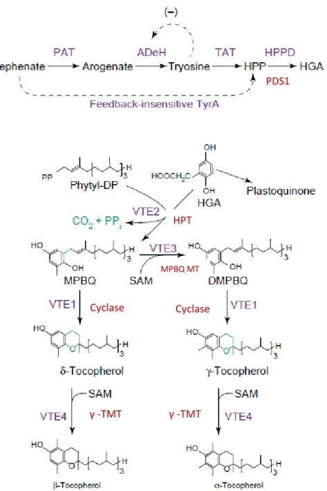
The first pathway of tocopherols biosynthesis consists in the production of the aromatic (phenolic) head group from Tyrosine (DellaPenna, 2005b; Matkowski, 2008). Indeed, a Tyr transaminase acts on tyrosine (Valentin et al., 2006) forming p-hydroxyphenylpyruvic acid
16
Fig 2: Tocopherol biosynthesis in plants and cyanobacteria (DellaPenna, 2005b). Compounds abbreviations: DMPBQ, 2,3-dimethyl-5-phytyl-1,4-benzoquinone; HGA, homogentisic acid; HPP, p-hydroxyphenylpyruvate; MPBQ, 2-methyl-6-phytyl-1,4-benzoquinone; phytyl-DP,
phytyl-diphosphate; SAM, S-adenosyl methionine. Enzyme abbreviations: AdeH, arogenate dehydrogenase; cyclase, tocopherol cyclase; HPPD, HPP dioxygenase; HPT, homogentisate phytyltransferase; MPBQ MT, MPBQ methyltransferase; PAT, prephenate amino transferase; TAT, tyrosine amino transferase; g-TMT, g-tocopherol methyltransferase.
HPT
MPBQ MT
Cyclase Cyclase
γ -TMT γ -TMT
17 In conclusion, HGA and PDP constitute the main precursors for tocopherols biosynthesis (Valentin, 2006). Once synthesized, tocopherols, tocotrienols or both compound classes, may be produced in an organism according to the substrate specificity of HPT (DellaPenna, 2005a). Actually, the level of tocopherols synthesis is highly dependent on the PDP availability, which, in turn, depends on the availability of its precursor. Thus, it has long been known that the hydrophobic phytyl tail is a geranylgeranyl tail because PDP is originated from the direct reduction of GGDP via geranylgeranyl diphosphate reductase (GGR) (DellaPenna & Pogson, 2006; Valentin, 2006). Although, an alternate pathway for the phytyl tail synthesis from chlorophyll-derived phytol was also demonstrated (Fig. 3) (DellaPenna & Pogson, 2006;
Valentin, 2006).
Fig3: Overview of tocopherols biosynthesis in plants showing the two pathways of PDP synthesis
19
4. Working plan
4.1. Mushroom species to be studied
A large variety of mushroom species from various habitats around the world has been studied for their bioactive properties, and explored as a source of interesting compounds for biotechnological applications mainly for the pharmaceutical and food industries. Indeed, filamentous fungi are already used for the production of food colorants, with the expectation that this technology will be applied for other food additives (Carocho et al., 2014), such as antioxidants for food preservation.
Ganoderma species, namely Ganoderma lucidum (Curtis) P. Karsten, are among the most cited in research publications for their cultivation, chemical analysis, pharmacology and medicinal effects (Saltarelli et al., 2009). They are the most sought medicinal mushrooms in the world market (Stojković et al., 2013) with an annual global market value over than $1.5 billion for their extracts (Ćilerdžić et al., 2014; Heleno et al., 2011, 2013). Actually, nowadays the market is growing rapidly, with a worldwide consumption estimated at thousand tons (Wachtel-Galor et al., 2011).
G. lucidum is a woody Basidiomycota mushroom, either parasitic of living hardwoods
(especially oaks) or saprobic of deadwood from hardwoods, belonging to the Polyporales order and Ganodermataceae family (Heleno et al., 2011; Wachtel-Galor et al., 2011). It is a large, dark mushroom with a glossy exterior and a woody texture (Wachtel-Galor et al., 2011). This species constitutes a potential source of important bioactive compounds with antioxidant potential (Heleno et al., 2011). Actually, its fruiting bodies, as well as its mycelium are already used, mainly as functional foods and in nutraceutical formulations (Heleno et al., 2011; Saltarelli et al., 2009). Several studies have been carried out in order to obtain the species by in vitro culture, and thus to obtain compounds of interest.
20 some studies reporting levels of 1.19 mg/g of extract in mushrooms from Taiwan (Mau et al., 2002). Other authors, studied G. lucidum from Serbia, reporting levels of 104.75 mg/100 g dw, consisting of 15.02 mg/100 g dw of α-tocopherol and 89.73 mg/100 g dw of δ-tocopherol
(Stojković et al., 2013).
The Northeastern region of Portugal is one of the European regions with higher mycological diversity (Heleno et al., 2009). Indeed, potentially interesting Portuguese Ganoderma species
were characterized in these recent years (Heleno et al., 2011) namely G. lucidum. In these
studies, G. lucidum fruiting bodies, spores and in vitro produced mycelia were studied regarding
their antioxidant potential (Heleno et al., 2011). However, its tocopherol content has not yet been reported.
4.2. Objectives
In the present work, Ganoderma lucidum from the Northeast of Portugal was studied regarding its tocopherols content. Furthermore, a tocopherol rich extract obtained from in vitro
cultured mycelia was incorporated in natural yogurt, in order to test it as a potential food additive, namely an antioxidant additive. Therefore, the main objectives consist of:
✓ Evaluation of the tocopherols content on the in vitro produced mycelium from G. lucidum, as well as on the fruiting bodies;
✓ Incorporation of the tocopherol rich extract in natural yogurt, in order to assess its antioxidant capacity and its potential as food additive.
22
MATERIALS AND METHODS
1. Standards and reagents
The solvents acetonitrile 99.9%, n-hexane 95% and ethyl acetate 99.8% were of high-performance liquid chromatography (HPLC) grade, obtained from Fisher Scientific (Lisbon,
Portugal). Tocopherols (α-, β-, γ- and δ-isoforms), as well as the fatty acids methyl ester (FAME)
reference standard mixture 37 (standard 47885-U) and other individual fatty acid isomers, and sugars [lactose and D(+)-raffinose pentahydrate] and trolox (6-hydroxy-2,5,7,8-tetramethylchroman-2-carboxylic acid) were purchased from Sigma (St. Louis, MO, USA). Racemic tocol, 50 mg/ml, was supplied from Matreya (Pleasant Gap, PA, USA) and 2,2-diphenyl-1-picrylhydrazyl (DPPH) was obtained from Alfa Aesar (Ward Hill, MA, USA). Potassium sorbate was acquired from Acros Organics (Geel, Belgium). Thiamine, casamino acids, malt extract and agar were obtained from Panreac AppliChem (Barcelona, Spain). PDA and PDB were acquired from Oxoid microbiology products (Hampshire, United Kingdom). Methanol and all other chemicals and solvents were of analytical grade and purchased from common sources. Water was treated in a Milli-Q water purification system (TGI Pure Water Systems, Greenville, SC, USA).
2. Samples and in vitro mycelium production
Ganoderma lucidum (Curtis) P. Karsten fruiting bodies were obtained from the herbarium of
the School of Agriculture from the Polytechnic Institute of Bragança, Portugal. These samples were used to analyze the tocopherols profile, since no results have been reported for G. lucidum
from the Northeastern region of Portugal.
Ganoderma lucidum (Curtis) P. Karsten mycelium was obtained from previous cultures maintained in the laboratory of Biology and Biotechnology of the School of Agriculture from the Polytechnic Institute of Bragança, Portugal.
23 culture chamber of the laboratory mentioned above, at 23 ° C / 18 ° C during the photoperiods of day and night (16h / 8h), respectively.
In order to verify the best culture medium for the growth of the species under study, small excised pieces from the previous cultures preserved in the laboratory of Biology and Biotechnology were transferred, under sterile conditions (laminar flow hood), to new Petri dishes containing different solid media (Fig 4): i) Potato Dextrose Agar medium (PDA) pH 5.6 ± 0.2;
ii) Melin-Norkans medium (MMN) pH 6.6 (NaCl 0.025 g/l; (NH4)2HPO4 0.25 g/l; KH2PO4 0.50
g/l; FeCl3 0.005 g/l; CaCl2 0.050 g/l; MgSO4.7H2O 0.15 g/l; thiamine 100 µg/l; malt extract 5 g/l;
casamino acids 1 g/l; glucose 10 g/l; agar 20 g/l); and iii) modified MMN medium (mMMN) pH 6.6 (NaCl 0.025 g/l; (NH4)2HPO4 0.25 g/l; KH2PO4 0.50 g/l; FeCl3 0.005 g/l; CaCl2 0.050 g/l;
MgSO4.7H2O 0.15 g/l; thiamine 100 µg/l; glucose 10 g/l; agar 20 g/l) (Marx, 1969).
Fig 4: Mycelium subculture under the laminar flow hood.
24
Fig 5: Radial growth measurements.
After reaching the maximum growth, the cultured Petri dishes were used for the inoculation of G. lucidum in the ideal culture medium, but in the liquid form [Potato Dextrose Broth (PDB)], in order to obtain higher yields to perform the chemical assays. Three excised fragments were inoculated in each flask with 30 ml of culture medium. The flasks were held in the above-mentioned in vitro culture chamber, until enough biomass was obtained for the subsequent assays
(≈ 32 days) (Fig. 6).
Fig 6: Mass production of Ganoderma lucidum mycelium in PDB liquid medium.
After approximately 32 days, the mycelia were recovered from the culture flasks using a sieve (particle size, 2 mm; Fig. 7). Afterwards, the recovered mycelia were weighted in order to
25
Fig 7: Mycelium recovery using sieves.
3. Tocopherols rich extract
3.1. Extraction procedure
The extracts were prepared following a procedure previously described by (Barros et al., 2008). Briefly, BHT (butylhydroxytoluene) (100 µl) and tocol (internal standard (IS) solution)
(250 µl) were added to the samples. The samples (≈ 500 mg) were homogenized with methanol
(4 ml) by vortex mixing (1 min). Subsequently, hexane (4 ml) was added and the mixture was vortex again (1 min). Saturated NaCl aqueous solution (2 ml) was added, and the mixture was homogenized (1 min), centrifuged (5 min, 4000g) and the clear upper layer was transferred to a
vial wrapped in aluminum paper and placed in the ice. The samples were re-extracted twice with hexane. The combined extracts were dehydrated with anhydrous sodium sulfate and taken to dryness under a nitrogen stream, and i) re-dissolved in 1 ml of n-hexane, dehydrated with anhydrous sodium sulphate, filtered through a 0.22 µm disposable LC filter disk and transferred into a dark injection vial for the analysis by HPLC; or ii) re-dissolved in the volume of methanol required to obtain a stock solution of 10 mg/ml.
3.2. Determination of tocopherols content
Tocopherols analysis was made by HPLC following a procedure previously optimized and described by (Heleno et al., 2010). The equipment of HPLC (Fig. 8) consisted of an integrated
26 (Jasco, Easton, MD) programmed for excitation at 290 nm and emission at 330 nm. Data were analysed using Clarity DataApex 2.4 Software. The column used was a normal-phase 250 mm × 4.6 mm i.d., 5 mm, Polyamide II, with a 10 mm × 4 mm i.d. guard column of the same material (YMCWaters, Dinslaken, Germany), operating at 30 °C. The mobile phase used was a mixture of
n-hexane and ethyl acetate (70:30, v/v) at a flow rate of 1 ml/min. The compounds were identified by chromatographic comparisons with authentic standards. Quantification was based on the fluorescence signal response, using the internal standard method. Tocopherols content in the samples was expressed in µg per g of dry weight (Barros et al., 2008; Heleno et al., 2009; Reis et al., 2011).
Fig 8: HPLC-UV equipment.
3.3. Evaluation of the antioxidant activity
For the evaluation of the antioxidant activity of the tocopherols rich extract, two different in vitro assays were performed, the 2,2-diphenyl-1-picrylhydrazyl (DPPH) radical scavenging assay
and the reducing power assay.
DPPH radical scavenging activity. DPPH is a stable radical characterized by an
absorbance, in concentrated ethanol solution, at about 515 nm. This assay consists of reducing the DPPH radical, through the donation of a hydrogen (H) atom from a scavenger molecule (i.e.,
antioxidants), resulting in the color alteration from purple to yellow, with a concomitant decrease in the absorbance at 515 nm (Mishra et al., 2012).
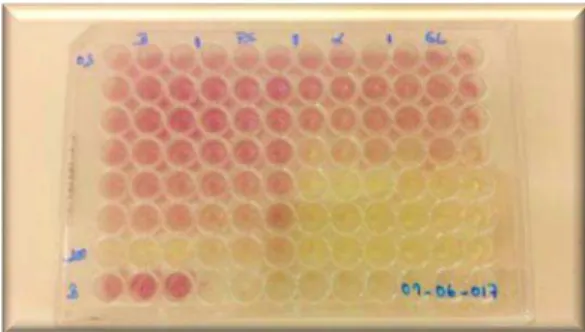
This methodology was performed using an ELX800 Microplate Reader (BioTek Instruments, Inc., Winooski, VT). The in each of the 96 wells (Fig. 9) consisted of different
27 × 105 mol/l). The mixture left to stand in the dark for 60 min and the reduction of the DPPH
radical was determined by measuring the absorption at 515 nm. The radical-scavenging activity (RSA) was calculated as a percentage of DPPH discoloration using the equation: %RSA= [(ADPPH-AS)/ ADPPH] × 100, where AS corresponds to the absorbance of the solution containing a
given extract concentration and ADPPH is the absorbance of the DPPH solution. The assays were
carried out in duplicate and the results were expressed as EC50 values, which correspond to the
extract concentration providing 50% of radicals-scavenging activity. This value was calculated by interpolation from the graph of RSA percentage against extract concentration(Ferreira et al., 2007; Heleno et al., 2009; Reis et al., 2011). Trolox was used as standard.
Fig 9: Microplates for the DPPH radical scavenging activity assay.
Reducing power assay. The present assay is based on the reduction of the yellow ferric
form (Fe3+) to the blue ferrous form (Fe2+) by the action of electron-donating antioxidants. The
resulting Perl’s Prussian blue color could be measured spectrophotometrically at 700 nm
(Ferreira et al., 2007).
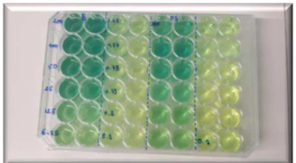
This methodology was performed using the same Microplate Reader described above. In eppendorf tube, different concentrations prepared from the stock solutions (0.5 ml) were mixed with sodium phosphate buffer (200 mmol/l, pH 6.6, 0.5 ml) and potassium ferricyanide (1% w/v, 0.5 ml). The mixture was incubated at 50 °C for 20 min, and then trichloroacetic acid (10% w/v, 0.5 ml) was added. The mixture (0.8 ml) was poured in the microplate wells (Fig. 10) with
28 were expressed as EC50 values, which correspond to the extract concentration providing 0.5 of
absorbance. These EC50 values were calculated from the graph of absorbance at 690 nm against
the extract concentration. Trolox was used as standard.
Fig 10: Microplates for the reducing power assay.
4. Incorporation of the tocopherols rich extract in natural yogurt
The natural yogurts were purchased from a local supermarket.
Four yogurt formulations were prepared (50 g of natural yogurt): i) control (yogurt without any type fortifying agent); ii) yogurt with potassium sorbate (E202; 20 mg); iii) yogurt with α -tocopherol (E307; 8 mg); and iv) yogurt with G. lucidum mycelium tocopherol enriched extract
(33 mg, according to the EC50 value obtained from the reducing power assay). (Fig. 11). All the
yogurts were prepared in duplicate.
29
4.1. Nutritional composition and evaluation of the antioxidant activity of the samples along
the shelf-life period
The samples were analyzed immediately after preparation and after seven days of storage at 4 ºC. All the analyses were performed in triplicate.
Nutritional parameters. The nutritional value of the samples was evaluated (moisture,
protein, fat, ash and carbohydrates) using the AOAC (2016) standard procedures (George & Latimer., 2016). The crude protein content (N × 6.38) of the samples was estimated by Kjeldahl method; the crude fat was determined by extracting a known weight of powdered sample with petroleum ether, using a Soxhlet apparatus; the ash content was determined by incineration at 600 ± 15 ºC and total carbohydrates were calculated by difference. Energy was calculated according to the Regulation (EC) No. 1169/ 2011 of the European Parliament and of the Council, of 25 October 2011, on the provision of food information to consumers, following the equation Energy (kcal/100g dw) = 4 × (g protein + g carbohydrates) + 9 × (g fat).
Soluble sugars were detected by HPLC coupled to refraction index (RI) detector.
Briefly, the lyophilized samples (≈ 1 g) were spiked with melezitose (internal standard; IS)
and were extracted with 40 ml of 80% aqueous ethanol at 80 °C for 90 min. The resulting suspension was centrifuged at 15,000g for 10 min. The supernatant was concentrated at 40 °C under reduced pressure and defatted three times with 10 ml of ethyl ether, successively. After concentration at 40 °C, the solid residues were dissolved in water to a final volume of 5 ml and filtered through 0.22 μm disposable LC nylon disk filters and transferred into an injection vial to be analyzed by HPLC-RI.
The HPLC equipment consisted of an integrated system with a Smartline 1000 pump (Knauer, Berlin, Germany), a Smartline manager 5000 degasser, an AS-2057 auto-sampler (Jasco, Easton, MD) and a Smartline 2300 refraction index (RI) detector (Knauer). Data were analysed using Clarity 2.4 Software (DataApex). The chromatographic separation was achieved with a Eurospher 100-5 NH2 column (4.6 × 250 mm, 5 mm, Knauer) operating at 30 °C. The
30 standards. Quantification was made by the internal standard method based on the RI signal response of each standard, using the internal standard (IS, melezitose) method and by using calibration curves obtained from commercial standards of each compound. The results are expressed in g/100 g of yogurt, calculated by internal normalization of the chromatographic peak area (Caleja et al., 2016; Heleno et al., 2015; Heleno et al., 2009; Reis et al., 2011).
Fatty acids were analyzed by gas chromatography (GC) coupled to a flame ionization detector (FID) detector.
Briefly, the fatty acids obtained after Soxhlet extraction, were subjected to a trans-esterification procedure (methylated with 5 ml of methanol:sulfuric acid 95%:toluene 2:1:1 (v/v/v) for, at least, 12 h in a bath at 50 °C and 160 rpm). Afterwards, 3 ml of deionised water were added in order to obtain phase separation; the fatty acids methyl esters (FAME) were recovered by shaking in a vortex with 3 ml of diethyl ether, and the upper phase was passed through a micro-column of anhydrous sodium sulfate to eliminate the water. The sample was recovered in a vial with Teflon and filtered through a 0.2 μm Whatman nylon filter.
Fatty acids were determined by gas–liquid chromatography with flame ionization detection (GC-FID)/capillary column as described previously by the authors (Heleno et al., 2009). The fatty acid profile was analyzed with a DANI model GC 1000 instrument equipped with a split/splitless injector, a flame ionization detector (FID) and a Macherey–Nagel column (30 m × 0.32 mm ID × 0.25 μm df). The oven temperature program was as follows: the initial temperature of the column was 50 °C, held for 2 min, then a 10 °C/min ramp to 240 °C and held for 11 min. The carrier gas (hydrogen) flow-rate was 4.0 ml/min (0.61 bar), measured at 50 °C. Split injection (1:40) was carried out at 250 °C. For each analysis 1 μl of the sample was injected in GC. Fatty acid identification was made by comparing the relative retention times from samples with FAME peaks from samples with standards. The results were recorded and processed using CSW 1.7 software (DataApex 1.7) and expressed in relative percentage of each fatty acid (Heleno et al., 2009; Reis et al., 2011).
Antioxidant activity.The lyophilized samples (≈ 1 g) were extracted with methanol at room
31 resulted combined extracts were evaporated at 40 ºC under reduced pressure (rotary evaporator Büchi R-210, Büchi, Flawil, Switzerland)) until complete removal of methanol (Fig. 12). Finally,
the evaporated extracts were dissolved in methanol at a concentration of 200 mg/ml.
DPPH radical-scavenging activity and reducing power were evaluated at 515 and 690 nm, respectively, using the ELX800 microplate Reader (Bio-Tek Instruments, Inc., Winooski, Vermont, USA), as previously described.
32
5. Statistical analysis
All statistical tests were performed at a 5% significance level using IBM SPSS Statistics for Windows, version 22.0. (IBM Corp., Armonk, NY, USA). Data were expressed as mean±standard deviation, maintaining the significant numbers allowed by the magnitude of the standard deviation.
The results were compared through an analysis of variance (ANOVA) with type III sums of squares using the general linear model (GLM) procedure. The dependent variables were analyzed using 2-way ANOVA with the factors “yogurt formulation” (YF) and “storage time” (ST). When a statistically significant interaction among these two factors was detected, their effects were evaluated simultaneously by the estimated marginal means plots for all levels of each factor. On the contrary, if no statistical significant interaction was found, means were compared using
Tukey’s multiple comparison test, after checking the equality of variances through a Levene’s
test.
In addition, a linear discriminant analysis (LDA) was used to compare the effect of YF over
the assayed parameters. A stepwise technique was applied, considering the Wilks’ ʎ test with the
usual probabilities of F (3.84 to enter and 2.71 to be removed) for variable selection. This
procedure is based in sequential forward selection and backward elimination steps, where the inclusion of a new variable requires verifying the significance of all previously selected variables (Zielinski et al., 2014). The main objective was estimating correlations between single categorical dependent variables (yogurt formulations) and quantitative independent variables (results obtained in the laboratorial assays). The LDA outputs identified the independent variables with highest contribution to the differences in the average score profiles of different yogurt formulations. To verify the significance of the canonical discriminating functions, Wilk’s
ʎ test was used. A leaving-one-out cross validation procedure was carried out to assess the model
33
RESULTS AND DISCUSSION
Nowadays, natural antioxidants are among the most searched compounds due to the several issues related to the oxidative stress and the increasing awareness of the possible side effects of the synthetic alternatives. Indeed, it has been reported that mushrooms constitute a valuable source of several antioxidants including phenolic compounds, vitamin C and E and carotenoids. Moreover, it has been established a direct relationship between the compounds present in mushrooms extracts and their verified bioactivity, making them potentially bioactive ingredients for use in the nutraceutical formulations (Reis et al., 2017).
Hence, in a continuous way of mushrooms valorization, and for the first time, this study aims to evaluate the antioxidant capacity of a tocopherols rich extract obtained from the mycelium of Ganoderma lucidum produced by in vitro culture, as well as evaluate its antioxidant effects when incorporated in natural yogurt.
As abovementioned, although several studies have reported that the antioxidant activity of
G. lucidum is correlated mainly with the phenolic compounds, polysaccharides, peptides and
polysaccharides-peptides complexes present in their chemical constitution (Ćilerdžić et al., 2014;
Heleno et al., 2011; Stojković et al., 2013), there are no information regarding the tocopherols
content and the antioxidant potential of tocopherol extracts from this species. Moreover, the inclusion of such extract, as an antioxidant additive, in foodstuff has not been tested yet.
In general, fruiting bodies seem to have highest antioxidant properties than the in vitro
produced mycelia. However, some studies proved that mycelia produced by in vitro culture may
have higher tocopherols content than the fruiting bodies (Reis et al., 2011).
1. Mycelium production
In the present work, the in vitro culture was exploited for the production of G. lucidum
34 After 7 days of the inoculation time, it was possible to measure the mycelium growth on both MMN and PDA media. Mycelium revealed a better growth on MMN medium, comparing with PDA (Fig 14), however, these differences were not statistically significant. Besides, Heleno
et al. (2012) reported, for this species, better results for mycelium growth, on PDA medium. Some slight differences on both studies may be explained by the fact that the culture was carried out after a long period without mycelia sub-culturing, therefore, the starting conditions of the mycelia were different.
The mycelium was sub-cultured several times and over a period of six months (Fig 13A).
During the first months, it was noticed that, on both media, the mycelium was covering the surface of the plates in a thinly layer and seemed to had a more dehydrated aspect mainly on the center of the Petri dish (where mycelium cells are older) (Fig 13B). During the last two months,
deep changes were noticed. Mycelium became colonizing the plate area in more dense layers with a clear cottony aspect, and without a dehydrated appearance (Fig 13C and D). After
covering all the surface of the petri dishes, the growth remains constant. Hence, the repeated sub-cultures of the mycelium seem “reactivate” and “refresh”G. lucidum mycelium cells.
It was also performed an experiment to test the mycelium growth in modified MMN medium (mMMN), but it showed a very poor and insufficient mycelium development due to the stress caused by the absence of some nutrients in the culture medium. These results are in agreement with the results published by Heleno et al. (2012) (Fig 13E).
PDB liquid medium was inoculated using the Petri dishes with the best growth rates. After four days of the inoculation time, it was possible to see the starting growth in the majority of the flasks. In five weeks, the entire medium surface became covered by the mycelium. Whereas, in many flasks the mycelia did not cover the entire surface medium and stopped growing at the end of the five weeks (Fig 13F). Overall, ≈ 250 flasks with 30 ml of PDB liquid medium were used.