ARTIGO ORIGINAL
Morphological Indexes: Can They Predict Lupus Nephritis
Outcomes? A Retrospective Study
Índices Morfológicos na Nefrite Lúpica: Orientação
Prognóstica? Um Estudo Retrospetivo
David NAVARRO1, Ana Carina FERREIRA1, Helena VIANA1, Fernanda CARVALHO1, Fernando NOLASCO1
Acta Med Port 2019 Oct;32(10):635–640 ▪ https://doi.org/10.20344/amp.11598
1. Nephrology Department. Hospital Curry Cabral. Centro Hospitalar Universitário de Lisboa Central. Lisbon. Portugal. Autor correspondente: David Navarro. [email protected]
Recebido: 28 de novembro de 2018 – Aceite: 18 de abril de 2019 | Copyright © Ordem dos Médicos 2019 ABSTRACT
Introduction: Lupus nephritis is a serious complication of systemic lupus erythematosus. Currently, therapy is guided by findings in the
renal biopsy, following the International Society of Nephrology / Renal Pathology Society classification. Austin and Hill’s histomorpho-logical indexes are not routinely obtained. In this retrospective single-centre study, we aimed to analyze the importance and applicability of the different morphological indexes in predicting response to treatment and prognosis.
Material and Methods: Patients with kidney biopsy demonstrating lupus nephritis from the 2010 – 2016 period were included. We
ana-lyzed their demographic data, comorbidities, clinical presentation and laboratorial evaluation at the time of renal biopsy. We evaluated the following outcomes: clinical remission, renal function and proteinuria at end of follow-up. Histologic analysis was performed using the International Society of Nephrology / Renal Pathology Society classification and the morphological indexes described by Austin (Activity and Chronicity) and Hill. Univariate and multivariate statistical analysis was performed using STATA software.
Results: We analyzed 46 biopsy-proven lupus nephritis cases, with a median follow-up of 31.9 (13.2 – 45.6) months. Based on
biopsy findings, 35 patients were started on immunosuppressive therapy. We observed that Class IV patients had, at presentation, lower estimated glomerular filtration rate (67.3 vs 94.6 mL/min; p = 0.02), higher proteinuria (4.26 vs 2.37 g/24 hours; p = 0.02) and a non-significantly higher C3 consumption (58.9 vs 77.4 mg/dL; p = 0.06). We did not observe correlations between International Society of Nephrology / Renal Pathology Society classification and the outcomes at the end of follow-up. In contrast, both the Hill biopsy index and Austin’s Chronicity index were correlated with renal function and proteinuria at the end of follow-up. Austin’s Activity index correlat-ed with the immunological findings (C3, C4 and anti-dsDNA) at presentation.
Discussion: Because clinical activity poorly correlates with histologic activity, histological findings are fundamental when assessing
patients with suspected lupus nephritis. The most recent International Society of Nephrology / Renal Pathology Society report supports the European League Against Rheumatism guidelines, encouraging the adoption of histomorphological indexes when evaluating lupus nephritis. Our data, showing a correlation between the renal outcomes and the indexes described by Austin and Hill, supports this view.
Conclusion: The histomorphological indexes in lupus nephritis are easily obtainable, can predict renal outcomes and may help in the
management of such patients.
Keywords: Biopsy; Kidney/pathology; Lupus Erythematosus, Systemic/complications; Lupus Nephritis Predictive Value of Tests;
Prognosis RESUMO
Introdução: A nefrite lúpica é uma complicação grave do lúpus eritematoso sistémica. Atualmente, a terapêutica dirigida é ditada
pelos achados histológicos da biópsia renal, através da classificação da International Society of Nephrology / Renal Pathology Society. Os índices histomorfológicos descritos por Austin e Hill não são rotineiramente realizados. Neste estudo retrospetivo unicêntrico, pro-curámos analisar a aplicabilidade e relevância dos índices morfológicos na predição da resposta à terapêutica e do prognóstico em doentes com nefrite lúpica.
Material e Métodos: Foram incluídos doentes cuja biópsia renal, realizada entre 2010 e 2016, documentava nefrite lúpica.
Analisá-mos os dados demográficos, comorbilidades, apresentação clínica e avaliação laboratorial destes doentes correspondente à altura da biópsia renal. Avaliámos os seguintes outcomes: remissão clínica, função renal e proteinúria no final do seguimento. A avaliação histológica foi realizada segundo a classificação da International Society of Nephrology / Renal Pathology Society e aplicando os índices morfológicos descritos por Austin (Actividade e Cronicidade) e Hill. A análise estatística univariada e multivariada foi realizada com software STATA.
Resultados: Foram revistos 46 casos de nefrite lúpica, com um follow-up mediano de 31,9 (13,2 – 45,6) meses. A partir dos achados
histológicos, 35 doentes foram submetidos a imunossupressão. Observámos que os doentes com nefrite lúpica Classe IV tinham, à apresentação, taxa de filtrado glomerular estimada mais reduzida (67,3 vs 94,6 mL/min; p = 0,02), proteinúria mais elevada (4,26 vs 2,37 g/24 horas; p = 0,02) e consumo de C3 mais elevado de modo não-significativo (58,9 vs 77,4 mg/dL; p = 0,06). Não se verificou correlação entre a classificação ISN/RPS e os desfechos no final do follow-up. Por outro lado, tanto o índice de Hill quanto o score de cronicidade de Austin correlacionaram-se com a função renal e a proteinúria no final do seguimento. Adicionalmente, o score de atividade de Austin correlacionou-se com os achados imunológicos à apresentação (C3, C4 e anti-dsDNA).
Discussão: Uma vez que a actividade clínica tem fraca correlação com a actividade histológica, os achados histológicos são
fun-damentais durante a avaliação na suspeita de nefrite lúpica. A mais recente revisão da International Society of Nephrology / Renal Pathology Society vai ao encontro das linhas de orientação da European League Against Rheumatism, encorajando a aplicação de índices histomorfológicos na avaliação da nefrite lúpica. Os dados da nossa população, onde verificámos uma correlação entre o prognóstico renal e os índices histomorfológicos descritos por Austin e Hill, apoiam essa sugestão.
ARTIGO ORIGINAL
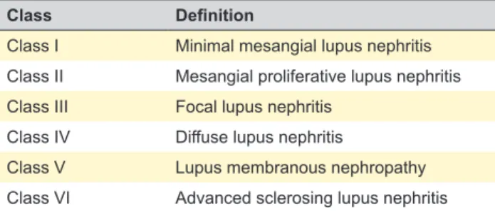
Table 1 – Abbreviated lupus nephritis ISN/RPS classification
Class Definition
Class I Minimal mesangial lupus nephritis Class II Mesangial proliferative lupus nephritis Class III Focal lupus nephritis
Class IV Diffuse lupus nephritis
Class V Lupus membranous nephropathy
Class VI Advanced sclerosing lupus nephritis
Conclusão: Os índices histomorfológicos na nefrite lúpica são de fácil aplicação, conseguem prever os outcomes clínicos e podem
representar uma ferramenta adicional na avaliação dos doentes com nefrite lúpica.
Palavras-chave: Biópsia; Lupus Eritematoso Sistémico/complicações; Nefrite Lúpica; Prognóstico; Rim/patologia; Valor Preditivo dos
Testes
INTRODUCTION
Systemic lupus erythematosus (SLE) is a systemic autoimmune disease associated with significant comorbid-ity and mortalcomorbid-ity.1 Lupus nephritis (LN) is one of its most serious complications and includes a wide spectrum of clinical presentations, ranging from isolated haematuria to end-stage renal disease. Immunosuppression can have an important role in controlling renal affection — with current therapies, a high remission rate is observed, which is the most important renal prognostic factor.2 For those reasons, lupus nephritis requires a well-weighted diagnostic and therapeutic approach for which renal biopsy currently occu-pies a central role.
The International Society of Nephrology/Renal Pathology Society (ISN/RPS) lupus nephritis classification is widely used and recommended in current guidelines.3 Histology findings allow the sub-classification into six class-es (I – VI), as shown in Table 1. The most recent report4 and EULAR guidelines5 additionally recommend the use of mor-phological indexes; these have been originally described by Austin et al,6 who defined two indexes (activity and chronic-ity), using a semi-quantification of findings. This was fur-ther studied by Hill et al,7 who modified previous indexes, improving its prognostic value. Subcategorization of class III and IV into active, chronic or both is also suggested to be replaced by this semi-quantification method.4
Histomorphology has been of paramount importance in defining the field of nephrology. The quantification of microscopy findings has found its place in renal transplan-tation through the semi-quantitative Banff criteria.8 In LN, while the ISN/RPS classification is routinely used, Austin and Hill indexes are not. The aim of this study was to ana-lyze the applicability and the efficacy of the different LN morphological indexes in predicting response to treatment and prognosis.
MATERIAL AND METHODS
This is a retrospective single-centre descriptive analysis of consecutive SLE and biopsy-proven LN patients, diag-nosed from 2010 to 2016. Patients whose kidney biopsies were found to be inadequate due to an insufficient num-ber of glomeruli (defined as less than five) were excluded.
Patients’ data were obtained from clinical files. We analyzed patients’ demographic data (age, gender, race), comorbid-ities and their duration (including SLE history and previous manifestations and medications), hypertension according to the most recent definition by the European Society of Cardiology,9 clinical presentation and laboratorial evaluation at the time of renal biopsy [including full blood count, serum creatinine, urinalysis, proteinuria (24 hours-collection or uri-nary protein to creatinine ratio in a random urine sample), C3, C4 and anti-dsDNA].
The following outcomes were evaluated: clinical remis-sion, renal function and proteinuria (g/24 hours) at end of follow-up (FUP). The LUNAR trial10 definition for clinical remission was used: complete remission was defined as reduction of proteinuria to < 0.5 g/24 hours, inactive urinary sediment and normal serum creatinine level if it was abnor-mal at baseline (or a serum creatinine level of < 115% of baseline if it was normal at baseline); partial remission was defined as a reduction in proteinuria to < 1 g/24 hours if initial level < 3 g/24 hours (or reduction to < 3 g/24 hours if initial level > 3 g/24 hours), inactive urinary sediment and a serum creatinine level of < 115% of baseline. All renal biopsies were performed after obtaining the patient's written consent. The histologic optic examination was performed with hematoxylin and eosin, periodic acid–Schiff, Jones’ silver and Masson’s trichrome stains; immunofluorescence study included studies with antibodies against IgG, IgA, IgM, C3, C4, C1q and fibrinogen.
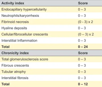
The studied predictors were the ISN/RPS LN classifi-cation and the morphological indexes described by Austin (activity and chronicity)6 and Hill,7 obtained after histomor-phological review of renal biopsies. Both scores describe findings in a quantitative fashion (zero to 3+). Austin’s activ-ity score measures active LN findings of glomerular prolifer-ation, leucocyte exudation/karyorrhexis, fibrinoid necrosis, cellular crescents, hyaline deposits and interstitial inflam-mation; Austin’s chronicity score contains chronic irrevers-ible LN findings: glomerular sclerosis, fibrous crescents, tubular atrophy and interstitial fibrosis.
Hill’s biopsy index slightly adapted Austin’s activity and chronicity scores and added histological signs of tubuloin-terstitial activity (including tubular cell necrosis and flatten-ing and the presence of macrophages or epithelial cells in tubular lumens) and immunofluorescence findings (includ-ing glomerular, tubulointerstitial and vascular deposition). Regarding the latter, in the review of our samples, we did not re-perform immunofluorescence and instead used the findings described in our original report. Table 2 summariz-es both indexsummariz-es.
Renal function was monitored by serum creatinine and estimated glomerular filtration rate (eGFR) using the Chronic Kidney Disease Epidemiology Collaboration (CKD-EPI) equation.
ARTIGO ORIGINAL
Data are presented as frequencies for categorical varia-bles; continuous variables are presented as median (inter-quartile range).
One-way ANOVA was used for comparisons between the different indications for kidney biopsy, the laboratory data and the morphologic data obtained after the applica-tion of the different morphological indexes; chi-square test of independence/Fisher’s exact test was performed when comparing the different indications for kidney biopsy and the ISN/RPS LN classes. Associations between the different LN classes and clinical and laboratory variables were investi-gated using Fisher’s exact test and Wilcoxon. Associations between Austin and Hill indexes and laboratory variables were investigated using Spearman test.
After this first analysis, we fitted a model of multivariate analysis using all subjects and defining the chronicity index-es as our main predictors and eGFR at the end of follow-up as our main outcome. Age, gender and proteinuria were the variables used as potential confounders (older patients may have a lower renal function, women tend to have lower GFR and proteinuria is associated with a rapid decline of renal function). All tests were performed using STATA software
version 13, and a p-value < 0.05 was considered statistical-ly significant.
Given the nature of this analysis (retrospective and observational) and the anonymization of the collected data, authorization from the ethics committee and the Portuguese Data Protection Authority was not required.
RESULTS
From 2010 to 2016, we found 46 biopsy-proven LN cases in our unit. The patients’ median age was 35 years (27 – 42), and 84.8% (n = 39) were women, in a mostly Caucasian population (56.5%; n = 26). In seven patients, SLE diagnosis was previously unknown, while the remain-ing 39 patients had SLE for a median of 3.96 (1.1 – 11.9) years, resulting in treatment with hydroxychloroquine in 46.1% and additional immunomodulation in 76.9% of cas-es. At presentation, median creatinine was 0.8 (0.6 – 1.4) mg/dL, corresponding to a median eGFR of 89 (46.9 – 117) mL/min; median level of proteinuria was 2.45 (1 – 4.4) g/day; 34.7% (n = 16) of patients were hypertensive and 58.7% (n = 27) had haematuria at presentation. We classified our patients according to the indication for renal biopsy: 18 had asymptomatic urinary abnormalities, 16 nephrotic-range proteinuria (proteinuria > 3.5 g/d) and 12 renal insufficiency (eGFR < 45 ml/min). The histomorphological findings are summarized in Table 3.
Regarding histology, sample quality was adequate, the minimum number of glomeruli per sample was five and we had a maximum of 30 glomeruli, with a median of 11 glo-meruli (7.75 – 14.25). The 46 patients were evaluated dur-ing a median follow-up of 31.9 months (13.2 – 45.6) months.
Table 2 – Morphological indexes scoring system. (A) Austin’s activity and chronicity indexes; (B) Hill biopsy index B:
Components Score
Glomerular Activity index 0 – 3
Tubulointerstitial Activity index 0 – 4
Chronic lesions index 0 – 2
Immunofluorescence index 0 - 3
Total 0 – 12
A:
Activity index Score
Endocapillary hypercellularity 0 – 3 Neutrophils/karyorrhexis 0 – 3 Fibrinoid necrosis (0 - 3) x 2 Hyaline deposits 0 – 3 Cellular/fibrocellular crescents (0 – 3) x 2 Interstitial Inflammation 0 – 3 Total 0 – 24
Chronicity index Score
Total glomerulosclerosis score 0 – 3
Fibrous crescents 0 – 3
Tubular atrophy 0 – 3
Interstitial fibrosis 0 – 3
Total 0 – 12
Table 3 – Renal clinical characteristics and histomorphology findings
Clinical presentation Patients Class II Class III Class IV Class V Activity IndexAustin’s Chronicity Austin’s Index Hill Biopsy Index Asymptomatic urinary abnormalities 18 44% 17% 28% 11% (0 – 6)3.03 (0 – 2.63)1.50 (0.14 – 1.53)0.86 Nephrotic syndrome (proteinuria > 3.5 g/d) 16 0% 25% 63% 31% (1.65 – 10)6.59 (1 – 3)2.66 (0.76 – 2.27)1.63 Renal insufficiency (eGFR < 45 ml/min) 12 0% 8% 75% 17% (0.5 – 13)6.95 (0.5 – 7)3.73 (1.56 – 2.56)1.96 p-value — < 0.001 NS 0.02 NS 0.07 0.08 0.007
eGFR: estimated glomerular filtration rate; NS: non-significant. LN Classes (from II to V) frequencies are expressed for each renal clinical characteristic (asymptomatic urinary abnormalities | nephrotic syndrome | renal disease). Median indexes (Austin activity and chronicity and Hill biopsy) are expressed for each renal clinical characteristic (asymptomatic urinary abnormalities | nephrotic syndrome | renal dis-ease). The analysis of the proportions between renal characteristics and LN classes was performed using Fisher exact test; the analysis of the variance of the different indexes was performed using one-way ANOVA.
ARTIGO ORIGINAL Based on biopsy findings, immunosuppressive therapy was started/intensified on 36 patients — induction therapy
with cyclophosphamide in 50% and mofetil mycophenolate (MMF) in the remaining 50%; MMF was used as mainte-nance therapy in 80.6% (n = 29) of patients, azathioprine being the choice for the other seven patients. Excluding patients with ongoing induction immunosuppression, clini-cal remission was obtained in 93.8% (30/32) of patients — we observed 15 complete, 15 partial remissions and four patients suffered LN recurrence.
By the end of follow-up, two patients (4.3%) died due to septic shock, one of them having developed end-stage renal disease secondary to LN. The remaining 95.7% (n = 44) had a median serum creatinine of 0.8 mg/dL (0.7 – 0.99), eGFR of 99.8 mL/min (71.2 – 116.8) and proteinuria of 0.6 g/24 hours (0.2 – 1.6).
Using Wilcoxon test, we observed that patients classi-fied as ISN/RPS class IV had, at presentation, lower eGFR (67.3 vs 94.6 mL/min; p = 0.01), higher proteinuria (4.26 vs 2.37 g/24 hours; p = 0.002) and non-significant reduction in C3 serum levels (58.9 vs 77.4 mg/dL; p = 0.06). We found no correlation between the different ISN/RPS LN classes and clinical outcomes, including end of follow-up renal func-tion or proteinuria. We did, however, find several correla-tions between clinical findings (both at presentation and at the end of follow-up) and the activity and chronicity histo-morphological indexes, as described in Table 4.
Using the Spearman test, we observed that the Activity index correlated with the immunological findings at pres-entation — C3, C4 and anti-dsDNA. More importantly, both the Hill biopsy index and the Chronicity index were correlat-ed with renal function and proteinuria at the end of follow-up, and hence a predictor of unsuccessful clinical remission. No individual histological finding of the indexes could, by itself, predict clinical remission.
In the multivariate model, the chronicity index was inversely correlated with eGFR, using age, gender, and initial proteinuria as potential confounders (p-value of the model 0.02).
DISCUSSION
Management of SLE patients can be complex, being especially challenging to attain disease control without causing iatrogenesis. For this reason, many efforts are put into the elaboration and refining of disease activity index-es.11 The same holds true for LN, especially since some of the clinical parameters we rely on for renal disease (serum creatinine and proteinuria) can represent both active dis-ease — that may be treated with further immunosuppres-sion — and chronic damage — which would not benefit from additional immunomodulation. For those reasons, clin-ical remission definitions in LN are many and rarely agreed upon.12 More importantly, clinical activity poorly correlates with histologic activity,13 further complicating clinical deci-sions. To mitigate this, some centers are routinely perform-ing repeat renal biopsies regardless of clinical indication.14
Nephrologists rely heavily on histomorphological find-ings in LN, and general agreement is that immunosuppres-sion should be started when the ISN/RPS classification suggests the patient has an active proliferative disease (class III or IV with activity signs) or a severe membranous LN (class V); treating renal affection with immunosuppres-sive drugs for other classes is typically not recommended. Some limitations to the ISN/RPS classification need to be considered, starting with the fact that it is, by definition, glo-merulocentric, leaving little to no space to the description of the full spectrum of histomorphological manifestations of LN. This confers an almost dichotomic-aura (i.e. treat / don’t treat) to the current classification and hampers the intro-duction of diagnostic subtleties such as lupus podocytopa-thy.15 Another issue is that while nephrologists are sensible to findings such as tubular atrophy and interstitial fibrosis, until the last update, ISN/RPS failed to account for tubuloin-terstitial lesions adequately. Injury to the tubulointubuloin-terstitial compartment appears to poorly correlate with glomerular lesions,16,17 suggesting independent immunological path-ways, which were oversimplified by the ISN/RPS classifica-tion — this raises the quesclassifica-tion if the tubulointerstitial activ-ity should itself be an indication for immunosuppressive
Table 4 – Correlations between clinical parameters and histomorphological indexes
Austin’s Activity Index Austin’s Chronicity Index Hill Biopsy Index
R p R p R p Presentation Anti-dsDNA (IU/mL) 0.3 0.05 –0.07 NS 0.2 NS C3 (mg/dL) –0.3 0.03 0.1 NS -0.2 NS C4 (mg/dL) –0.3 0.04 0.1 NS –0.2 NS Serum creatinine (mg/dL) 0.1 NS 0.3 0.03 0.3 0.03 eGFR (ml/min) –0.3 NS –0.4 0.004 –0.5 0.001 Proteinuria (g/24h) 0.4 0.009 –0.3 0.006 0.6 0.0001 End of follow-up Serum creatinine (mg/dL) 0.07 NS 0.4 0.002 0.3 0.046 eGFR (mL/min) 0.09 NS –0.5 0.002 –0.2 NS Proteinuria (g/24h) 0.2 NS 0.3 0.03 0.4 0.02
ARTIGO ORIGINAL
therapy. Furthermore, and perhaps most importantly, there is an increasing body of evidence showing that tubuloin-terstitial lesions are the most significant prognostic indi-cator in LN.17,18 It is therefore not surprising that, despite being extensively used, the ISN/RPS classification could not predict clinical outcomes. This has led to the develop-ment of the indexes described by Austin and later by Hill, which have proven capable of estimating renal outcomes from the renal biopsy findings. Their integration in the most recent ISN/RPS classification reflects its importance. In an era of a continuous search for more sophisticated therapeu-tic strategies,19,20 it’s surprising that so little emphasis had been given to improve upon our current clearly insufficient diagnostic methods. Despite being ‘old news’, morpholog-ical indexes have the potential to introduce a more refined approach to diagnosis and treatment, similar to what the Banff criteria offer to the renal transplantation field.
Our first goal was to evaluate the applicability of such scores: despite not being familiar with them and requiring an additional effort, we had no substantial difficulty in their application. The second goal was to evaluate their useful-ness in predicting response to treatment and prognosis. As shown in Table 3, we observed an association between clinical presentation and the ISN/RPS classification. The same is valid for Hill’s biopsy index and Austin’s Activity and Chronicity indexes. However, when considering out-comes at the end of follow-up, the ISN/RPS classification added little, while the morphological indexes can predict outcomes (Table 4).
Based on these findings, although we feel morphologi-cal indexes are useful in LN, some limitations are observ-able: i) we only reviewed light microscopy findings and did not re-perform immunofluorescence; ii) it is a retrospective single-centre study, with a fairly low number of patients lim-iting our conclusions — for instance, we would have liked to further examine the role of the indexes with different
immunosuppressive drugs, which were not apparent in our population. Further analysis with a larger number of patients could shed some light on this subject, potential-ly identifying the patients who require a more aggressive therapeutic approach.
CONCLUSION
In summary, the most recent ISN/RPS consensus rec-ognizes the efficacy of histomorphological indexes in the evaluation of LN, and our data supports that. We observed a significant correlation between the renal outcomes and the indexes described by Austin and Hill. We encourage other units to include morphological indexes in their evalu-ation of such patients, as they are easily applicable scores and may provide a useful and individualized quantitative measure of clinical outcomes.
PROTECTION OF HUMANS AND ANIMALS
The authors declare that the procedures were followed according to the regulations established by the Clinical Research and Ethics Committee and to the Helsinki Declaration of the World Medical Association.
DATA CONFIDENTIALITY
The authors declare having followed the protocols in use at their working centre regarding patients’ data publication. CONFLICTS OF INTEREST
The authors declare no potential conflicts of interest with respect to the research, authorship, and/or publication of this article.
FUNDING SOURCES
The authors received no financial support for the research, authorship, and/or publication of this article.
REFERENCES
1. Cervera R, Khamashta MA, Font J, Sebastiani GD, Gil A, Lavilla P, et al. Morbidity and mortality in systemic lupus erythematosus during a 10-year period. Medicine. 2003;82:299–308.
2. Korbet SM, Lewis EJ, Schwartz MM, Reichlin M, Evans J, Rohde RD, et al. Factors predictive of outcome in severe lupus nephritis. Lupus. 2000;35:904–14.
3. Kidney Disease Improving Global Outcomes. KDIGO Clinical practice guideline for glomerulonephritis. Kidney Int Suppl. 2012;2:1–274. 4. Bajema IM, Wilhelmus S, Alpers CE, Bruijn JA, Colvin RB, Cook HT, et al.
Revision of the International Society of Nephrology/Renal Pathology Society classification for lupus nephritis: clarification of definitions, and modified National Institutes of Health activity and chronicity indices. Kidney Int. 2018;93:789–96.
5. Bertsias GK, Tektonidou M, Amoura Z, Aringer M, Bajema I, Berden JH, et al. Joint European League Against Rheumatism and European Renal Association–European Dialysis and Transplant Association (EULAR/ ERA-EDTA) recommendations for the management of adult and paediatric lupus nephritis. Ann Rheum Dis. 2012;71:1771–82.
6. Austin HA, Muenz LR, Joyce KM, Antonovych TA, Kullick ME, Klippel JH, et al. Prognostic factors in lupus nephritis. Contribution of renal histologic data. Am J Med. 1983;75:382–91.
7. Hill GS, Delahousse M, Nochy D, Tomkiewicz E, Remy P, Mignon F, et al. A new morphologic index for the evaluation of renal biopsies in lupus nephritis. Kidney Int. 2000;58:1160–73.
8. Loupy A, Haas M, Solez K, Racusen L, Glotz D, Seron D, et al. The
Banff 2015 Kidney Meeting Report: Current Challenges in Rejection Classification and Prospects for Adopting Molecular Pathology. Am J Transplant. 2017;17:28–41.
9. Williams B, Mancia G, Spiering W, Agabiti Rosei E, Azizi M, Burnier M, et al. 2018 ESC/ESH Guidelines for the management of arterial hypertension. Eur Hear J. 2018;39:3021–104.
10. Rovin BH, Furie R, Latinis K, Looney RJ, Fervenza FC, Sanchez-Guerrero J, et al. Efficacy and safety of rituximab in patients with active proliferative lupus nephritis: the Lupus Nephritis Assessment with Rituximab study. Arthritis Rheum. 2012;64:1215–26.
11. Rao V, Gordon C. Advances in the assessment of lupus disease activity and damage. Curr Opin Rheumatol. 2014;26:510–9.
12. Corapi KM, Dooley MA, Pendergraft WF. Comparison and evaluation of lupus nephritis response criteria in lupus activity indices and clinical trials. Arthritis Res Ther. 2015;17:110.
13. Malvar A, Pirruccio P, Alberton V, Lococo B, Recalde C, Fazini B, et al. Histologic versus clinical remission in proliferative lupus nephritis. Nephrol Dial Transplant. 2017;32:1338–44.
14. Zickert A, Sundelin B, Svenungsson E, Gunnarsson I. Role of early repeated renal biopsies in lupus nephritis. Lupus Sci Med. 2014;1:e000018.
15. Bomback AS, Markowitz GS. Lupus podocytopathy: a distinct entity. Clin J Am Soc Nephrol. 2016;11:547–8.
16. Clark MR, Trotter K, Chang A. The Pathogenesis and therapeutic implications of tubulointerstitial inflammation in human lupus nephritis.
ARTIGO ORIGINAL
Semin Nephrol. 2015;35:455–64.
17. Hsieh C, Chang A, Brandt D, Guttikonda R, Utset TO, Clark MR. Predicting outcomes of lupus nephritis with tubulointerstitial inflammation and scarring. Arthritis Care Res. 2011;63:865–74.
18. Pagni F, Galimberti S, Galbiati E, Rebora P, Pietropaolo V, Pieruzzi F, et al. Tubulointerstitial lesions in lupus nephritis: International multicentre study in a large cohort of patients with repeat biopsy. Nephrology.
2016;21:35–45.
19. Dall’Era M. Treatment of lupus nephritis. Curr Opin Rheumatol. 2017;29:241–7.
20. Dobronravov V, Dooley MA, Haq SA, Adzerikho I, Bugrova O, Isenberg D, et al. LB0002 48 week complete remission of active lupus nephritis with voclosporin. Ann Rheum Dis. 2017;76:153 LP.