Carole Lara Veiga de Sousa
Universidade do Minho
Escola de Ciências
Characterization of the in vitro cytokine
response of phagocytes to mycobacteria
Tese de Mestrado
Escola de Ciências
Trabalho efectuado sob a orientação de:
Doutor Jorge Manuel Rolo Pedrosa
Professor Associado da Escola de Ciências da
Saúde da Universidade do Minho, Braga, Portugal
e co-orientação de:
Doutora Margarida Sofia da Silva Santos
Saraiva
Investigadora Auxiliar da Escola de Ciências da
Saúde da Universidade do Minho, Braga, Portugal
Carole Lara Veiga de Sousa
Universidade do Minho
Escola de Ciências
Characterization of the in vitro cytokine
response of phagocytes to mycobacteria
Caracterização da resposta in vitro de
fagócitos a micobactérias
DECLARAÇÃO
Nome: Carole Lara Veiga de Sousa
Endereço electrónico: [email protected] Telefone: +351 9 14980607
Número do Bilhete de Identidade: 12239264
Título da dissertação:
Characterization of the in vitro cytokine response of phagocytes to mycobacteria Caracterização da resposta in vitro de fagócitos a micobactérias
Orientador:
Doutor Jorge Manuel Rolo Pedrosa Co-orientadora:
Doutora Margarida Sofia da Silva Santos Saraiva
Ano de conclusão: 2009
Designação do Ramo de Conhecimento do Mestrado: Ciências – Genética Molecular
É AUTORIZADA A REPRODUÇÃO INTEGRAL DESTA TESE/TRABALHO APENAS PARA EFEITOS DE INVESTIGAÇÃO, MEDIANTE DECLARAÇÃO
ESCRITA DO INTERESSADO, QUE A TAL SE COMPROMETE
Universidade do Minho, 30/10/2009
The work presented in this thesis was done in the Laboratory of Immunology of Infection in the Life and Health Sciences Research Institute (ICVS), Minho University. The financial support was given by Ciência 2007.
ACKNOWLEDGMENTS
Quero expressar a minha sincera gratidão e o meu respeito a todas as pessoas que de alguma forma contribuíram para a realização deste trabalho, especialmente aquelas que me deram a oportunidade de aprender e trabalhar com elas.
Assim, começo por agradecer às duas pessoas que me receberam no seu laboratório e permitiram a realização deste trabalho: ao Doutor Jorge Pedrosa e ao Doutor António Gil Castro.
Ao Doutor Jorge Pedrosa, meu orientador de mestrado, obrigada pela confiança e incentivo depositado em mim, pelo seu acompanhamento e partilha de ensinamentos, a sua atitude crítica, o seu apoio e simpatia.
À Doutora Margarida Saraiva, minha co-orientadora de mestrado, sinceramente não sei como desdecrever todo o apoio, dedicação, disponibilidade, paciência e amizade que me dedicou durante este tempo. Obrigada por toda a orientação prática e científica, por tudo o que me ensinou e pelo incentivo a aprender cada vez mais e a não desistir face às dificuldades. Obrigada por ter eceite ser a minha mentora.
Ao Doutor Gil Castro, agradeço a sua orientação, acompanhamento e transmissão de conhecimento ao longo deste período de trabalho. Obrigada pela sua disponibilidade, atitude crítica e simpatia.
À Doutora Andrea Cruz, agradeço toda a sua orientação, ajuda, disponibilidade e amizade. Andrea, obrigada por estares sempre presente quendo precisei.
À Doutra Paula Sampaio, minha supervisora de mestrado, agradeço a sua disponibilidade e supervisão durante o meu mestrado.
A todos os meus colegas no I.3.02 – Daniela, Alexandra, Jenny, Maria, Teresa, Bernado, Nuno, Diogo, Palmira, Cláudia, Susana, Cláudio e Margarida Correia-Neves, obrigada pela vossa amizade, alegria, disponibilidade e por toda a ajuda que me dedicaram.
Aos meus amigos… agradeço ter partilhado convosco todos os momentos de alegria e tristeza ao longo destes anos. Obrigada pelo vosso ânimo, a vossa ajuda, e pelos momentos de partilha. Vocês são demais!
Aos meus pais, obrigado pela coragem e pelo apoio incondicional sem os quais nada disto seria possível! Simplesmente obrigada!
ABBREVIATIONS
APC Antigen presenting cells
AraLam Arabinosylated lipoarabinomannan BCG Bacille Calmette-Guérin
CFU Colony forming units DCs Dendritic cells
DC-SIGN Dendritic cell-Specific intercellular adhesion molecule-3-grabbing non-integrin
DNA Deoxyribonucleic acid
ERK Extracellular signal-regulated kinase
GM-CSF Granulocyte macrophage colony stimulating factor
HIV Human immunodeficiency virus IFN Interferon
IL Interleukin IFN- Interferon-
iNOS inducible NOS
IRF Interferon regulatory factor KDa Kilodalton
KO Knockout
LAM Lipoarabinomannan
LAMP-1 Lysosomal-associated membrane protein – 1
ManLam Mannose-capped lipoarabinomannan MAPK Mitogen-activated protein kinase MDR-TB Multidrug-resistant tuberculosis
MHC Major histocompatibility complex MyD88 Myeloid differentiation protein 88 NF-kB Nuclear factor-kB
NK Natural killer NO Nitric oxide
NODs Nucleotide-binding oligomerization domain
NOS2 Nitric oxide synthase 2 ORF Open reading frame PGLI Phenolic glycolipid I
PIM Phosphatidylinositol mannoside PRR pattern recognition receptors R Receptor
RD Region of deletion
RNI Reactive nitrogen intermediates ROI Reactive oxygen intermediates
TACO Tryptophan-aspartate containing coat protein
TB Tuberculosis
TGF- Transforming growth factor
Th T helper
TLRs Toll-like receptors TNF Tumour necrosis factor WHO World health organization
ABSTRACT
Tuberculosis, caused by Mycobacterium tuberculosis, remains one of the main threats to mankind. Despite the intense research on the immune response to tuberculosis, major questions remain unsolved, one of which relates to the fact that the efficacy of the current vaccine, Mycobacterium Bovis BCG, is variable. However, no other experimental vaccines against tuberculosis developed in the last 100 years were proven to be better than BCG. Therefore BCG remains the only vaccine available to date to prevent tuberculosis. A possible explanation for the variability of BCG efficacy relies on the fact that the immune response triggered by BCG and M. tuberculosis is different. Nevertheless, M. tuberculosis-based experimental vaccines tend not to protect better than BCG. We therefore hypothesized that the basic protective response triggered by BCG must be appropriate, although it needs to be improved in order to confer higher and longer lasting protection.
In this work, we performed a comparative study of M. tuberculosis versus BCG infection on dendritic cells (DCs) and macrophages to better understand how these pathogens interact with cells of the innate immune system and how that might translate into an effective, or not, T helper (Th) cell response. We found that macrophages and DCs respond differently to M. tuberculosis or BCG stimulation in what concerns cytokine expression. The expression of IL-12p40, TNF and IL-6 is significantly higher in DCs than in macrophages stimulated with either mycobacteria. Nevertheless, the expression of IL-12p35 and IL-10 was similar in both cell types. We also found that the same cell type respond differently to M. tuberculosis or BCG. M. tuberculosis-stimulated DCs induced higher levels of p40 and p19 monomers and of the bioactive cytokines IL-12 and IL-23, respectively. BCG-stimulated DCs produced higher amounts of TNF. The amounts of IL-6 and IL-10 secreted upon stimulation of DCs with M. tuberculosis or BCG were similar. The differential expression of IL-12 and IL-23 might be correlated in part to a higher activation of the MAP kinase ERK1/2 in DCs in response to M. tuberculosis than to BCG. Although macrophages were in general poorly induced to produce cytokines, when compared to DCs, the threshold of ERK1/2 activation was higher in stimulated macrophages. A consequence of the differential activation of DCs was reflected on the distinct type of Th responses developed when M. tuberculosis- or BCG-infected DCs presented OVA peptide to TCR-transgenic CD4+ T cells. M. tuberculosis-infected DCs were able to induce the development of both
Th1 and Th17 responses, whereas BCG-infected DCs presented a shift towards Th17 responses. These differences are of interest considering the importance of Th1/Th17 balance during vaccination. Further understanding the molecular mechanisms dictating this differential Th response will be used for the development of new vaccines.
RESUMO
A tuberculose é causada pelo Mycobacterium tuberculosis e constitui um grave problema a nível mundial. Apesar de todo o esforço dedicado ao estudo desta infecção, questões como a variabilidade da eficácia da corrente vacina em uso, M. bovis BCG, permanecem inexplicáveis. No entanto, nos últimos 100 anos, não foi experimentalmente desenhada outra vacina mais eficiente do que o BCG. Sendo assim, a única vacina disponível até ao momento para combater a tuberculose é o BCG. Uma possivel explicação para a variabilidade da vacina consiste no facto de o M. tuberculosis e o M. bovis BCG desencadearem uma resposta imunológica diferente.Contudo, vacinas experimentais baseadas no M. tuberculosis não conferem maior protecção do que o BCG. A nossa hipótese, é que a resposta desencadeada pelo BCG deve ser adequada, embora necessite de ser melhorada de modo a conferir uma protecção maior e mais prolongada.
Neste trabalho foram comparadas as infecções por M. tuberculosis versus BCG em células dendríticas (DCs) e em macrófagos, com os objectivos de compreender melhor a interacção entre estes agentes patogénicos e o sistema imunológico inato, e como é que isso se traduz, ou não, numa reposta T de ajuda (Th) eficaz. Os nossos resultados mostram que as respostas induzidas pelos macrófagos e pelas DCs estimulados por M. tuberculosis ou por BCG, em termos de expressão de citocinas, são diferentes. A expressão de IL-12p40, TNF e IL-6 induzida por ambas as micobactérias foi significativamente maior em DCs do que em macrófagos. Contudo, a expressão de IL-12p35 e IL-10 foi semelhante em ambos os tipos celulares. Observamos, também, que o mesmo tipo celular responde de modo diferente ao M. tuberculosis ou ao BCG. As DCs estimuladas pelo M. tuberculosis induziram níveis elevados de p40 e p19, bem como das respectivas citocinas bioactivas IL-12 e IL-23. As DCs estimuladas pelo BCG produziram elevados níveis de TNF. A produção de IL-6 e IL-10 foi semelhante quer nas DCs estimuladas pelo M. tuberculosis quer pelo BCG. A expressão diferencial de IL-12 e de IL-23 poderá estar correlacionada, em parte, com uma maior activação da MAP cinase ERK1/2 pelas DCs em resposta ao M. tuberculosis relactivamente ao BCG. Embora a indução de citocinas pelos macrófagos fosse menor do que pelas DCs, o threshold de activação do ERK1/2 induzido pelos macrófagos foi maior. A diferente activação induzida pelo M. tuberculosis ou BCG em DCs reflectiu-se no tipo de resposta Th diferenciada. Em DCs estimuladas com M. tuberculosis ou BCG, que apresentam o péptido OVA a células T CD4+ cujo TCR é transgénico,
observamos que as DCs estimuladas com M. tuberculosis induziram o desenvolvimento de respostas Th1 e Th17, enquanto que DCs estimuladas com BCG induziram uma resposta Th17 superior à observada com M. tuberculosis. Considerando a importância do balanço Th1/Th17 durante a
vaccinação, as diferenças observadas são de extrema relevância, dado que a compreensão dos mecanismos moleculares que ditam esta resposta diferencial consiste numa estratégia para o desenvolvimento de novas vacinas contra a tuberculose.
TABLE OF CONTENTS
Abbreviations ix Abstract xi Resumo xii 1. Introduction 1 1.1. Epidemiology 11.2. Prevention and Therapy of Tuberculosis 2
1.3. Mycobacterium tuberculosis complex – M. tuberculosis and M. bovis BCG features
3
1.4. The Innate Immune Response to M. tuberculosis 4
1.4.1. M. tuberculosis and Host Phagocyte Interplay 4 1.4.2. Role of TLR Signalling for M. tuberculosis Recognition 8
1.5. Role of CD4+ T Cells and Cytokine in the infection by M. tuberculosis 10
2. AIMS 15
3. MATERIAL & METHODS 17
3.2. Bacteria 17
3.3. Culture of Bone marrow Derived Macrophages 17
3.4. Culture of Bone marrow Derived Dendritic Cells 17
3.5. Quantitative real Time-PCR (RT-PCR) Analysis 18
3.6. ELISA Assay 18
3.7. Protein Analysis 19
3.8. In vitro CD4 T Cell Activation 19
3.9. In vivo infection 19
3.10. Elispot Assay 19
3.11. Bacterial Load Determination 20
3.12. Nitrites Quantification 20
3.13. Immunofluorescence 20
3.14. Statistical Analysis 20
4. RESULTS 21
4.1. DCs respond to M. tuberculosis and to BCG stronger than macrophage responses, and .tuberculosis is a stronger stimulus than BCG to DCs
4.2. M. tuberculosis-stimulated DCs are stronger producers of IL-12 and IL-23 than BCG-stimulated DCs
24
4.3. M. tuberculosis induces earlier and stronger activation of ERK phosphorylation in macrophages as compared to DCs
25
4.4. Differential Th responses are induced in vitro by M. tuberculosis- or BCG-infected DCs
27
4.5. .M. tuberculosis and BCG induce a differential Th response kinetics in vivo
28
4.6. The growth of M. tuberculosis in infected macrophages is faster than the growth of BCG
29
4.7. The induction of NO by M. tuberculosis or BCG in infected macrophages is similar
30
4.8. M. tuberculosis induces high rates of caspase 3-mediated apoptosis at early time points post-infection
31
5. DISCUSSION 33
6. CONCLUSION 41
I. INTRODUCTION
1.1. EPIDEMIOLOGYIn 1993, the World Health Organization (WHO) declared tuberculosis (TB) a global emergency. Since then, TB remains one of the main threats to mankind. Nowadays, TB is the world’s second commonest cause of mortality and morbidity from infectious diseases, despite the improvements in the health care services and all the efforts devoted to the understanding of this disease in the last decades (Corbett et al., 2003).
The WHO has estimated that one-third of the world’s population (2 billion people) is infected with Mycobacterium tuberculosis, the etiologic agent of TB. Of these, 1 in 10 people will become sick with active TB in their lifetime. The latest estimates of the global burden of TB show that there were 9.27 million new cases of TB in 2007 (including 1.37 million cases among human immunodeficiency virus (HIV)-positive people). There were also approximately 0.5 million new cases of multidrugresistent-TB (MDR-TB), of which around 0.3 million were among people not previously treated for TB. South-East Asia and Western Pacific regions account for 55% of global cases and the African Region for 31%; the other three regions (the Americas, European and Eastern Mediterranean regions) account for small fractions of the global cases. Among the 15 countries with the highest estimated TB incidence rates, 13 are in Africa, a phenomenon linked to high rates of HIV coinfection (WHO, 2009). The estimates of cases and deaths in HIV-positive individuals in 2007, as well as in previous years are substantially higher than those published previously by WHO (1.32 million deaths from TB in HIV-negative people with an additional 0.46 million TB deaths in HIV-positive people) (WHO, 2009).
Regarding the incidence rate of TB in Western Europe, the situation in Portugal is considered one of the most severe (DGS, 2006; EuroTB, 2006). Nearly 3 000 of new cases of all forms of TB were notified in 2007, being 20% of all TB cases in HIV-positive people, and 0.9% of all new cases MDR-TB (Hollo et al., 2009; WHO, 2009).
Collectively, these statistics show that TB remains a major global health problem and that there is an urgent need for more effective control and prophylactic resources to fight TB worldwide.
1.2. PREVENTION AND THERAPY OF TUBERCULOSIS
Mycobacterium bovis BCG is the only vaccine currently available against TB. BCG is an attenuated strain of M. bovis (the etiological agent of cattle TB), derived from a virulent strain at the start of the last century, after more than 13 years of continuous in vitro passage (Andersen and Doherty, 2005). After almost a century from its discovery and more than 3 billion administrations, BCG is still in use today (Fine, 1995). However, BCG vaccination did not match all the expectations it evoked, because although it prevents disseminated TB in newborns (Colditz et al., 1995; Lanckriet et al., 1995; Murhekar et al., 1995; Trunz et al., 2006; Zodpey et al., 2005; Zodpey et al., 1998) its protection against the most common form of the disease, pulmonary TB in adults, can range anywhere from below zero to over 80% (Fine, 1995; Kaufmann, 2000; Sterne et al., 1998). This difference in protection is not well understood, however some aspects might account for it, such as the interference with the immune response to BCG vaccination by previous exposure to environmental mycobacteria (Brandt et al., 2002; Demangel et al., 2005; Roche et al., 1995); differences in BCG sub-strains (Behr, 2001a, b; Fine, 1995; Fine et al., 1994); deletion of protective antigens from BCG; failure of BCG to stimulate adequate immunity (Aagaard et al., 2009); differences in the route of administration (Skeiky and Sadoff, 2006), age of administration (Skeiky and Sadoff, 2006). Furthermore, the protection afforded by BCG is not life-long lasting, and it is believed that BCG is protective for only 10-20 years, which implies that protection wanes just as the risk of getting pulmonary TB increases (Sterne et al., 1998). The need to develop a new vaccine against TB is urgent and efforts are being made to do so. The most promising strategies for the generation of vaccine candidates are subunit protein vaccines, attenuated live vaccines or the combination of both. While attenuated live vaccines provide prolonged exposure of the host immune system to newly synthesized antigens, the advantage of subunit vaccines is the possibility that their efficacy may not be compromised by exposure to environmental mycobacteria or by prior BCG vaccination (Andersen, 2007; Andersen and Doherty, 2005).
Although it is clear that BCG is limited to confer protection against TB, the search for a better vaccine has so far failed. The efforts underlying the search of a new vaccine against TB should perhaps not only focus on the understanding of M. tuberculosis infection, but also on the understanding of BCG immunobiology, the way it activates innate immunity and further induces T cell responses. Additionally, the way that M. tuberculosis and BCG interfere with the host immunity should be a target of investigation and an issue to be taken into account in the
development of a new vaccine against TB. Therefore, a comprehensive analysis of the interaction of both M. tuberculosis and BCG with the immune system is important and pertinent.
1.3. Mycobacterium tuberculosis COMPLEX - M. tuberculosis AND M. bovis BCG FEATURES
TB, in humans and in animals, results from exposure to bacilli within the M. tuberculosis complex (i.e., Mycobacterium tuberculosis, Mycobacterium bovis, Mycobacterium africanum, Mycobacterium pinnipedi, Mycobacterium microti, Mycobacterium caprae, or Mycobacterium canettii (Cousins et al., 2003). Mycobacteria from M. tuberculosis complex share more than 99% homology for some loci (Brosch et al., 2002; Mostowy and Behr, 2005; Smith et al., 2006). The Mycobacterium genus comprises several bacteria, including non-pathogenic environmental mycobacteria (Dormans et al., 2004; Kremer et al., 1998; Mostrom et al., 2002; van Soolingen et al., 1998); but also virulent mycobacteria, in addition to M. tuberculosis, such as M. leprae and M. ulcerans, the causative agents of leprosy and Buruli ulcer, respectively (Debacker et al., 2004; WHO, 2000). Another pathogenic mycobacteria is M. avium, although it only causes disease in immunocompromised individuals, such as HIV-positive patients (Pozniak, 2002; Primm et al., 2004). M. tuberculosis and M. bovis cause a similar course of infection and pathology in humans (Cosma et al., 2003). Although M. tuberculosis has no natural reservoir outside humans, several TB experimental animal models exist (Boshoff and Barry, 2005; Cosma et al., 2003; Flynn, 2006; Kaufmann, 2003; North and Jung, 2004; Young, 2009). In contrast, M. bovis has a broad range of natural hosts, from humans to cattle (O'Reilly and Daborn, 1995). Mycobacteria share a characteristic cell wall, composed by mycolic acids, that makes up more than 50% of its dry weight. The lipid content of this cell wall enables the retention of basic dyes in the presence of acid alcohol, a hallmark characteristic of mycobacteria (Brennan, 2003; Cosma et al., 2003; Kaufmann, 2006).
The genome of M. tuberculosis has been sequenced and is 4.41 Mb in size. It contains near 4000 protein-coding genes of which 52% have known function (Cole et al., 1998). Only 376 putative proteins share no homology with known proteins and presumably are unique to M. tuberculosis (Camus et al., 2002; North and Jung, 2004).
Whole genome DNA microarray techniques have identified 129 M. tuberculosis specific open reading frames (ORFs) that are absent in the genome of BCG vaccine strains (Behr et al., 1999). These ORFs are clustered in 16 regions of deletion (RDs). A total of 61 ORFs (clustered in
9 RDs) are missing in all M. bovis strains, including BCG, and 29 ORFs are missing in some BCG strains only. There are 39 ORFs (clustered in 3 RDs) that are missing in all BCG strains. Clearly, the M. tuberculosis ORFs that are absent in BCG represent candidates not only for virulence factors, but also for protective antigens. For instance, three genomic RDs were identified to be present in virulent M. bovis, M. tuberculosis and BCG - RD1, RD2 and RD3 (Mahairas et al., 1996). Among these, the RD1 region seems to be most interesting for the specific diagnosis of TB because the genes predicted in this genomic DNA segment are deleted from all the vaccine strains of BCG, while they are conserved in all of the tested virulent laboratory and clinical isolates of M. bovis and M. tuberculosis (Mahairas et al., 1996). The RD1 region of M. tuberculosis encodes two low molecular weight secretory proteins, the 10-kDa culture filtrate protein - CFP-10, and the 6-kDa early-secreted target antigen - ESAT-6, two-major T-cell antigens of M. tuberculosis (Arend et al., 2000a; Arend et al., 2000b; Mustafa et al., 1998). Not only the deletion of the RD1 locus attenuates the virulence of M. bovis, but also, conversely, re-introduction of the M. tuberculosis-RD1 in the genome of M. bovis BCG increases the latter’s virulence and immunogenicity (Behr, 2002; Demangel et al., 2005; Pym et al., 2002).
1.4. THE INNATE IMMUNE RESPONSE TO M. tuberculosis 1.4.1. M. tuberculosis AND HOST PHAGOCYTE INTERPLAY
Infection of a host with M. tuberculosis follows the inhalation of droplets (aerosols) containing a small number of bacilli (Kaufmann, 2001b). Since the main route of entry of the causative agent is the respiratory route, the resident macrophages of the lung (alveolar macrophages) are the primary cell type involved in the initial uptake of mycobacteria (Orme and Cooper, 1999). After inhalation of tubercle bacilli, alveolar macrophages ingest the bacilli through phagocytosis and often destroy them. Phagocytosis of mycobacteria was shown to be an active process, mediated by an array of different receptors expressed on the surface of phagocytes (Brightbill et al., 1999; Cambi et al., 2005; DesJardin et al., 2002; Ernst, 1998; Greenberg, 1999; Kang et al., 2005; Swanson and Hoppe, 2004).
Both in natural and experimental infections mycobacteria are found and proliferate essentially inside macrophages and dendritic cells (DCs), even though neutrophils and eosinophils have also been shown to phagocytose mycobacteria (Castro et al., 1991; Kisich et
al., 2002). Macrophages and DCs are the main cells harbouring mycobacteria, functioning simultaneously as host and effector cells.
Following phagocytosis, mycobacteria are contained inside phagosomes, membrane-bound intracellular vesicles in which microorganisms can be killed and digested. The phagosome-containing ingested bacterium is then fused to lysosomes that contain numerous hydrolytic enzymes and are very acidic organelles. Phagosome-lysosome fusion is a highly regulated event and constitutes a significant antimicrobial mechanism of phagocytes (Flynn and Chan, 2001). Mechanisms involved in killing of M. tuberculosis within the phagolysosomes of activated macrophages include the production of reactive oxygen intermediates (ROI) and nitrogen oxides (Flynn and Chan, 2001). Hydrogen peroxide (H2O2), one of the ROI generated by macrophages via the oxidative burst, was the first identified effector molecule that mediated mycobactericidal effects of mononuclear phagocytes (Walker L, 1981). The ability of ROI to kill M. tuberculosis, although well demonstrated in mice, remains to be confirmed in humans. Several studies have demonstrated that M. tuberculosis infection induces the accumulation of macrophages in the lung, accompanied by H2O2 production (North and Medina, 1998). Moreover, different strains of M. tuberculosis induce the production of different amounts of ROI, which was suggested to be associated with the virulence of the strains (Firmani and Riley, 2002; Laochumroonvorapong et al., 1997).
Upon activation, phagocytes also generate nitric oxide (NO) and related reactive nitrogen intermediates (RNI) via inducible nitric oxide synthase (iNOS) using L-arginine as substrate. The role of RNI in defence against mycobacteria has been demonstrated following the observation that in genetically altered NOS gene knock-out mice, M. tuberculosis replicates much faster than in wild type animals (MacMicking et al., 1997). High levels of NOS2 expression have been detected in macrophages from broncho alveolar lavage of patients with active pulmonary TB (Nicholson et al., 1996).
Following M. tuberculosis infection, programmed cell death also constitutes an effector mechanism of the macrophage (Lee et al., 2009b). Apoptosis contributes to host defence by eliminating a protected intracellular environment favourable to bacterial replication, forcing the infecting pathogen to re-establish residence in a naive host cell, and by packaging M. tuberculosis bacilli and specific molecules in apoptotic bodies. The subsequent engulfment of these apoptotic bodies by newly recruited macrophages and DCs promotes the control of infection and the induction of the adaptive immune response (Lee et al., 2009b). Phagocytosis of apoptotic bodies
derived from M. tuberculosis-infected macrophages by DCs could lead to the presentation of mycobacterial lipid and peptide antigens and subsequent activation of specific T-cells (Schaible et al., 2003), a process defined as “crosspriming” (Guermonprez and Amigorena, 2005). Remarkably, apoptotic bodies containing mycobacterial antigens have the capacity to protect mice from challenge by virulent M. tuberculosis (Winau et al., 2006). The importance of apoptosis in the host's innate immune response was underlined by a report showing that apoptotic cell death reduced mycobacterial viability, whereas necrotic cell death had no effect on bacterial viability (Fratazzi et al., 1997; Keane et al., 2002; Molloy et al., 1994). In line with these findings is a report demonstrating that the susceptibility of different mouse strains to mycobacterial infections could be linked to the capacity of infected macrophages to either undergo necrotic or apoptotic cell death upon infection, with the former imparting a susceptible phenotype and the latter a resistant phenotype (Pan et al., 2005).
In order to survive in its host, M. tuberculosis has evolved several mechanisms to overcome the host macrophage defence mechanisms (Flynn and Chan, 2001; North and Jung, 2004). These immune evasion strategies allow M. tuberculosis to survive inside the host cells, thus contributing to the virulence of this pathogen. M. tuberculosis interferes with host trafficking pathways by modulating eventsbased mainly on the arrest of phagosome maturation (Houben et al., 2006). The non-fusogenicity of mycobacterial phagosomes is believed to be a major factor in the capacity of pathogenic mycobacteria to survive within the potentially hostile environment of the macrophages (Nguyen and Pieters, 2005; Vergne et al., 2004). By blocking its delivery to lysosomes, M. tuberculosis is able to avoid the acidic proteases of the lysosomes, avoid exposure to the bactericidal mechanisms that operate within lysosomes, prevent degradation and hence processing and presentation of mycobacterial antigens to the adaptative immune system (Pancholi et al., 1993; Pieters, 2001). Another strategy apparently used by mycobacteria to modulate host immune responses in order to avoid the bactericidal activity of phagocytes (Chan J., 1994) is the production of superoxide dismutase and catalase that detoxify ROI (Andersen et al., 1991). In addition, mycobacterial components such as sulphatides, lipoarabinonannan (LAM) and phenolic-glycolipid I (PGLI) are potent oxygen radical scavengers (Chan et al., 1991; Chan et al., 1989).
M. tuberculosis was also shown to inhibit host cell apoptosis (Balcewicz-Sablinska et al., 1998; Fratazzi et al., 1999; Sly et al., 2003; Spira et al., 2003). Upon infection, M. tuberculosis was demonstrated to induce the up-regulation of anti-apoptotic genes that encode for Bcl-2-like
proteins (Spira et al., 2003). Other studies have shown that the expression of anti-apoptotic proteins is upregulated in cells infected with virulent strain of M. tuberculosis H37Rv, but not with the avirulent strain M. tuberculosis H37Ra (Spira et al., 2003), while pro-apoptotic proteins are inactivated following M. tuberculosis-H37Rv infection (Maiti et al., 2001). In order to inhibit tumor necrosis factor (TNF)-induced apoptosis, M. tuberculosis-infected macrophages have been reported to exhibit increased secretion of soluble TNFR2 (sTNFR2). The sTNFR2 binds to TNF in the extracellular milieu and thus inhibits its binding to the TNFR1 (Balcewicz-Sablinska et al., 1998; Fratazzi et al., 1999).
The interaction of DCs with the infectious agents plays a vital role in the initiation of the immune response against the pathogens (Lopez-Bravo and Ardavin, 2008). Although M. tuberculosis is able to grow equally well within DCs and macrophages, and activated DCs and macrophages were equivalent in their ability to inhibit replication of M. tuberculosis in an NOS2-dependent manner (Bodnar et al., 2001), DCs interact with live M. tuberculosis bacilli in a manner different from that of macrophages. DCs are considered to be the professional antigen presenting cells (APC), due to their ability to endocytose antigens and express abundant quantities of MHC class II, co-stimulatory molecules, and cytokines (Giacomini et al., 2001), that all together drive T helper (Th) cell differentiation. Previous studies have revealed that human and murine DCs can ingest M. tuberculosis, and that DCs exposed to M. tuberculosis in vitro undergo a typical maturation program and upregulate their antigen-presenting activities (Bodnar et al., 2001; Demangel et al., 1999; Demangel and Britton, 2000; Giacomini et al., 2001; Gonzalez-Juarrero and Orme, 2001; Tascon et al., 2000). In addition, it was shown that DCs, but not macrophages, infected with M. tuberculosis are capable of driving Th1 polarization of naive CD4+ T cells (Hickman et al., 2002). Therefore, it is likely that DCs play an important role in initiating the acquired immune response to M. tuberculosis. An additional property of DCs that contributes to their effectiveness in initiating immune responses in vivo is their ability to migrate from peripheral tissues to secondary lymphoid tissues after acquiring antigens and in the presence of proinflammatory stimuli (Alvarez et al., 2008).Infection of DCs by M. tuberculosis results in the expression of CCR7 and subsequent migration of these cells to the lymph node (Bhatt et al., 2004). Maturation and migration of DCs from the lung to the draining lymph nodes is a key step for the initiation of naive T cell activation (Bhatt et al., 2004; Chackerian et al., 2002; Demangel et al., 2002; Humphreys et al., 2006; Khader et al., 2006; Skold and Behar, 2008; Winslow et al., 2008; Wolf et al., 2008).
In summary, upon infection of the host, alveolar macrophages become activated and initiate several effector mechanisms that aim at eliminating M. tuberculosis. Soon after, DCs become exposed to M. tuberculosis in the lung, maturate and migrate to the draining lymph nodes where the initiation of the T cell response takes place. The understanding of the interaction of the pathogen with both macrophages and DCs is therefore important to provide clues on possible ways to modulate both the innate and the acquired immune responses. These steps of pathogen recognition, phagocytosis by macrophages and presentation by DCs are of particular importance for the understanding of the immune response to M. tuberculosis versus BCG.
1.4.2. ROLE OF TLR SIGNALLING FOR M. tuberculosis RECOGNITION
Early recognition of M. tuberculosis or mycobacterial products is a crucial step for the initiation of an effective host response. Recognition of infectious agents depends on a variety of pattern recognition receptors (PRRs) (Akira et al., 2006; Bhatt and Salgame, 2007; Geijtenbeek et al., 2003; Rothfuchs et al., 2007). Several PRRs have been involved in the recognition of M. tuberculosis by macrophages and DCs. This is the case of toll-like receptors (TLRs) 2, 4 and 9 (Bafica et al., 2005; Pai et al., 2004; Quesniaux et al., 2004); dectin-1 (Lee et al., 2009a; Rothfuchs et al., 2007; Yadav and Schorey, 2006); the mannose receptor (Desjardins et al., 1994; Kang et al., 2005); DC-Specific Intercellular adhesion molecule-3-Grabbing Non-integrin (DC-SIGN) (Maeda et al., 2003; Tailleux et al., 2003) and nucleotide-binding oligomerization domain (NODs) (Ferwerda et al., 2005). Of these, the better characterised interaction is the one mediated by TLRs. Accumulating data indicate that M. tuberculosis expresses a large repertoire of TLR2 ligands. The 19-kDa lipoprotein (LpqH), a secreted antigen of M. tuberculosis, was the first M. tuberculosis ligand shown to interact specifically with TLR2 and to induce TNF and nitric oxide production from both murine and human macrophages (Brightbill et al., 1999). In addition, the 19-kDa lipoprotein is a major inducer of IL-12 production in human monocytes (Brightbill et al., 1999). Abel et al. demonstrated that phosphatidylinositol mannoside (PIM) structures can also elicit cellular activation via TLR4 (Abel et al., 2002), with the induction of nuclear factor-kB (NF-kB) activation in a dose dependent manner. Interestingly, mannose-capped lipoarabinomannan (ManLam) derived from virulent M. tuberculosis fails to activate either TLR2- or TLR4-transfected cells (Means et al., 1999). In contrast, arabinosylated lipoarabinomannan (AraLAM) purified from fast-growing mycobacteria is capable of TLR2-mediated cellular activation (Means et al., 1999).
Binding of TLR ligands to TLRs activate downstream signalling cascades through the adaptor protein myeloid differentiation protein 88 (MyD88), which links to IL-1R-associated kinase (IRAK), a serine kinase that activates transcription factors like NF-kB to signal the production of inflammatory cytokines and chemokines (O'Neill and Bowie, 2007), needed to promote the attraction of innate immune cells and the initiation and polarization of adaptive immune responses (Akira et al., 2006). The mitogen-activated protein kinases (MAPK) family, composed of the ERK1/2, p38 and SAPK/JNK pathways, has have been implicated in the mediation of TLRs signalling and activation of cytokine gene transcription (Liu et al., 2007). Several reports have shown that mycobacteria in general activate the MAPK pathway (Chan et al., 2001; Cobb, 1999; Jones et al., 2001a; Jones et al., 2001b). For instance, Jones and colleagues demonstrated that AraLAM, isolated from avirulent mycobacteria, and PIM, isolated from M. tuberculosis, stimulated ERK1/2 phosphorylation and activated the transcription factors NF-kB and AP-1 in a murine macrophage cell line, in a TLR2-dependent manner (Jones et al., 2001a; Jones et al., 2001b). In addition, Chan and colleagues shown that, although ERK1/2 and p38 were phosphorylated, activation of ERK1/2 was sufficient for the induction of the NOS2 gene following the stimulation of a macrophage cell line with both ManLAM and interferon (IFN)-
(Chan et al., 2001). Mycobacteria are able to modulate MAPK signalling to promote their survival in the host cell. Several studies show that virulent strains of mycobacteria caused greater inhibition of MAPK, particularly the ERK1/2 pathway, as compared to avirulent strain (Florido et al., 1999; Hasan et al., 2003; Roach and Schorey, 2002).
The relevance of TLR signalling for the development of the immune response to M. tuberculosis has been addressed in several studies in vitro and in vivo. MyD88 was found to be essential for M. tuberculosis-induced macrophage activation (Fremond et al., 2004; Scanga et al., 2004; Shi et al., 2003). In addition, M. tuberculosis-infected MyD88-deficient mice have increased numbers of bacteria in the lung in comparison to wild type controls (100 to 1000-fold) (Fremond et al., 2004; Scanga et al., 2004; Sugawara et al., 2003b). As for TLR2, its role in the infection by M. tuberculosis remains controversial. In a model of low-dose aerosol infection, TLR2 deficiency did not affect host defence against M. tuberculosis infection (Reiling et al., 2002; Sugawara et al., 2003a). However, with high-dose aerosol infection, a role for TLR2 in host resistance was revealed (Reiling et al., 2002; Sugawara et al., 2003a). TLR2-deficient mice were not compromised in their ability to induce Th1 immunity, but on the contrary, exhibited exaggerated immunopathology (Reiling et al., 2002). In vitro studies have shown that
engagement of TLR2 with M. tuberculosis ligands induces inhibition of macrophage MHC class II antigen presentation (Noss et al., 2001) and also blocks macrophage responsiveness to IFN-γ (Banaiee et al., 2006; Fortune et al., 2004). Together with the in vivo studies, these in vitro findings suggest that TLR2 signalling negatively modulates macrophage functions. Recent data indicate that TLR9 cooperates with TLR2 to recognize M. tuberculosis in macrophages as well as splenic DCs (Bafica et al., 2005). When murine TLR9_/_ splenic DCs were stimulated with live M. tuberculosis, there was a partial reduction in IL-12p40 (Bafica et al., 2005). However, in TLR2/9 double deficient cells, there was further inhibition of cytokine production to background levels, suggesting that the majority of TLRs-mediated mycobacterial signalling is through these two receptors (Bafica et al., 2005). Moreover, TLR2/9 double deficient mice displayed markedly enhanced susceptibility to infection (Bafica et al., 2005). Interestingly, TLR2/4 double deficient mice have been found to display unimpaired resistance to M. tuberculosis (Shi et al., 2005) as well as to BCG infection (Nicolle et al., 2004). In what regards BCG infection, in some studies TLR2, TLR4 and TLR6 were shown to be redundant for the control of infection (Fremond et al., 2003; Heldwein et al., 2003; Nicolle et al., 2004). However, in another study TLR2 appears to be necessary for the expansion of effector T cells and for the induction of IFN- secretion by these cells, while, TLR4 was shown to be necessary for the development of a normal Th1 response against BCG, however only when larger bacterial numbers are encountered by the host (Heldwein et al., 2003).
1.5. ROLE OF CD4+ T CELLS AND CYTOKINES IN THE INFECTION BY M. tuberculosis
The protective response against TB requires cell-mediated immunity (Boom, 1996; Cooper, 2009a; Flynn and Chan, 2001; Kaufmann, 2001a). Among T lymphocytes, the CD4+ T-cell subset is of primary importance in the protection against M. tuberculosis (Saunders et al., 2002). Studies in mouse models deficient in CD4+ T cells clearly demonstrated that these cells are required for the control of infection (Saunders et al., 2002). In addition, other studies demonstrated that adoptive transfer of CD4+ T cells enhanced protection against TB (Orme and Collins, 1984). Moreover, the high numbers of individuals co-infected with HIV and M. tuberculosis strongly suggest that the loss of CD4+ T cells greatly increases the susceptibility of human hosts to both acute TB and to reactivation of TB (Jones et al., 1993; Lawn et al., 2002).
Upon aerosol infection with M. tuberculosis, the acquired cellular response is show to be induced in the lung, and dissemination of the mycobacteria from the lung to the draining lymph node has been suggested to be required for the activation of antigen-specific T cells and the induction of effector function (Bhatt et al., 2004; Chackerian et al., 2002; Demangel et al., 2002; Humphreys et al., 2006; Khader et al., 2006; Skold and Behar, 2008; Winslow et al., 2008; Wolf et al., 2008). This migration of DCs to the lymph nodes during mycobacterial infection appears to be promoted by IL-12p40 homodimers (Khader et al., 2006) and limited by IL-10 (Demangel et al., 2002). Following infection by mycobacteria, two subsets of CD4+ Th cells have been shown to differentiate in the lymph node and to subsequently migrate to the infected tissue to exert their effector activities. These subsets are Th1 (Cooper et al., 1995; Flynn et al., 1995) and Th17 (Khader et al., 2005) cells. The differentiation of Th1 cells is determined mainly by the presence of IL-12, and results in the production of high levels of IFN-γ (O'Garra and Robinson, 2004). In the presence of transforming growth factor (TGF)- and IL-6, naive T cells differentiate into a Th17 phenotype that produces high levels of IL-17 and requires IL-23 for survival (Veldhoen and Stockinger, 2006).
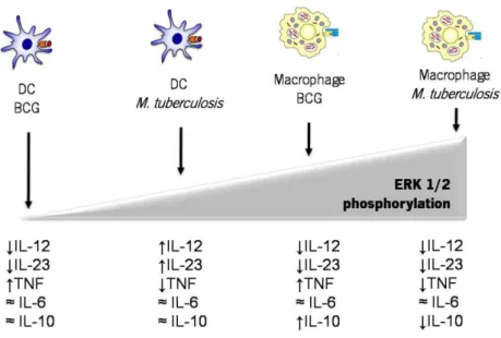
The expression of IL-12, IL-6 and IL-23 is mainly associated with cells of the innate immune response. IL-12 is a heterodimeric cytokine consisting of the two subunits IL-12p40 (p40) and IL-12p35 which are covalently linked (Trinchieri, 2003). IL-12 expression is induced following phagocytosis of M. tuberculosis bacilli in macrophages and DC (Henderson et al., 1997; Ladel et al., 1997). Two studies comparing murine macrophages and DC demonstrated that DC release significantly higher amounts of IL-12 than did macrophages in response to live M. tuberculosis (Giacomini et al., 2001; Hickman et al., 2002). In vitro, M. tuberculosis-infected DC also primed naive T cells toward Th1 development, while macrophages did not (Verreck et al., 2004). IL-23 is another cytokine of the IL-12 family (Hunter, 2005) and, as IL-12, is a heterodimeric cytokine composed by the p40 subunit covalently linked to a p19 subunit . IL-23 secretion in response to TLRs activation appears to be more pronounced in DC than in macrophages (Gerosa et al., 2008; Jang et al., 2008; Lyakh et al., 2008). Interestingly, secretion of IL-12 and IL-23 by M. tuberculosis stimulation of DC can, in addition to TLRs, be also dependent on signals mediated by Dectin-1 (Gerosa et al., 2008; Rothfuchs et al., 2007; Zenaro et al., 2009).
Several studies highlight the role of IL-12, IL-23, IL-6 and of IFN- and IL-17 during the course of infection. A role for IL-12 and cytokines of the Th1 axis, predominantly IFN-, is
established in protection against mycobacterial infections in both mouse models (Cooper, 2009a; Flynn et al., 1993) and human disease (Casanova and Abel, 2002; Flynn and Chan, 2001) (Cooper, 2009b). In particular, IFN- that in addition to Th1 cells can also be produced by natural killer (NK) cells (Scharton and Scott, 1993) and CD8+ T cells (Barnes et al., 1993; Lalvani et al., 1998; Orme et al., 1992) is also the hallmark molecule for protection against TB (Chackerian et al., 2002; Cooper et al., 1993; Ellner et al., 2000; Flynn et al., 1993; Ottenhoff et al., 1998). The role of IL-23 and Th17 responses in the infection by M. tuberculosis is yet not fully understood. Studies performed in mice lacking p40, p35, p19 or combinations of these genes, showed that IL-23 is less critical than IL-12 for protection against M. tuberculosis, and only provides a moderate level of protection to the host in the absence of biologically active IL-12 (Khader et al., 2005). IL-17, secreted not only by CD4+ Th17 cells (Khader et al., 2007), but also by T cells (Lockhart et al., 2006), has been shown to have a limited role in host defense against M. tuberculosis during primary infection (Aujla et al., 2007). However, vaccination has been shown to trigger an IL-17-dependent accelerated IFN- response by CD4 T cells in the lung during
subsequent M. tuberculosis infection (Khader et al., 2007). Importantly, IL-17 promotes neutrophil recruitment to the site of infection (Fossiez et al., 1996; Jones and Chan, 2002; Ye et al., 2001), but this ability of IL-17 to induce chemokine production and cell recruitment to the infected tissue can in certain situations be associated with the development of immune pathology (Cooper, 2009a) (Cruz et al, umpublished data).
Another important cytokine with a key role in the immune response to M. tuberculosis is TNF. M. tuberculosis induces TNF secretion by macrophages, DC and T cells (Barnes et al., 1993; Henderson et al., 1997; Ladel et al., 1997; Serbina and Flynn, 1999). The requirement for TNF for the control of M. tuberculosis infection is complex, but it clearly is an important component for macrophage activation, as TNF, in synergy with IFN-, induces NOS2 expression (Chan et al., 1992; Liew et al., 1990). The importance of this cytokine in granuloma formation in TB and other mycobacterial diseases has been significantly documented (Flynn and Chan, 2001; Miller and Ernst, 2008).
The proinflammatory response which is initiated by M. tuberculosis is antagonized by anti-inflammatory cytokines that contribute to the control of the magnitude of the anti-inflammatory responses. TGF- was found to be present in granulomatous lesions of TB patients and is produced by human monocytes after stimulation with M. tuberculosis (Toossi et al., 1995) or M. tuberculosis lipoarabinomannan (Lam) (Dahl et al., 1996). TGF- has important
anti-inflammatory effects, including deactivation of macrophage production of ROI and RNI (Ding et al., 1990), inhibition of T cell proliferation (Toossi and Ellner, 1998), interference with NK and CTL function and downregulation of IFN- (Ruscetti et al., 1993), TNF and IL-1 release (Ruscetti et al., 1993). TGF-β is also an important mediator of immune-suppression by regulatory T cells (Gorelik and Flavell, 2002). However, together in the presence of IL-6, TGF- induces Th17 differentiation (Veldhoen et al., 2006).
The anti-inflammatory cytokine IL-10 is also expressed during M. tuberculosis infection (Barnes et al., 1993; Boussiotis et al., 2000; Gerosa et al., 1999; Shaw et al., 2000). IL-10 is needed to limit tissue damage by controlling the immune response (Moore et al., 2001). However, an excess of IL-10 most likely prevents pathogen clearance. Indeed, transgenic mice constitutively expressing IL-10 were less capable of clearing a BCG infection, although T cell responses including IFN- production were unimpaired (Murray et al., 1997), thus suggesting that IL-10 may counter the macrophage activating properties of IFN- . Interestingly, IL-10-/- mice were not more resistant to acute M. tuberculosis, compared to wild type mice (North, 1998). However, lack of IL-10 was recently link to a decreased control over the inflammatory response that eventually resulted in progression of disease, bacterial multiplication and morbidity (Higgins et al., 2009).
An appropriate immune response to M. tuberculosis is the result of a balance between inflammation and regulation. In what extent does the vaccination with BCG change the parameters of this response is still not fully known. This knowledge is however important in order to improve the efficacy of BCG vaccination or to develop new vaccination strategies.
II. AIMS
BCG is the only approved vaccine used for TB prevention. Although BCG is used worldwide, the cellular and molecular mechanisms by which this vaccine acts are still poorly understood. Although BCG is protective against tuberculous meningitis, it shows great variability in the prevention of pulmonary disease.
Several experimental vaccines are being developed against TB. Some are able to induce the same level of protection as BCG, although none were demonstrated to induce better protection than BCG. Therefore it is fundamental to understand how BCG modulates the innate and cellular immunity and where it fails, in order to improve vaccination strategies to TB.
The main goal of this work was to perform a comparative study of BCG versus M. tuberculosis infection on DCs and macrophages to better understand how these pathogens interact with cells of the innate immune system, what molecular pathways are triggered, and how that interaction translates into an effective, or not, Th cell responses
Specifically, the following aims were addressed:
1. Investigation of the magnitude of macrophage and DCs responses to M. tuberculosis or BCG, in terms of cytokine production by these phagocytes;
2. Elucidation of the molecular events that might explain differences in the cytokine response of macrophages and DCs to M. tuberculosis or BCG;
3. Understanding the type of Th cell responses developed in the presence of M. tuberculosis- or BCG- infected DCs;
4. Clarification of the effector activity of macrophages in what regards the control of M. tuberculosis or BCG growth.
Dissecting the cellular and molecular events that occur upon mycobacterial challenge will help to reveal possible weak points of both the host and the bacteria that can be targeted and will subsequently provide clues for the design of new and better vaccines.
III.
MATERIAL & METHODS
3.1. AnimalsEight-week-old female C57BL/6 and Balb/c mice were obtainned from Charles River Laboratory (Barcelona, Spain). Mice transgenic for the DO11.10 / TCR were backcrossed on a RAG-deficient (RAG 2/2) BALB/c background and were kindly provided by Dr. Anne O’Garra (NMIR, London, England).
3.2. Bacteria
The H37Rv strain of M. tuberculosis and M. bovis BCG Pasteur were grown in Proskauer Beck medium containing 0.05% Tween 80 to mid-log phase and frozen in 1-ml aliquots at -80ºC. Bacterial viability was determined by counting the number of CFU (colony forming units) on Middlebrook 7H11 agar plates. M. bovis BCG Pasteur were obtained from Trudeu Institute Mycobacterium Collection (TMC 1011).
3.3. Culture of Bone Marrow Derived Macrophages
Primary mouse bone marrow derived macrophages were generated from WT C57BL/6 animals as described elsewhere(Saraiva et al., 2009). Petri dishses were initially seeded with 1x10⁵ cells in complete medium with 20% of L-929 conditioned medium (LCCM) and incubated at 37ºC in 5% CO2 humidified air chamber. On day 4, the medium was renewed and cultures were used at day 7. Macrophages were stimulated with M. tuberculosis or BCG at a multiplicity of infection (MOI) 2:1 (bacteria/macrophage) for different periods of time. Some cultures received 100 U/ml of mouse IFN- (R&D Systems).
3.4. Culture of Bone Marrow Derived Dendritic Cells
Primary mouse bone marrow derived dendritic cells (DCs) were differentiated from WT C57BL/6 mice as described previously (Saraiva et al., 2009). Cells were culture in 6-well plates, containing 5x10⁶ cells in complete medium with 20% of granulocyte-macrophage colony-stimulating factor (GM-CSF) and incubated at 37ºC in 5% CO2 humidified air chamber. On day 2, 4 and 6, the medium was renewed and cultures were used at day 7. DCs were exposed to live M. tuberculosis or BCG at a MOI of 2 for different periods of time.
3.5. Quantitative Real Time-PCR (RT-PCR) Analysis
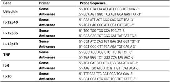
Total RNA from cultured macrophages was extracted with TRIzol® Reagent (Invitrogen, San Diego, CA) according to the manufacturer’s instructions. Reverse transcription was done with whole RNA in a final volume of 20 μl using SuperScript II (Invitrogen) and Oligo(dT) (Roche) according to the manufacturer’s instructions. The cDNA was then subjected to real-time PCR for quantification of IL-12p40, IL-12p35, IL23p19, TNF, IL-6, IL-10 and Ubiquitin (used as house-keeping gene) in 10 µl with SYBR Green Supermix (Bio-Rad)in an CFX 96 Real-Time (Bio-Rad) system. The specific conditions of each PCR are listed in the table 1. All reaction were performed using the following cycling parameters: 1 cycle of 95ºC for 15 min, followed by 30 cycles of 95ºC for 15 min, 58ºC for 20 min and 72ºC for 15 min, and 2 amplification cycles of 65ºC for 1.3 min and 95ºC for 15min; and 1 cooling cycle of 35ºC for 1.3 min. Relative mRNA expression was calculated accordingly with the following equation: 1.8 ^ (ubiquitin mRNA expression – specific cytokine gene mRNA expression) x 100000.
Table 1 - Sequence of the primers specific for genes and conditions used in RT-PCR reaction.
3.6. ELISA Assay
Supernatants from M. tuberculosis- or BCG-infected DCs were collected at 24h post-infection and screened for IL-12p70, IL-12p40, IL-23p19, TNF, IL-6 or IL-10 Ready Set-Go ELISA kit (eBioscience) by sandwich ELISA.
Gene Primer Probe Sequence
Ubiquitin Sense
Anti-sense
5’- TGG CTA TTA ATT ATT CGG TCT GCA -3’ 5’- GCA AGT GGC TAG AGT GCA GAG TAA -3’
IL-12p40 Sense
Anti-sense
5’- CAA ATT ACT CCG GAC GGT TCA -3’ 5’- AGA GAC GCC ATT CCA CAT GTC -3’
IL-12p35 Sense
Anti-sense
5’- TGC TGG TGG CCA TCG AT -3’ 5’- GCA GAG TCT CGC CAT TAT GAT TC-3’
IL-12p19 Sense
Anti-sense
5’- CGT ATC CAG TGT GAA GAT GGT TGT -3’ 5’- GCT CCC CTT TGA AGA TGT CAG A-3’
TNF Sense
Anti-sense
5’- GCC ACC ACG CTC TTC TGT CT -3’ 5’- TGA GGG TCT GGG CCA TAG AAC -3’
IL-6 Sense
Anti-sense
5’- ACA CAT GTT CTC TGG GAA ATC GT -3’ 5’- AAG TGC ATC ATC GTT GTT CAT ACA -3’
IL-10 Sense
Anti-sense
5’- TTT GAA TTC CCT GGG TGA GAA -3’ 5’- GCT CCA CTG CCT TGC TCT TAT T -3’
3.7. Protein Analysis
Macrophages and DCs were cultured in medium containing 1% FBS overnight before stimulation with M.tuberculosis or BCG and washed in PBS before lysis (1% NP-40, 0.1% SDS, 0.5% deoxycholate acid, 50 mM Tris HCl, pH 8.0, 50 mM NaCl, 2 mM EDTA, 2 mM sodium-pyrophosphate, 50 mM sodium fluoride, 100 mM vanadate [all from Sigma-Aldrich], and complete EDTA-free protease inhibitor cocktail (Roche). Immunoblotting of proteins was performed as previously described (Saraiva et al., 2005) and visualized with ECL (GE Healthcare) or SuperSignal West Femto Substrate (Thermo Fisher Scientific). Using specific antibodies that exclusively recognize the bi-phosphorylated forms (activated) of ERK1/2 or the total form of the same enzyme, the ratio between the Western Blot signals obtained for the phosphorylated versus the total form, allow the quantification of the amount of ERK1/ERK2 activated within the cells upon M. tuberculosis or BCG stimulation. Antibodies used: rabbit (polyclonal) anti-ERK1/2 [pTpY185/187) phosphospecific (Biosource), rabbit (polyclonal anti-ERK1/2 pan (Biosource).
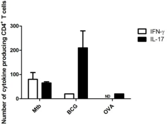
3.8. In vitro CD4 T Cell Activation
Naive CD4+ T cells were generated from OVA-TCR transgenic DO.11.10 mice as described (Cruz et al., 2006) and cultured (1x10⁶ cells/ml) with M. tuberculosis- or BCG- infected DCs (1x10⁶ cells/ml) for 72h at 37°C in 5% CO
2, and in 10 ng/ml IL-2 and 5 M OVA323–339 peptide. Stimulated T cells were washed and counted, and the frequency of IFN-- and IL-17-producing CD4+ T cells determined by ELISPOT.
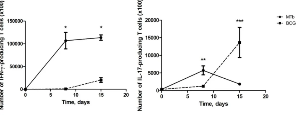
3.9. In vivo Infection
C57BL/6 animals were infected by the intravenous route with M. tuberculosis strain H37Rv or with BCG (1x10⁶ CFU) and at different time points the animals were sacrificed and the spleens removed. The ability of the splenocytes to produce IFN- or IL-17 in response to Antigen 85 was assessed by ELISPOT.
3.10. ELISPOT Assay
ELISPOT was performed as described previously (Cruz et al., 2006). Briefly, a total of 1x10⁵ cells was added to Ab-coated wells, 2-fold dilutions were made, and irradiated splenocytes from Balb/6 or C57/BL6 mice were added at 1x10⁶ cells per well. A peptide representing an I-Ab -restricted epitope of antigen85A (Cole et al., 1998) were used to stimulate cells from infected
mice, whereas Ova323–339 stimulated DO.11.10 T cells (Camus et al., 2002); all wells contained 10 ng/ml IL-2. After 24 h, plates were washed and the number of IFN-- or IL-17-producing CD4+T cells determined as described (Cruz et al., 2006). Cells from mice infected with M. tuberculosis or BCG, but cultured in the absence of antigen, did not produce spots.
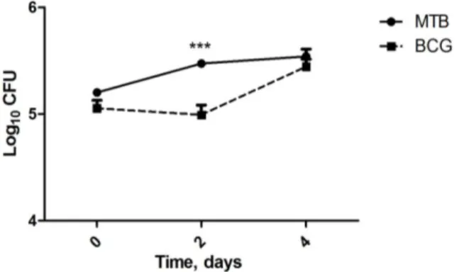
3.11. Bacterial Load Determination
To determined the number of viable bacteria, macrophage monolayers were lysed with 0.1% (final concentration) saponin and the bacterial suspensions were serially diluted and plated onto Middlebrook 7H11 agar medium. Bacterial colony formation was counted after 3 weaks of incubation at 37°C.
3.12. Nitrites Quantification
Nitrite production by macrophage monolayers was determined by the colorimetric Griess assay as described elsewhere (Turner et al., 2001). Briefly, supernatants from macrophage cultures were placed into 96-well enzyme-linked immunosorbent assay plate in duplicates, and equal volume of Griess reagent (1% sulfanilamine, 0.1% nathylethylenediamine, 2,5% H3PO4) was added. The absorbance was measured at 550nm on a spectrophotometer, and the concentrationof nitrite was calculated by comparing optimal density values to a standard curve of NaNO2.
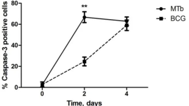
3.13. Immunofluorescence
Macrophages were cultivated as described above except that lamellae were placed on the bottom of the wells of the 24-wells incubation plaques. At specific time points after infection, the lamellae were collect and fixed in 2% PFA. Next, the lamellae were incubated with the primary antibody affinity-purified rabbit anti-human/mouse caspase 3 active (R&B Systems) and was detected with goat anti-rabbit IgG (H+L) Alexa Fluor 568 (Molecular Probes A11011). DAPI was used to counter stained and to detect nuclei. Pictures were observed with Olympus BX61 microscope and images were recorded with Olympus DP70 camera.
3.14. Stastistical Analysis
The results are given as means ± SE of the mean. Statistical significance was performed by using Studant’s t test. Values of p < 0.05 were considered significant.
IV.
RESULTS
4.1. DC responses to M. tuberculosis and to BCG are stronger than macrophageresponses, and M. tuberculosis is a stronger stimulus than BCG to DCs
To understand the early steps of the immune response to M. tuberculosis and BCG and since DCs and macrophages are the central sensory components of the immune system, being within the first cells to recognize the presence of pathogens and to respond to it, we decided to compare the response of both cells types to either M. tuberculosis or BCG in a systematic and extensive fashion.
The first point to be addressed in our comparison was the response of macrophages and DCs to M. tuberculosis or BCG in terms of cytokine production. To do this, we prepared primary cultures of macrophages and DCs derived from mouse bone marrow and stimulated with live M. tuberculosis or BCG at a MOI of 2. At different time points post-infection, we extracted RNA from the stimulated cells, prepared cDNA and measured the expression of several cytokines by RT-PCR. We were particularly interested in studying whether the expression of cytokines with a role in T cell differentiation induced by M. tuberculosis or BCG was different. Since 12p70 and IL-23 are important for Th1 and Th17 responses, respectively, we started by measuring the transcription of the monomers that compose IL-12 and IL-23 (p40-p35 and p40-p19, respectively). We also measured the expression of other immune mediator cytokines such as TNF, which contributes to the initial control of the infection, for example by activating the infected macrophages in an autocrine way (Chan et al., 1992; Liew et al., 1990); IL-6 described to be required for the development of Th17 cells (Veldhoen and Stockinger, 2006); and IL-10, an anti-inflammatory cytokine known to inhibit macrophage and DCs functions (Demangel et al., 2002; Madura Larsen et al., 2007).
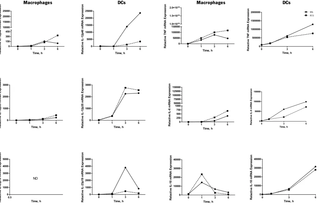
As shown in Fig.1, we observed that both macrophages and DCs express various cytokines in response to M. tuberculosis or BCG. Both cell types showed similar patterns of expression for p35, TNF, IL-6 and IL-10 in response to either mycobacterium. However, major differences were identified in terms of the IL-12 subunits p40 and p19. Indeed, whereas macrophages stimulated with M. tuberculosis or with BCG did not express detectable amounts of IL-12p19 monomer, DCs did express this molecule, but the levels obtained in response to M. tuberculosis stimulation were higher than those obtained with BCG stimulation. In what regards the expression of IL-12p40, the pattern was similar to the one observed for p19. Again, maximum p40 expression was induced
by M. tuberculosis in DCs. BCG induced only modest amounts of p40 in DCs and the expression of this molecule by macrophages was low, but detectable. Interestingly, M. tuberculosis-stimulated DCs, always expressed higher levels of the tested cytokines, suggesting that M. tuberculosis is stronger than BCG as a stimulus to DCs. As for macrophages, the two agents yielded similar responses, thus suggesting that different regulatory pathways are in place in macrophages versus DCs. Overall, our data show a differential cytokine expression by macrophages and DCs when stimulated with M. tuberculosis or BCG.
0 1 3 6 0 150 300 450 10000 15000 20000 25000 Time, h R ela ct iv e IL -1 2p 40 m R N A E xp re ss io n 0 1 3 6 0 5000 10000 15000 20000 25000 Time, h R ela ct iv e IL -1 2p 40 m R N A E xp re ss io n 0 1 3 6 0 50000 100000 150000 1.0×1006 1.5×1006 2.0×1006 Time, h R ela ct iv e T N F m R N A E xp re ss io n 0 3 6 0 500000 1000000 1500000 2000000 Mtb BCG Time, h R el ac ti ve T N F mR N A E xp re ss io n 0 1 3 6 0 1000 2000 3000 Time, h R ela ct iv e IL -1 2p 35 m R N A E xp re ss io n 0 1 3 6 0 1000 2000 3000 Time, h R ela ct iv e IL -1 2p 35 m R N A E xp re ss io n 0 1 3 6 0 250 500 750 1000 50000 75000 100000 125000 150000 Time, h R el ac ti ve I L -6 mR N A E xp re ss io n 0 3 6 0 50000 100000 150000 Time, h R el ac ti ve I L -6 mR N A E xp re ss io n 0.5 0 1000 2000 3000 4000 5000 ND Time, h R ela ct iv e IL -2 3p 19 m R N A E xp re ss io n 0 1 3 6 0 1000 2000 3000 4000 5000 Time, h R ela ct iv e IL -2 3p 19 m R N A E xp re ss io n 0 1 3 6 0 10000 20000 30000 40000 Time, h R ela ct iv e IL -1 0 m R N A E xp re ss io n 0 3 6 0 10000 20000 30000 40000 Time, h R ela ct iv e IL -1 0 m R N A E xp re ss io n
Figure 1 - M. tuberculosis and BCG are stronger stimuli to DCs than to macrophages, and M. tuberculosis is a stronger stimulus than BCG to DCs.
4.2. M. tuberculosis-stimulated DCs are stronger producers of IL-12 and IL-23 than BCG-stimulated DCs.
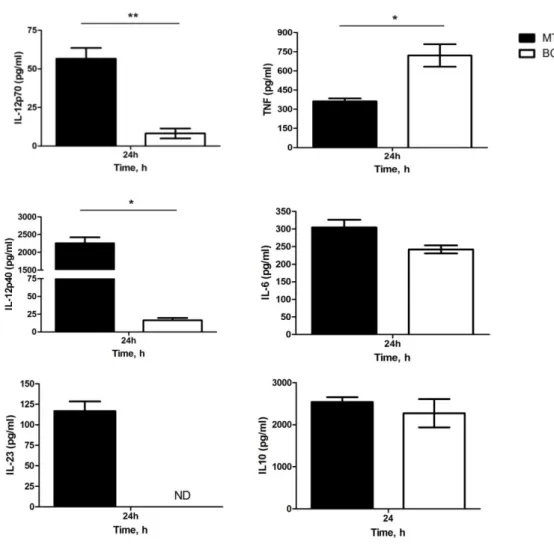
From our mRNA analysis, we concluded that the transcription of p40 and p19 by DCs was only induced in relevant levels by M. tuberculosis stimulation. Taking into consideration the role of IL-12 (p40-p35) and IL-23 (p40-p19) molecules in Th cell differentiation, we decided to assess the secretion of IL-12p70 and IL-23 by M. tuberculosis or BCG stimulated DCs. For that, we collected DCs culture supernatants 24 hours post-stimulation and measured by immunoassay the amounts of cytokines secreted. Consistently with the observed gene expression pattern, the analysis of supernatants from stimulated DCs showed that both IL-12p70 and IL-12p40 release was higher in DCs stimulated with M. tuberculosis comparing to stimulation with BCG (Fig. 2). IL-23 production was only detectable in M. tuberculosis-stimulated DCs. We also measured the secretion of TNF, IL-6 and IL-10. We found that IL-6 and IL-10 production was similar in both M. tuberculosis- and BCG-stimulated DCs, but TNF production was higher in DCs stimulated with BCG (Fig.2). Thus our data suggest that, in terms of protein expression, M. tuberculosis-stimulated DCs are potent producers of IL-12 and IL-23 whereas BCG-tuberculosis-stimulated DCs are not. Importantly, our data also show that, despite the difference observed for IL-12 and IL-23, BCG is able to induce the production of certain cytokines by DC, thus suggesting that activation signals can be generated by BCG. We are currently performing the ELISA assays for macrophages supernatants to further validate the results obtained by PCR.
Figure 2 - M. tuberculosis infected DCs produce higher amounts of IL-12 and IL-23 than BCG-infected DCs. DCs were differentiated from WT mice and exposed to M. tuberculosis or BCG at an MOI of 2. DCs culture supernatants were harvested 24 hours post stimulation and analyzed by ELISA for IL-12p70, IL-12p40, IL-23, TNF, IL-6 and IL-10 concentration. Each time point represents the mean of three wells. Results are representative of three independent experiments with similar results and show mean ± SD; *, **, p<0.05 and 0.01, respectively. Mtb (M. tuberculosis). ND (not detected)
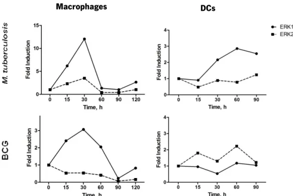
4.3. M. tuberculosis induces earlier and stronger activation of ERK phosphorylation in macrophages as compared to DCs
From the results shown in the previous sections, macrophages and DCs respond differently to M. tuberculosis or BCG stimulation in terms of cytokine expression, particularly in what regards the expression of IL-12 and IL-23 (Fig.1 and Fig.2). We hypothesised that these differences might be a consequence of a differential activation of intracellular signalling cascades. Previous studies have suggested the activation of various signalling cascades following mycobacterial infection, such as the MAPK pathway (Cobb, 1999) that includes three main