INTRODUCTION
Address to: Dra. Inês Aparecida Tozetti. FM/UFMS. Caixa Postal 549,
79070-900 Campo Grande, MS, Brasil.
Phone: 55 67 3345-7387
e-mail: [email protected]
Received 7 February 2013
Accepted 4 June 2013
Glucocorticoid-induced tumor necrosis factor
receptor expression in patients with cervical
human papillomavirus infection
Cacilda Tezelli Junqueira Padovani
[1],
Camila Mareti Bonin
[2],
Inês Aparecida Tozetti
[3],
Alda Maria Teixeira Ferreira
[2],
Carlos Eurico dos Santos Fernandes
[2]and Izaias Pereira da Costa
[1][1].Programa de Pós-Graduação em Saúde e Desenvolvimento da Região Centro-Oeste, Faculdade de Medicina, Universidade Federal de Mato Grosso do Sul, Campo Grande, MS. [2]. Centro de Ciências Biológicas e da Saúde, Universidade Federal de Mato Grosso do Sul, Campo Grande, MS. [3]. Programa de Pós-Graduação em Doenças Infecciosas e Parasitárias, Faculdade de Medicina, Universidade Federal de Mato Grosso do Sul, Campo Grande, MS.
ABSTRACT
Introduction: The progression of human papillomavirus (HPV) infection in the anogenital tract has been associated with the involvement of cells with regulatory properties. Evidence has shown that glucocorticoid-induced tumor necrosis factor receptor (GITR) is an important surface molecule for the characterization of these cells and proposes that GITR ligand may constitute a rational treatment for many cancer types. We aimed to detect the presence of GITR and CD25 in cervical stroma cells with and without pathological changes or HPV infection to better understand the immune response in the infected tissue microenvironment. Methods: We subjected 49 paraffi n-embedded cervical tissue samples to HPV DNA detection and histopathological analysis, and subsequently immunohistochemistry to detect GITR and CD25 in lymphocytes. Results: We observed that 76.9% of all samples
with high GITR expression were HPV-positive regardless of histopathological fi ndings. High GITR expression (77.8%) was predominant in samples with ≥1,000 RLU/PCB. Of the HPV-positive samples negative for intraepithelial lesion and malignancy,
62.5% had high GITR expression. High GITR expression was observed in both carcinoma and high-grade squamous intraepithelial
lesion (HSIL) samples (p = 0.16). CD25 was present in great quantities in all samples. Conclusions: The predominance of high
GITR expression in samples with high viral load that were classifi ed as HSIL and carcinoma suggests that GITR+ cells can exhibit
regulatory properties and may contribute to the progression of HPV-induced cervical neoplasia, emphasizing the importance of GITR as a potential target for immune therapy of cervical cancer and as a disease evolution biomarker.
Keywords: Human papillomavirus. Immune response. Immunohistochemistry.
Human papillomavirus (HPV) infects the basal and parabasal cells of squamous epithelium in the female anogenital tract, and HPV types 16, 18, 31, 33, and 45 in particular are believed to put patients at high risk for the development of high-grade
cervical intraepithelial neoplasia (CIN) and cervical carcinoma1.
Infection progression has been associated with several factors, including the persistence of HPV, the presence of high-risk oncogenic HPV types, high viral load, integration of
viral DNA, and E6 and E7 viral oncoprotein activity1. Evidence
shows that regulatory T cells (Treg) are also involved in the progression to cervical neoplasia in HPV-infected patients2-5. HPV-specifi c CD4+ regulatory cells isolated from lymph node
biopsies of patients with cervical carcinoma were found to
suppress proliferation and cytokine (interferon-γ, interleukin [IL]-2) production by responder T cells5.
Treg cells play a crucial role in modulating the elimination of pathogens and tumor antigens and perform their function through immunosuppressive cytokine production and immunosuppression induction mediated by cell-to-cell contact, having the ability to suppress the activation, proliferation, and effector function of different cell types contributing to the immune response6,7.T
reg cells are subdivided into several
subpopulations, one being the natural Treg cells (CD4+CD25+T reg),
which numerically represent the largest group of cells with suppressor activity8.
Studies show that Treg cells are activated with greater sensitivity than naïve effector T cells after antigenic stimulation, which has been attributed mainly to their semi-activated state that is thought to be due to the increased expression of CD25
(α-chain of the IL-2 receptor), glucocorticoid-induced tumor
necrosis factor receptor (GITR) markers, and others9-11.
METHODS RESULTS
DISCUSSION FOXP3 (forkhead box p3) is highly specific and that its
transduction into naïve T cells increases the molecular expression associated with Treg cells, such as that of CD25 and GITR12,13.
Evidence shows that another characteristic surface molecule of cells with regulatory properties, Treg cells in particular, is the GITR14 — a tumor necrosis factor receptor superfamily
member — which is predominantly expressed in CD25+ CD4+
Treg cells and plays an important role in the regulation of mucosal immune responses15-19. Recent studies have demonstrated that in vivo GITR ligation using an agonist anti-GITR monoclonal antibody, DTA-1, can augment anti-tumor T-cell responses by modulating Treg cells, which makes targeting GITR a potential immunotherapeutic approach to cancer treatment20-22.
Given the fi ndings that indicate the involvement of cells with
regulatory properties, especially Treg cells, in the progression of cervical malignant lesions3,4,23,24, this study aimed to detect both
CD25 and GITR markers in lymphocytes of cervical stroma to better understand the immune response in the microenvironment of HPV infection, which may shed light on novel therapeutic interventions against intraepithelial neoplasia and cervical cancer of viral etiology, and perhaps also make GITR a possible candidate biomarker for disease evolution.
Samples
Forty-nine patient cervical samples embedded in paraffi n and
selected on a non-probabilistic form by convenience sampling from 2000 to 2002 in the Cancer Prevention Center of Campo Grande, Mato Grosso do Sul, Brazil, were used. These samples previously underwent a Hybrid Capture II reaction (Digene, Gaithersburg, MD, USA) to quantify the viral load for group B - high
oncogenic risk types that were classifi ed into scores from 0 to 3: 0
(HPV-negative samples); 1 (1 to < 100U of light released for
probe; relative light units/positive control to group B (RLU/PCB); 2 (100 to <1,000 RLU/PCB); and 3 (≥1,000 RLU/PCB). On the basis of histopathological analysis, the samples were classifi ed as low-grade squamous intraepithelial lesions (LSIL) (CIN I); high-low-grade squamous intraepithelial lesions; (HSIL) (CIN II, III); carcinoma, and negative for intraepithelial lesion and malignancy (NILM).
Immunohistochemistry of CD25 and GITR
The Immunohistochemistry (IHC) reaction was developed using antigen retrieval with wet heat and 0.05M ethylenediaminetetraacetic acid (EDTA) pH 8.0 for the detection of CD25 marker25 and 10mM Tris and 1mM EDTA for the GITR marker. The primary antibodies used included anti-human IL-2R
(eBioscience®, San Diego, CA, USA; clone B-B10/cod.BMS134)
and anti-GITR (R&D systems®, Minneapolis, MN, USA; goat
IgG/cod.AF689).
The detection system was a Universal LSAB + Kit/HRP
(Dako®, Carpinteria, CA, USA), and diaminobenzidine (Dako®)
was used as a chromogen. Counterstaining was performed in hematoxylin, and the slides were observed under common optical microscopy with 10× and 40× objective lenses. Samples
showing brown staining on characteristic cells were considered positive. Human tonsil tissue was used as the external control of the reaction over which the primary antibody (positive control) and phosphate buffer pH 7.4 containing 1% albumin (negative control) was applied.
Quantitative analysis
According to the presence of immunomarked cells, the
histological sections were classifi ed in low (small quantities)
and high (large quantities) scores. The slides were analyzed by
2 independent observers, previously cal ibrated (κ = 0.98), and the fi nal result of discordant cases was obtained by common analysis to produce a consensus.
Statistical analysis
Analysis of the frequencies among the histopathological
fi ndings and viral load according to GITR expression intensity
were compared using Fisher’s exact test.
Ethical considerations
This study was approved by the Research Ethics Committee of the Universidade Federal de Mato Grosso do Sul, protocol number 975, July 31, 2007.
We observed a predominance of GITR in large quantities
(7/9; 77.8%) in the samples with ≥1,000 RLU/PCB (viral load
3), although increases in viral load did not have a statistical
correlation with high GITR expression (p=0.40). Regardless of histopathological fi ndings, among all samples with high
GITR expression, 76.9% (20/26) were HPV-positive (viral load, 1-3). Among the NILM samples, 40% (8/20) were HPV-positive (viral load, 1-3) and 62.5% (5/8) of these showed high GITR expression, while among NILM-HPV negative samples (viral load, 0), only 33.3% (4/12) showed high GITR expression (Table 1).
A frequency analysis of the histopathological findings according to GITR expression intensity is shown in Table 2. High GITR expression was predominant in the carcinoma and
HSIL samples (p = 0.16) (Figure 1).
All samples showed intense staining for CD25 regardless of
the result of viral load or histopathological fi ndings (Figure 2).
In the present study, we observed that among the high GITR expression samples 76.9% were HPV-positive and 23.1% were HPV-negative. The expression of this marker was predominant in samples with high viral load as well as high-grade lesions and carcinoma.
A number of surface and secreted molecules have been associated with Treg, and GITR has been recognized as CD4+
Treg markers in mice and humans22,26,27. In this context, it is of
interest that GITR+ T
TABLE 1 - Distribution of histopathological fi ndings according to viral load and GITR expression (n=49).
NILM LSIL HSIL CA
GITR GITR GITR GITR
Viral load low high low high low high low high Total
0 8 4 0 0 0 1 0 1 14
1 1 2 5 2 3 4 1 1 19
2 1 1 0 0 2 2 0 1 7
3 1 2 1 1 0 1 0 3 9
Total 11 9 6 3 5 8 1 6 49
GITR: glucocorticoid-induced tumor necrosis factor receptor; NILM: negative for intraepithelial lesion and malignancy; LSIL: low grade squamous intraepithelial lesion; HSIL: high grade squamous intraepithelial lesion; CA: carcinoma. Viral load - 0 (negative); 1 (1 to < 100 RLU/ PCB); 2 (100 to < 1,000 RLU/PCB); 3 (> 1,000 RLU/PCB); RLU/PCB: relative light unit/positive controls to group B; GITR - low: small quantities of immunomarked cells; GITR - high: large quantities of immunomarked cells.
TABLE 2 - Frequence of histopathological fi ndings according to the intensity of GITR expression.
Histopathological (n/%)
NILM LSIL HSIL CA
GITR n % n % n % n %
low 11 55.0 6 66.7 5 38.5 1 14.3
high 9 45.0 3 33.3 8 61.5 6 85.7
Total 20 100.0 9 100.0 13 100.0 7 100.0
GITR: glucocorticoid-induced tumor necrosis factor receptor; NILM: negative for intraepithelial lesion and malignancy; LSIL: low grade squamous intraepithelial lesion; HSIL: high grade squamous intraepithelial lesion; CA: carcinoma. GITR - low: small quantities of immunomarked cells; GITR - high: large quantities of immunomarked cells. (p=0.16).
the immune system to control the development of HPV-induced cancer. Studies have demonstrated increased frequencies and suppressive activity of Treg cells in patients with high-grade lesions and cervical cancer. In addition, compared to colorectal cancer, skin melanoma, and bronchial carcinoma, HPV-derived
CIN lesions and cervical carcinomashave higher numbers of Treg cells23,24.
One study that investigated the infl uence of tumor-infi ltrating
Treg cells on tumor-specifi c T cell responses found that Treg cells in patients with liver cancer upregulated GITR expression compared with Treg cells in tumor-free liver tissue and blood28. Another study identifi ed increased numbers of Treg GITR
+ cells in
tumor-positive lymph nodes compared with tumor-negative nodes in the same patient29. Both studies propose that GITR ligand could
be a promising treatment for cancer and that GITR and GITR ligand are good candidates for disease evolution biomarkers. Studies investigating the natural history of HPV infection have shown that viral clearance may vary from 4-16 months according to the virus’ oncogenicity30-32. However, it has been
observed that persistent infection with a higher likelihood of progression to high-grade lesions and invasive carcinoma can occur in the face of an ineffective immune response. In this context and considering that HPV infection is restricted to epithelial cells, the importance of the local immune response is highlighted,
making the components present in the microenvironment crucial for lesion development or regression33-36. The role of GITR has
been unclear until now, emphasizing the importance of the present study to clarify the immune response in the cervical microenvironment.
The presence of high GITR and CD25 expression levels
found in HPV-derived CIN lesions and cervical carcinomas
indicates that these cells may play an important role in the downregulation of immune responses37. A strong association
between these markers and Treg cells was demonstrated in a study that found GITR expression in only those cells that also expressed CD4 and CD25, and most of them co-expressed FOXP338. The association of GITR and CD25 with negatively
regulated T helper-activated lymphocytes has been demonstrated
in experiment with C57BL/6 GITR+/+ mice (wild type), which showed decreased IL-2 expression compared to C57BL/6
GITR-/-39.
The relevance of cells expressing the studied markers in immune response suppression is emphasized by studies that evaluate in vitro regulatory activity through cytokine expression by CD4+ T cells, CD4+CD25+GITR+ cells, and CD4+CD25
-GITR+ cells. These studies showed that the fi rst produced
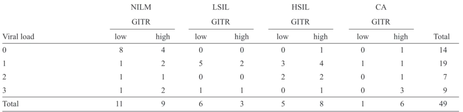
FIGURE 1 - GITR expression in HPV positive cervical lesions. a: positive control human tonsil stained with anti-human GITR antibody; b: negative control human tonsil - omitting primary antibody, showed no staining; c: CIN II and d: NILM, with GITR expressing cells in small quantities; e: CIN III and f: CIN I, with GITR expressing cells in large quantities. All fi gures are presented in the same magnifi cation (400X). Black arrows indicate positive-staining cells with anti-human GITR antibody. GITR: glucocorticoid-induced tumor necrosis factor receptor; HPV: human papillomavirus; CIN: cervical intraepithelial neoplasia.
A
B
C
D
E
F
FIGURE 2 - CD25 expression in HPV positive cervical lesions. A: Positive control human tonsil stained with anti-human IL-2R antibody; B: negative control
human tonsil - omitting primary antibody, showed no staining; C: CIN II, with CD25 expressing cells in large quantities. All fi gures are presented in the same magnifi cation (400X). Black arrows indicate positive-staining cells with anti-human IL-2R antibody. HPV: human papillomavirus; IL-2R: IL-2 receptor; CD25: α-chain of the IL-2 receptor.
It is unclear whether increased frequencies of regulatory cells are a cause or consequence of high viral load and chronic infection2,40,41. The predominant expression of GITR in samples with high viral load and classifi ed as HSIL and carcinoma in this
study suggest that GITR+ cells can exhibit regulatory properties.
The lack of a correlation between GITR and viral load or GITR and
histopathological fi ndings can be explained by the small sample size. Additional studies are required to confi rm these observations.
Further longitudinal studies are required to assess the true association between HPV persistence and immunoregulatory cell involvement in lesion progression and the development of neoplasia. Studies have demonstrated increased frequencies and suppressive activity of Treg cells in HPV-infected patients with
cervical cancer and its precursor lesions (CIN) and suggest that
Treg cells may be a potential marker of cervical disease persistence. One longitudinal analysis of Treg cell frequencies showed a modest decline 1 year after curative surgery or chemoradiation3,4. Finally, on the basis of the fi nding that GITR confi gures
a surface molecule characteristic of cells with a regulatory
profi le, our results suggest that GITR+ cells may play a role
in the development of a favorable microenvironment for the progression of HPV-induced cervical neoplasia that omits proper activation of the immune response for antigen elimination. Additional studies have been made by the same group including the characterization of FOXP3+/CD25+, CD4+/transforming growth factor-β+and IL-10 - secreting cells in HPV-infected
samples by using IHC to help elucidate the role of Treg cells
in cervical intraepithelial neoplasia (CIN)and cervical cancer (manuscript in preparation).
The authors declare that there is no confl ict of interest.
CONFLICT OF INTEREST
FINANCIAL SUPPORT
Fundação de Apoio ao Desenvolvimento do Ensino,
REFERENCES
1. International Agency for Research Cancer (IARC). Working group on the evaluation of carcinogenic risks to humans. IARC monographs on
A
B
C
Ciência e Tecnologia de Mato Grosso do Sul (FUNDECT/ MS) and Conselho Nacional de Desenvolvimento Científi co e
the evaluation of carcinogenic risk to humans. Human Papilomaviruses.
Lyon: IARC Monographs; 2007.
2. Molling JW, Gruijl TD, Glin J, Moreno M, Rozedaal L, Meijer CJLM, et al. CD4+ CD25 high regulatory T cell frequency correlates with persistence of human papillomavirus type 16 and T helper cell responses in patients
with cervical intraepithelial neoplasia. Int J Cancer 2007; 121:1749-1755.
3. Visser J, Nijman HW, Hoogenboom BN, Jager P, Van Baarle D, Schuuring E, et al. Frequencies and role of regulatory T cells in patients with (pre) malignant cervical neoplasia. Clin Exp Immunol 2007; 150:199-209. 4. Adurthi S, Krishna S, Mukherjee G, Bafna UD, Devi U, Jayshree RS.
Regulatory T cells in a spectrum of HPV-induced cervical lesions:
cervicitis, cervical intraepithelial neoplasia and squamous cell carcinoma. Am J Reprod Immunol 2008; 60:55-65.
5. Van der Burg SH, Piersma SJ, Jong A, Van der Hulst JM, Kwappenberg
KM, Van den Hende M, et al. Association of cervical cancer with the presence of CD4+ regulatory T cells specifi c for human papillomavirus antigens. Proc Natl Acad Sci USA 2007; 104:12087-12092.
6. Von Boehmer H. Mechanisms of suppression by suppressor T cells. Nat
Immunol 2005; 6:338-344.
7. Cruvinel WM, Mesquita DJ, Araújo JAP, Salmazi KC, Kállas EG,
Andrade LEC. Natural Regulatory T cells in Rheumatic Diseases. Rev Bras Reumatol 2008; 48:342-355.
8. Sakaguchi S, Sakaguchi N, Asano M, Itoh M, Toda M. Immunologic
self-tolerance maintained by activated T cells expressing IL-2 receptor β-chains (CD25): breakdown of a single mechanism of self tolerance causes various autoimmune diseases. J Immunol 1995; 155:1151-1164.
9. Itoh M, Takahashi T, Sakaguchi N, Kuniyasu Y, Shimizu J, Otsuka F, et al.
Thymus and autoimmunity: production of CD25+CD4+ naturally anergic
and suppressive T cells as a key function of the thymus in maintaining
immunologic self-tolerance. J Immunol 1999; 162:5317-5326.
10. Takahashi T, Tagami T, Yamazaki S, Uede T, Shimizu J, Sakaguchi N, et al. Immunologic self-tolerance maintained by CD25(+ CD4(+) regulatory T cells constitutively expressing cytotoxic T lymphocyte-associated
antigen 4. J Exp Med 2000; 192:303-310.
11. Yamaguchi T, Hirota K, Nagahama K, Ohkawa K, Takahashi T, Nomura
T, et al. Control of immune responses by antigen-specifi c regulatory T cells expressing the folate receptor. Immunity 2007; 27:145-159.
12. Rudensky AY. Regulatory T cells and Foxp3. Immunol Rev 2011;
241:260-268.
13. Ohkura N, Sakaguchi S. Regulatory T cells: roles of T cell receptor for their development and function. Semin Immunopathol 2010; 32:95-106. 14. Bushell A, Wood K. GITR ligation blocks allograft protection by induced
CD25+CD4+ regulatory T cells without enhancing effector T-cell function. Am J Transplant 2007; 7:759-768.
15. Shimizu J, Yamazaki S, Takahashi T, Ishida Y, Sakaguchi S. Stimulation of CD25+ CD4+ regulatory T cells through GITR breaks immunological selftolerance. Nat Immunol 2002; 3:135-142.
16. McHugh RS, Whitters MJ, Piccirillo CA, Young DA, Shevach EM, Collins M, et al. CD4+CD25+ immunoregulatory T cells: gene expression
analysis reveals a functional role for the glucocorticoid-induced TNF
receptor. Immunity 2002;16:311-323.
17. Uraushihara K, Kanai T, Ko K, Totsuka T, Makita S, Iiyama R, et al.
Regulation of murine infl ammatory bowel disease by CD25+ and CD25-CD4+ glucocorticoid-induced TNF receptor family-related gene+ regulatory T cells. J Immunol 2003; 171:708-716.
18. Sakaguchi S. Naturally arising CD4+ regulatory T cells for immunologic self-tolerance and negative control of immune responses. Annu Rev Immunol 2004; 22:531-562.
19. Negrini S, Fenoglio D, Balestra P, Fravega M, Filaci G, Indiveri F. Endocrine regulation of suppressor lymphocytes: role of the glucocorticoid-induced TNF-like receptor. Ann N Y Acad Sci 2006;
1069:377-385.
20. Cohen AD, Schaer DA, Liu C, Li Y, Hirschhorn-Cymmerman D, Kim SC, et al. Agonist anti-GITR monoclonal antibody induces melanoma tumor immunity in mice by altering regulatory T cell stability and intra-tumor
accumulation. PLoS One 2010; 5:e10436.
21. Hoffmann C, Stanke J, Kaufmann AM, Loddenkemper C, Schneider A, Cichon G. Combining T-cell vaccination and application of agonistic anti-GITR mAb (DTA-1) induces complete eradication of HPV oncogene expressing tumors in mice. J Immunother 2010; 33:136-145.
22. Bianchini R, Bistoni O, Alunno A, Petrillo MG, Ronchetti S, Sportoletti P, et al. CD4(+) CD25(low) GITR(+) cells: a novel human CD4(+) T-cell population with regulatory activity. Eur J Immunol 2011; 41:2269-2278.
23. Loddenkemper C, Hoffmann C, Stanke J, Nagorsen D, Baron U, Olek S, et al. Regulatory (FOXP3+) T cells as target for immune therapy of cervical intraepithelial neoplasia and cervical cancer. Cancer Sci 2009;
100:1112-1117.
24. Visser J, Nijman HW, Hoogenboom BN, Jager P, Van Baarle D, Schuuring E, et al. Frequencies and role of regulatory T cells in patients with (pre) malignant cervical neoplasia. Clin Exp Immunol 2007; 150:199-209.
25. Santos ALF, Derchain SFM, Martins MR, Nonogaki S, Pinto GA. Procedimentos laboratoriais em imunohistoquímica e hibridização in situ. In: Alves VAF, Bacchi C, Vassalo J, editors. Manual de imunohistoquimica.
1ª ed. São Paulo: Sociedade Brasileira de Patologia; 1999 p. 237-259.
26. McHugh RS, Whitters MJ, Piccirillo CA, Young DA, Shevach EM, Collins M, et al. CD4(+)CD25(+) immunoregulatory T cells: gene expression analysis reveals a functional role for the
glucocorticoid-induced TNF receptor. Immunity 2002; 16:311-323.
27. Shimizu J, Yamazaki S, Takahashi T, Ishida Y, Sakaguchi S. Stimulation of CD25(+) CD4(+) regulatory T cells through GITR breaks immunological self-tolerance. Nat Immunol 2002; 3:135-142.
28. Gonzalez AP, Verhoef C, Ijzermans JNM, Peppelenbosch MP,
Kwekkeboom J, Verheij J, et al. Activated tumor-infi ltrating CD4+
regulatory T cells restrain antitumor immunity in patients with primary or
metastatic liver cancer. Hematology 2013; 57:183-194.
29. Krausz LT, Fischer-Fodor E, Major ZZ, Fetica B. GITR-expressing regulatory T-cell subsets are increased in tumor-positive lymph nodes from advanced breast cancer patients as compared to tumor-negative lymph nodes. Int J Immunopathol Pharmacol 2012; 25:59-66.
30. Schiffman M, Kjar S. Natural history of anogenital human papillomavirus
infection and neoplasia. J Natl Cancer Inst Monogr 2003; 31:14-19.
31. Trottier H, Franco EL. The epidemiology of genital human papillomavirus
infection. Vaccine 2006; 24 (suppl I):1-15.
32. Tota JE, Chevarie-Davis M, Richardson LA, Devries M, Franco
EL. Epidemiology and burden of HPV infection and related
diseases: implications for prevention strategies. Prev Med 2011; 53
(supl I):12-21.
33. Bais AG, Beckmann I, Lindemans J, Ewing PC, Meijer CJ, Snijders PJ, et al. A shift to a peripheral Th2-type cytokine pattern during the
carcinogenesis of cervical cancer becomes manifest in CIN III lesions. J Clin Pathol 2005; 58:1096-1100.
34. Fernandes Jr PC, Garcia CB, Micheli DC, Cunha FQ, Murta EF, Tavares-Murta BM. Circulating neutrophils may play a role in the host response in
cervical cancer. Int J Gynecol Cancer 2007; 17:1068-1074.
35. BalkwilL F, Mantovani A. Infl ammation and cancer: back to Virchow?
Lancet 2001; 357:539-545.
36. Fine JS, Byrnes HD, Zavodny PJ, Hipkin RW. Evaluation of signal transduction pathways in chemoattractant-induced human monocyte
chemotaxis. Infl ammation 2001; 25:61-66.
37. Brusko TM, Wasserfall CH, Hulme MA, Cabrera R, Schatz D, Atkinson MA. Infl uence of membrane CD25 stability on T lymphocyte activity: implications for immunoregulation. PLoS One 2009; 4:e7980.
38. De Boer OJ, Van Der Loos CM, Teeling P, Van Der Wal AC, Teunissen MB. Immunohistochemical analysis of regulatory T cell markers FOXP3
and GITR on CD4+CD25+ T cells in normal skin and infl ammatory
dermatoses. Journal of Histochemistry & Cytochemistry 2007; 55: 891-898.
39. Ronchetti S, Nocentini G, Riccardi C, Pandolfi PP. Role of GITR in
activation response of T lymphocytes. Blood 2002; 100:350-352.
40. Sugimoto K, Ikeda F, Stadanlick J, Nunes FA, Alter HJ, Chang KM. Suppression
of HCV-specifi c T cells without differential hierarchy demonstrated ex vivo in
persistent HCV infection. Hepatology 2003; 38:1437-1448.
41. Boettler T, Spangenberg HC, Neumann-Haefelin C, Panther E, Urbani S, Ferrari C, et al. T cells with a CD4+ CD25+ regulatory phenotype suppress
in vitro proliferation of virus-specifi c CD8+ T cells during chronic