Filipa Oliveira Gomes
Setembro de 2011Escola de Ciências
U M in ho |2 01 1 F ili pa O liv ei ra G om esBone Cements – development of partially
degradable ionomer cements.
B o n e C e m e n ts – d ev e lo p m e n t o f p a rt ia ll y d e g ra d a b le i o n o m e r ce m e n ts .
Filipa Oliveira Gomes
Setembro de 2011
Escola de Ciências
Trabalho realizado sob a orientação de:
Supervisor: Professor Doutor Rui Luís Gonçalves dos Reis
Co-supervisor: Doutor Ricardo Alexandre Rodrigues Pires
Orientador do Departamento de Química: Doutora Maria
Isabel Pontes Correia Neves
Endereço eletrónico: [email protected]
Telefone: 914192367
Número do Bilhete de Identidade: 13249166
O título da tese: “Bone Cements – development of partially degradable ionomer
cements.”
Orientadores
Supervisor: Professor Doutor Rui Luís Gonçalves dos Reis Co-supervisor: Doutor Ricardo Alexandre Rodrigues Pires
Orientador do Departamento de Química: Doutora Maria Isabel Pontes Correia Neves
Mestrado em Técnicas de Caracterização e Análise Química
É AUTORIZADA A REPRODUÇÃO PARCIAL DESTA TESE APENAS PARA EFEITOS DE INVESTIGAÇÃO, MEDIANTE DECLARAÇÃO ESCRITA DO INTERESSADO, QUE A TAL SE COMPROMETE.
Universidade do Minho, 29-09-2011
iii
“Quero dedicar a minha tese a duas pessoas muito importantes para mim: à minha mãe que passou momentos muito complicados durante o meu Mestrado e que continuou a lutar devido à sua força e à minha madrinha que foi o meu pilar a todos os níveis principalmente a nível emocional, sem este pilar tudo seria muito complicado.”
iv
Gostaria de agradecer ao Professor Rui Reis por me ter dado a oportunidade de desenvolver a minha tese de mestrado no centro de excelência em biomateriais, biodegradáveis e biomiméticos (3B’s). Agradeço também a disponibilidade demonstrada e as ideias/opiniões sugeridas ao longo da tese. É muito gratificante trabalhar num grupo com um nível tão elevado de conhecimento e com pessoas de diferentes nacionalidades e áreas.
Agradeço também ao Doutor Ricardo Pires pelo planeamento do trabalho e pelo apoio nas várias etapas. É gratificante encontrar uma pessoa com humildade, alegria e vontade de trabalhar e ajudar como o Doutor Ricardo Pires. Muito obrigada pela compreensão e pela motivação, foi uma das pessoas mais importantes ao longo deste trabalho.
Gostaria de agradecer à minha orientadora da Universidade do Minho, a Professora Isabel Neves, pela sua disponibilidade e conhecimento sobre esta área de estudo. Decidi fazer a minha tese na área dos materiais devido ao incentivo e entusiasmo que a Professora Isabel Neves demonstrou.
Agradeço à Doutora Elsa Ribeiro pela ajuda nas análises de SEM e EDS e pela discussão dos resultados obtidos e também ao Doutor Stanislav Ferdov pela ajuda nas análises de XRD.
Quero deixar o meu agradecimento ao grupo dos 3B’s em geral e especialmente a algumas pessoas que me marcaram pela discussão de temas relacionados com a minha área e também sobre outros assuntos mas sobretudo pela amizade.
Agradeço particularmente à Maria que dispensou muito do seu tempo para me ensinar e pela preocupação demonstrada sobre o meu trabalho.
Aos meus colegas e professores de Mestrado, muito obrigada pela amizade e pelo conhecimento que adquiri.
Um agradecimento especial a toda a minha família principalmente ao meu pai por toda a força e inteligência que demonstrou no decorrer do meu trabalho. Mesmo com todas as adversidades nunca demonstraste fraqueza, obrigada pela força pai! Quero agradecer ao meu irmão o facto de sempre poder contar com ele, apesar de estar a viver em França.
As pessoas que mencionei contribuíram de forma diferente para o meu trabalho mas todas juntas foram essenciais para o resultado final.
v
The first glass-ionomer cement (GIC) was developed by Wilson and Kent in 1971. GICs are usually prepared through the mixing of a fluoroaluminosilicate glass powder, polyacrylic acid (PAA) and water. The PAA attacks the glass particles that leach some of its cations (e.g. Al3+ and Ca2+) to the cement matrix. These cations cross-link the PAA chains yielding the final cement structure. GICs possess as main advantage the ability to bind to hydroxyapatite present in the dentin and bone. These systems have been mainly used in the dentistry field (non-systemic application). Applications that induce a systemic uptake of the cement components (e.g. bone cements) have been discarded due to the presence of aluminium (a known neurotoxin) on the GIC formulations.
The present thesis targets the development of new glass-ionomer cement (GIC) formulations with potential to be applied as bone cements. To this purpose, new aluminium-free glass compositions of general formula 0.340SiO2 : 0.300ZnO :
(0.250-x-y)CaO : xSrO : yMgO: 0.050Na2O : 0.060P2O5 (where x and y = 0.000 or 0.125) were
synthesised and tested in the formulation of GICs through their mixing with PAA and water. The different parameters that influence the GIC mechanical performance (e.g. glass particle size, molecular weight of PAA, proportion of the constituents, etc.) were optimized. The GIC prepared with the developed glass compositions where in vitro tested for their bioactivity. To this purpose, GIC samples were immersion in SBF and their ability to form a surface apatite layer was evaluated by: 1) determination of the concentration of the calcium and phosphorous in the SBF (executed by ICP); 2) quantification of the calcium and phosphorous present at the surface of the cements (executed by EDS) and 3) morphological analysis (executed by SEM). Micro-CT was also used to evaluate the spatial distribution of the polymeric and inorganic phases. Finally, in an attempt to enhance the GIC biodegradability it was incorporated starch in the cement formulations, at different weight percentages (5% and 25%).
The results obtained under this thesis proved the suitability of some of the developed glass compositions (e.g. 0.34SiO2: 0.30ZnO: 0.125CaO: 0.125SrO: 0.05Na2O:
0.06P2O5) to prepare GICs in accordance with its use as bone cements, including:
suitable mechanical performance (compressive strength, CS=25 MPa; compressive modulus, CM=492 MPa) for non-load bearing applications; bioactivity; and 35 % porosity. Moreover, after the 8th week of degradation under enzymatic medium it was detected reducing sugars in the degradation solutions of the starch-containing formulations confirming its biodegradation potential at a longer timeframe.
vii
O primeiro cimento de ionómero de vidro (GIC) foi desenvolvido por Wilson e
Kent em 1971. Estes cimentos são normalmente preparados através de uma mistura
de um pó de vidro geralmente fluoroaluminosilicatos com ácido poliacrilico (PAA) e água. O PAA ataca as partículas de vidro que liberta alguns dos seus catiões (e.g. Al3+ e Ca2+) para a matriz do cimento que vão ligar-se às cadeias do PAA. Os cimentos possuem como vantagens a capacidade para se ligarem à hidroxiapatite presente nos dentes e ossos. Estes sistemas têm sido usados principalmente na área dentária (aplicações não sistémicas). Aplicações que induzem uma absorção sistémica dos componentes do cimento têm sido rejeitadas devido à presença de alumínio (uma neurotoxina conhecida).
A presente tese tem como principal objetivo o desenvolvimento de novas formulações de cimentos de ionómero de vidro para aplicação como cimentos ósseos. Para este propósito, foram sintetizadas novas composições de vidros sem alumínio com a fórmula geral 0.340SiO2: 0.300ZnO: (0.250-x-y)CaO: xSrO: yMgO: 0.050Na2O:
0.060P2O5 (onde x e y = 0.000 ou 0.125) e utilizadas na formulação de cimentos
através da mistura com PAA e água. Os parâmetros que influenciam a performance mecânica dos cimentos (e.g. tamanho de partícula, peso molecular do PAA, proporção dos constituintes, etc.) foram otimizados. Os cimentos foram analisados in vitro para obter informação acerca da sua bioactividade. Para este estudo, amostras de cimentos foram imersas em SBF e a sua capacidade de formar uma superfície de apatite foi avaliada através da: 1) determinação da concentração de cálcio e fósforo presente em SBF (efetuado por ICP); 2) quantificação de cálcio e fósforo presente na superfície dos cimentos (efetuado por EDS); 3) análise morfológica (efetuado por SEM). Micro-CT foi utilizado para avaliar a distribuição de fases poliméricas e inorgânicas. Finalmente, para obtenção de cimentos biodegradáveis foi incorporado amido na sua formulação, com diferentes percentagens (5% e 25%).
Os resultados obtidos nesta tese demonstraram a possibilidade de algumas composições de vidro (e.g. 0.340SiO2: 0.300ZnO: 0.125CaO: 0.125SrO: 0.050Na2O:
0.060P2O5) contribuírem para a preparação de cimentos com interesse para aplicação
como cimentos ósseos, incluindo uma adequada performance mecânica (Compressive
strength, CS= 25 MPa; Compressive modulus, CM= 492 MPa) para zonas de carga
não permanente, bioactividade e 35 % de porosidade. Além disso, após a oitava semana de degradação em condições enzimáticas foram detetados açúcares redutores nas formulações contendo amido confirmando o seu potencial de biodegradação.
ix
Acknowledgments iv
Abstract v
Resumo vii
List of Abbreviations xiii
List of Figures xv
List of Tables xvii
Chapter I: General Introduction
1. General remarks 3
2. Bone regeneration/treatment 3
2.1 Bone – What is it? 3
2.2 Constitution of bone 4
2.3 Mechanical properties of bone 5
2.4 Bone repair 7
2.5 Self regeneration of bone 9 3. Biomaterials relevant to bone regeneration/repair 9 3.1 Ceramic-based biomaterials 12 3.2 Biocompatibility and bioactivity 16 3.3 The relevance of biodegradability 16
4. Glass ionomer cements (GICs) 18
4.1 The development of GICs 18
4.2 The importance of glass composition 19 4.3 Methodologies to synthesize the glass component 20 4.3.1Melt-quenching methodology 20 4.3.2 Sol-gel methodology 20
4.4 The polymeric component 22
4.5 GIC curing reactions 22
4.6 Properties, applications and importance of GICs and its constituents 23
4.6.1 Properties of GICs 23
4.6.2 Applications of GICs 25 4.6.3 Biocompatibility of GICs 25 4.6.4 The importance of GIC cations in the regeneration of tissue 25 a) Aluminium and its toxicity 26 b) Other relevant cations 27
x
Chapter II: Materials and Methods
1. Materials 39
2. Materials synthesis and processing 40
2.1 Glass synthesis 40
2.2 Cement preparation 41
3. Characterization methodologies 42
3.1 Mechanical performance under compression loading 42 3.2 Fourier Transform Infrared Spectroscopy 43 3.3 X-ray powder Diffraction 43 3.4 Scanning Electron Microscopy 44 3.5 Micro-Computed Tomography 44 3.6 Water uptake and weight loss 45
3.6.1 Water uptake 45
3.6.2 Weight loss 46
4. Bioactivity assays 46
4.1 Preparation of Simulated Body Fluid and the cements samples 46
4.2 Bioactivity assay 47
4.3 Analysis of calcium and phosphorous concentration by inductive coupled plasma – optical emission spectroscopy
48
4.4 Energy-dispersive X-ray spectroscopy 48
5. Degradation studies 49
5.1 Enzymatic degradation – reducing sugars 49
6. Statistical methods 50
6.1. Dixon test (Q-test) 50
6.2. Normality test 51
6.3. t-test 52
Bibliography 53
Chapter III: Aluminum-free glass ionomer bone cements with enhanced bioactivity and biodegradability
Abstract 58
Keywords 58
1. Introduction 59
2. Materials and methods 60
2.1 Materials – glass synthesis 60
xi
2.3.2 Fourier Transform Infrared spectroscopy 61
2.3.3 Mechanical testing 61
2.4 Bioactivity tests 62
2.4.1 In vitro bioactivity 62 2.4.2 Inductive coupled plasma – optical emission spectroscopy 62 2.4.3 Energy-Dispersive x-ray Spectroscopy 62 2.4.4 Scanning Electron Microscopy 62 2.5 Water uptake and weight loss 63
2.6 Degradation tests 63
2.7 Micro-Computed Tomography 64
3. Results and discussion 64
3.1 Glass characterization 64
3.1.1 X-ray diffraction 64
3.2 Cement characterization 65
3.2.1 Chemical characterization 65
3.2.2 Mechanical testing 66
a) Influence of glass particle size and PAA molecular weight on the cement mechanical behaviour
66
b) Influence of the composition of each cement in the mechanical behaviour
67
3.2.3 In vitro bioactivity 68 3.2.4 3D distribution of glass, PAA and porosity of C5 cement 70 3.2.5 Water uptake and weight loss 71 3.3 Addition of starch to the cement formulation 71
3.3.1 Mechanical testing 71
3.3.2 3D distribution of the glass, polymers and porosity on the starch-containing cements
72
3.3.3 Water uptake and weight loss 72
3.3.4 Degradation tests 74
4. Conclusions 75
Bibliography 77
Chapter IV: General conclusions & Future research 81
xiii
CM – Compressive Modulus CPC – Calcium phosphate cement CS – Compressive Strength DNS – Dinitrosalicylic acid ECM – Extra cellular matrix
EDS – Energy dispersive x-ray spectroscopy FTIR – Fourier Transform Infrared Spectroscopy GIC – Glass ionomer cement
ICP – Inductive Coupled Plasma
ICP-OES – Inductive Coupled Plasma – Optical Emission Spectroscopy Micro-CT – Micro-Computed Tomography
Mw – Molecular weight PAA – Polyacrylic acid
PBS – Phosphate Buffer Saline
RMGIC – Resin Modified glass ionomer cement SBF – Simulated Body Fluid
SEM – Scanning Electron Microscopy TCP –Tricalcium phosphate
UV-VIS – Ultraviolet visible w/w – Weight/weight WL – Weight loss WU – Water uptake XRD – X-ray Diffraction
xv Chapter I
Figure 1: Hierarchical structural organization of bone 4 Figure 2: Stages of mechanical deformation: the elastic range (E), the continuum
damage mechanics range (CDM) and the fracture mechanics (FM)
6
Figure 3: (a) Primary and secondary mineralized tissue repair; (b) Primary and secondary bone remodelling
8
Figure 4: Structure of starch (amylose and amylopectin) 18
Figure 5: Sol-gel process 21
Figure 6: Polymers already tested in the development of GIC formulations 22 Figure 7: GICs curing reactions during the gelation stage 23 Figure 8: Aluminium inflammatory response of Al-based GICs resulting in an irregular
density at the specific positions (arrowheads)
27
Chapter II
Figure 1: Illustration the melt quenching process 40
Figure 2: Glass powder preparation and sieving 41
Figure 3: Example of the preparation of a moulded cement formulation 41 Figure 4: Instron 5540, Universal mechanical testing machine 42
Figure 5: Preparation of the KBr pellets 43
Figure 6: Gold deposition, microscope chamber and overall view of SEM microscope 44
Figure 7: Micro-Computed Tomography equipment 45
Figure 8: ICP-OES analysis of the developed cements 48 Figure 9: Carbon deposition of the analysed samples and EDS equipment 49
Figure 10: Scheme of normal distribution 51
Chapter III
Figure 1: X-ray powder patterns of the synthesised glass formulations 65 Figure 2: FTIR spectra of PAA, glass G5 and cement C5 65 Figure 3: Compressive modulus (CM) and compressive strength (CS) of the cements
prepared with the different particle sizes of glass powder and PAA Mws
66
Figure 4: Compressive modulus (CM) and compressive strength (CS) of the developed cements
67
Figure 5: EDS spectra the surface of cement C5 immersed in SBF for 7 and 14 days (spectrum of non-immersed cement shown as reference – 0 days)
69
xvi
as a function of the percentage of starch in the formulations
Figure 8: Micro-CT bidimensional image and 3D image of C5 cement without starch and with 5 % and 25 % of starch
72
Figure 9: Water uptake (WU) of the cements under PBS (a) and PBS + α-amylase (b)
during 12 weeks
73
Figure 10: Weight loss (WL) of the cements under PBS (a) and PBS + α-amylase (b) during 12 weeks
74
Figure 11: Concentration of reducing sugars in the cements (with 5% and 25% of starch) after immersion in PBS during 12 weeks
xvii Chapter I
Table 1: Mechanical properties of cortical and cancellous bone 7 Table 2: Fields of knowledge relevant for the development of biomaterials 10 Table 3: Different areas where the use of biomaterials has been exploited 10
Table 4: Different types of biomaterials 11
Table 5: The main calcium phosphates used in the biomedical field 14 Table 6: Applications of biodegradable biomaterials in biomedical devices 17
Chapter II
Table 1: Specifications of all reagents used for the glass and cement preparation 39 Table 2: Composition of the synthesized glasses formulations 40 Table 3: Ionic concentration of the human blood plasma and the SBF solution 46 Table 4: Quantities and sequence of the addition of each reagent in the protocol
followed for the preparation of the SBF solution
47
Chapter III
Table 1: Composition of the synthesized glass formulations (mol %) 60 Table 2: Ca and P concentrations in the SBF solutions (after 7 and 14 days of
immersion). Values are presented as percentage of the concentrations present in the original SBF, used as reference
68
Chapter I
3
1.
G
eneral remarks
This thesis addresses developments and optimizations on glass-ionomer cements (GICs) targeting their application in the repair of bone. Under this perspective, this introductory chapter will comprise an initial section that includes a series of considerations on the bone tissue, namely, its structure, properties, repair steps and self-regeneration. Afterwards, it will be presented a section on the biomaterials relevant to the bone regeneration and repair, namely, the inorganic-based bone cements - the group of materials that present properties and characteristics more similar to the system under study. This section also includes relevant concepts in the biomedical field, such as, biodegradability and bioactivity. Finally, a third section will focus on the GIC system, its components, curing reactions, properties, applications, advantages and disadvantages.
2.
B
one regeneration/treatment
2.1 Bone – What is it?
Bone is one of the main constituents of the skeleton that acts as a support structure for the vertebrates. In the materials point of view, it has been considered as a heterogeneous bioceramic composite exhibiting variations on its chemical composition in between species. In fact the most noted differences in its mineral constitution are observed between two types of bones: rat and fish bone. It was also detected differentiated chemical composition in the skeletons of the hamster, monkey, pig, man and other vertebrates [1-5].
In order to better understand the relationships between bone chemical composition and its properties it is relevant to understand the mechanisms involved in bone related chemical processes, e.g. chemistry of calcification, bone as an ion reservoir, among others. Biltz and co-workers also concluded
4
that bone possesses chemical differences that are dependent on the age, exerted strength, metabolic activity and sex of the individual. Depending on these factors, it is observed a variation on the quantity of water and organic components present in the bone. In this perspective, it was also established a relationship between the bone water content and its degree of mineralization [3].
Each individual presents different types of bone within the body. The main differences are related with its density, from a more compact structure (cortical bone) to a less dense material (cancellous bone). Based in the study of the bone individual components it was concluded that its main properties (stiffness, elasticity, hardness and toughness) are derived from their combination in an heterogeneous structure [1]. It is also known that systematic loading affects bone mass and size. This is one of the reasons for the observed variations on bone density with the age of the individual [5].
2.2 Constitution of bone
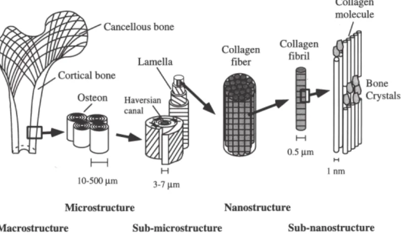
Bone is a metabolically active tissue composed by a mineral and an organic phase that contribute to two thirds and one third of its weight, respectively [4]. As shown in Figure 1 the bone exhibits a structure that has elements of several length scales and which, together, perform various mechanical, chemical and biological functions, e.g. it acts as a support structure, reservoir of mineral ions, among others [6]
5
Has stated before, bone is a heterogeneous bioceramic that combines an inorganic with an organic phase. In terms of its chemical composition, its main component is in the inorganic phase: carbonated hydroxyapatite (60-70 % w/w) of low crystallinity. The organic phase is mainly composed by fibrils of Type I collagen, although, other components are present, namely: growth factors and glycosaminoglycans. Finally, approximately 10 % of bone is composed by water [2].
The different types of bones (e.g. cortical or cancellous) are histologically different, although, their chemical composition is similar. Cortical bone represents, approximately, 80 % of the skeleton. It is dense and compact and possesses a high resistance to bending. Most of this type of bone is calcified and its main function is mechanical support and protection. Cancellous bone represents 20 % of skeletal mass and, approximately, 80% is found at the ends of long bones and in the interior of the vertebrae and pelvis. It is less dense, more elastic, and has a higher turnover rate than compact bone [7].
Bone is also constituted by cells. In fact there are three main types of cells in the bone tissue: osteoblasts, osteocytes and osteoclasts. Osteoblasts are the cells responsible for synthesis and deposition of minerals and, therefore, the mineralization of bone extracellular matrix. During their activity osteoblasts can become isolated in a cavity surrounded by bone matrix as a result of the deposition of minerals. Under these circumstances they differentiate into osteocytes, one of the most abundant types of cell in bone. Finally, osteoclasts are responsible for the bone remodelling process and their principal function is to resorb mineralized bone [8].
2.3 Mechanical properties of bone
In the context of mechanical evaluation it is relevant to describe the different data that can be extracted from the uniaxial mechanical tests. In this perspective, Figure 2 presents a stress-strain curve obtained from a mechanical test. The curve is the result of applied stress and the following data can be collected [1]:
6
• Beginning of the elastic behaviour;
• Elastic range;
• Range of plastic deformation;
• Breaking point;
• Amount of energy absorbed by the material;
• Elastic modulus (measuring the slope at the elastic range).
Figure 2: Stages of mechanical deformation: the elastic range (E), the continuum
damage mechanics range (CDM) and the fracture mechanics (FM).
The mechanical behaviour of a material can be divided in three phases (I, II and III). Under I (the elastic regime) the material deforms reversibly and only residual damage occurs. Phase II corresponds to the plastic regime where the material absorbs enough energy to develop microcracks. In phase III it occur mechanical failure and the amount of energy that the material is able to absorb reduces drastically.
Under mechanical loading, the bone can suffer of different types of ageing that can lead to fractures. These ageing mechanisms can be derived from creep (with prolonged load) or fatigue (repetitive) [9]. It has become recently clear that bone and other biological hard tissues show weak interlamellar interfaces, which are able to absorb energy and/or divert a crack. In this way, bone has a
7
limited capacity to detain the onset and growth of fracture. Furthermore, it is now increasingly clear that initiation of cracks in biomineralized tissues is far less important than their propagation since biological tissues use a number of processes (e.g. crack diversion/deflection, fibre pull-out, crack and/or matrix bridging) to increase the required amount of energy for fracture to occur [9]. The mechanical behaviour of bone depends on its type (cortical or cancellous). Additionally, as with any biological sample its variability is significant representing a reasonably large interval of expectable values. Table 1 resumes the most relevant mechanical properties of the two main types of bone.
Table 1: Mechanical properties of cortical and cancellous bone [10].
Property Cortical bone Cancellous bone
Compressive strength (MPa) 100-230 2-12
Flexural, tensile strength (MPa) 50-150 10-20
Strain to failure (%) 1-3 5-7
Young’s (tensile) modulus (GPa) 7-30 0.5-0.05
2.4 Bone repair
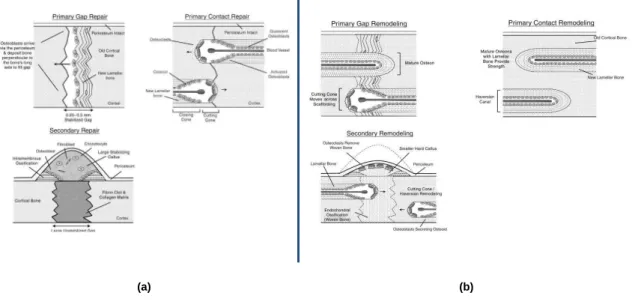
When the ends of fractured bone are held in place, there are two types of primary mineralized tissue healing that can occur: gap repair and contact repair. In gap repair, healing begins by the formation of blood vessels and connective tissue fills the empty spaces. After 2 weeks osteoblasts fill the gaps in the tissue by secreting osteoid. Upon 10 more days osteoids became mineralized and the osteoblasts that remained in the matrix become entrapped. The new bone acts as a scaffold for remodelling promoted by osteoclasts and osteoblasts. In contact repair, there is no gap between the bone ends, although, necrotic bone must be removed before new bone can be deposited to repair the fracture. Osteoclasts are responsible to resorb the necrotic bone. Afterwards, osteoblasts attach to the tissue matrix, creating a ruffled cell border between the
8
cell and the bone surface promoting mineralization of the bone and subsequent re-linking of the bone surfaces.
In bone repair, secondary healing occurs when bones are not rigidly supported after the injury. The first step is to create an ECM-rich bridge to support the fracture and the unstabilized mineralized tissue will undergo secondary repair. Most compact bone surfaces that make up the outer layer are covered by osteoblasts that promote mineralization. The wound site will re-gain some mechanical strength after 4 days of healing (Figure 3 (a)).
(a) (b)
Figure 3: (a) Primary and secondary mineralized tissue repair; (b) Primary and
secondary bone remodelling.
The mineralized bone remodelling is a dynamic process and occurs throughout the life of the individual. It produces new bone to be able to handle the demands of mechanical stresses. Primary gap remodelling also occurs during the repair process and lamellar bone (that presents collagen in a lamellar structure) is used as scaffold. After ca. 4 weeks osteogenesis stops leaving behind a vascularized cavity called an osteon that runs parallel to the long axis. A greater ultimate strength is obtained when a more osteons cross the injury site.
In primary gap healing, remodeling is important for restoring tissue strength. However, in primary contact healing, remodeling is coupled to the repair
9
process. During contact remodeling, the cutting cones mature, depositing lamellar bone centripetally to form ring-shaped structures.
The remodelling can last until 6 months and the healed bone is similar to the non-injured tissue in terms of robustness, although, it appears less organized. In this step it is also required to remove the excess of callus produced during the bone repair, being this step one of the main differences between the two reparation processes: primary and secondary. This callus removal is executed by the osteoclasts (Figure 3 (b)) [11].
2.5 Self-regeneration of bone
Bone is a dynamic tissue that is being formed and resorbed in a continuous cycle. These activities are executed in response to hormonal and physical factors. Bone is able to regenerate under different circumstances, e.g. when a fracture occurs in the bone. Unfortunately, this ability for self-regeneration is limited when the size of the trauma is too large. These cases are known as critical size defects due to the inability of the bone to self-regenerate. In order to repair these defects it is mandatory to execute a bone replacement. This can be achieved using a series of different biomaterials [12].
3.
B
iomaterials relevant to bone regeneration/repair
A biomaterial is a synthetic or natural material used to: replace part of living tissue; or to restore a specific function of the living tissue. From this definition it is clear to understand that the development of biomaterials usually comprise different fields of knowledge (Table 2), from materials science, biology and medicine. The effective influence of these areas is dependent on the specific tissue that is targeted and the function that the biomaterial is required to execute. In Table 3 is shown the application areas of the biomaterials. These can range from assistance in the diagnostic and treatment of diseases to tissue replacement or correction and improvement of tissue function [13].
10
Table 2: Fields of knowledge relevant for the development of biomaterials [13].
Knowledge domain Examples
Materials science and engineering
Structure-property relationship of synthetic and biological materials including metals, ceramics, polymers, composites, tissues (blood and connective tissues), etc.
Biology and physiology
Cell and molecular biology, anatomy, animal and human physiology, histopathology; experimental surgery,
immunology, etc
Clinical sciences All the clinical specialties: dentistry, maxillofacial,
neurosurgery, obstetrics and gynecology, ophthalmology, orthopaedics, otolaryngology, plastic and reconstructive surgery, thoracic and cardiovascular surgery, veterinary
medicine, and surgery, etc.
Table 3: Different areas where the use of biomaterials has been exploited [13].
Area of intervention Examples
Replacement of diseased or damaged part
Artificial hip joint, kidney dialysis machine
Assist in healing Sutures, bones plates, and screws
Improve function Cardiac pacemaker, intraocular lens
Correct functional abnormality Cardiac pacemaker
Correct cosmetic problem Augmentation mammoplasty, chin
augmentation
Aid to diagnosis Probes and catheters
11
The specific biological responses that the different biomaterials promote when in contact with the cells and body fluids are an important factor to be considered when designing new biomaterials [14-16]. In fact, the biomaterial-cell interaction is governed by the biomaterial surface and composition. Moreover, the different biomaterial properties (e.g. mechanical, biocompatibility, etc.) are governing their application area.
Table 4: Different types of biomaterials [13].
Biomaterial Advantages Disadvantages Examples
Polymers (nylon, silicone
rubber, polyester, poytetrafuoroethylene, etc.) Resilient Easy to fabricate Not strong Deforms with time, may degrade Sutures, blood vessels, hip socket, ear, nose, other soft tissues,
sutures
Metals (Ti and its alloys,
Co-Cr alloys, stainless steels, Au, Ag, Pt, etc.)
Strong, tough, ductile May corrode, dense, difficult to make Joint replacements, bone plates and screws, dental root
implants, pacer and sutures wires
Ceramics (aluminium oxide, calcium phosphates including hydroxyapatite, carbon) Very biocompatible, inert, strong in compression Brittle, not resilient, difficult to make Dental; femoral head of hip replacement, coating of dental and orthopaedic implants Composites (carbon-carbon, fibre-reinforced bone cement) Strong, tailor-made
Difficult to make Joint implants, heart valves
As can be seen from Table 4, in general, biomaterials can be produced from a variety of different materials (e.g. polymers, metals, ceramics and composites), although, their selection is dependent on their specific properties. As an example, while ceramics, metals and composites are usually used to repair
12
tissues that require high mechanical modulus, polymers that present lower mechanical modulus are usually selected to develop biomaterials for the repair or substitution of soft tissue. In the following subsection it will be presented examples of ceramic-based biomaterials due to the fact that is the type of biomaterials that presents more similarities with the systems studied under this thesis (GICs).
3.1 Ceramic-based biomaterials
Calcium phosphate cements (CPCs) were introduced in the field of bioceramics more than two decades ago and represented a real change in the medical applications. The possibility to have an injectable material with mouldable behaviour represented benefits for several clinical applications, e.g. minicracks, maxillofacial deformities and defects, vertebroplasty, among many others.
The formation of CPCs is based on the combination of one or more calcium orthophosphates. These phosphates are mixed with the liquid phase forming a paste that is able to set and harden after being implanted within the body. The CPC setting occurs through dissolution and precipitation of its components throughout the curing reactions. Its hardening is based on the entanglement of the precipitated crystalline phases. The most stable crystalline phases formed during the CPC curing are hydroxyapatite and brushite at pH >4.2 and pH<4.2, respectively [17].
The understanding of the mechanism of interaction of CPCs, allows accessing their long-term potential. It has been shown the occurrence of a gradual modification at the ceramic surface due to dissolution, precipitation and ion-exchange reactions. These events results in the production of a carbonate-containing, calcium-deficient hydroxyapatite with small crystal sizes. These changes are the beginning of a series of events that promotes bioactivity and that induces parallel reactions in cellular activity, bone mineralization and organic matrix deposition.
13
Absorption of proteins and other biological molecules occurs and the surrounding cells attach to the CPC surface. All these phenomena lead to the gradual incorporation of the ceramic into the regenerated bone tissue. Calcium phosphate ceramics include several materials which differ not only in their chemical composition, but also in their specific surface area, macro- and microporosity and crystal structure. There are differences due to variations in the calcium to phosphate ratio; as examples, tricalcium phosphate, hydroxyapatite and tetracalcium phosphate have Ca/P ratios of 1.50, 1.67 and 2.00 respectively, and there are other materials with ratios in between these (Table 5). Furthermore, hydroxyl ions may be missing from the structure, as in oxyhydroxyapatite, and other trace ions may be present. The importance of these compositional variations is not merely academic but they affect the biological response [18].
14
Table 5: The main calcium phosphates used in the biomedical field [8].
Compounds Chemical Formula (Ca/P) Molar
Ratio
Abbreviation
Precipitated CaP
Dicalcium phosphate CaHPO4 1.00 DCP
Monocalcium phosphate monohydrate Ca(HPO4)2.H2O 0.50 MCPM Dicalcium phosphate dihydrate (Brushite) Ca(HPO4)2.2H2O 1.00 DCPD Octacalcium phosphate Ca8H2(PO4)6.5H2O 1.33 OCP Precipitated hydroxypatite (tricalcium phosphate) Ca10-x(HPO4)x(PO4) 6-x(OH)2-x0≤x≤2 1.50-1.67 PHA Amorphous calcium phosphate Ca3(PO4)2.nH2O n = 3-4.5; 15-20 % H2O 1.50 ACP High-Temperature CaP Monocalcium phosphate Ca(HPO4)2 0.50 MCP α-Tricalcium phosphate α-Ca3(HPO4)2 1.50 α-TCP β-Tricalcium phosphate β-Ca3(PO4)2 1.50 β-TCP
Sintered hydroxypatite Ca5(PO4)3OH 1.67 HA
Oxyapatite Ca10(PO4)6O 1.67 OXA
Tetracalcium phosphate
15
Different types of bone cements are available for filling bone defects originated by illness or traumatic accident. More recently, bone cements have been tested for tendon-bone healing. In this perspective, Osteocrete, a magnesium-based injectable bone cement, has been reported to possess tensile strength 3 times higher than the calcium-based CPCs in both tendon-bone attachments and bone-bone structures. It has also been shown to enhance the formation of bone callous in an osteotomy model when compared with CPCs. While these preliminary studies are encouraging, the ability of Osteocrete to improve the healing of the bone tissue has not been studied [19].
Synthetic hydroxyapatite is a calcium-phosphate bioceramic of general chemical formula Ca10(PO4)6(OH)2. It has been tested in the development of
different types of biomaterials mainly targeting the substitution/regeneration of bone. This approach follows a biomimetic path derived from the fact that hydroxyapatite is one of the main constituents of natural bone. In fact, the inclusion of hydroxyapatite in biomaterials usually enhances their bioactivity and biocompatibility [20-23].The advances in the ceramic technology generated a significant number of ceramic materials for medical purposes. As an example, tricalcium phosphate (TCP) was first proposed in 1920 as a bioresorbable substance to fill bone defects. However, TCP is a weak ceramic, unable to sustain significant loading. The need for tougher and stronger ceramics was not met before 1965, when the fillers alumina-based materials were proposed for the substitution of hip joints. Zirconia and synthetic calcium phosphate ceramics (together with other calcium and/or phosphorus containing ceramics and glasses) were then proposed as alternatives to alumina and TCP, respectively. After roughly 100 years of clinical use, we come to the conclusion that there is, so far, no ceramic biomaterial able to create a strong and biologically relevant interface with bone. On the other hand, ceramics and glasses are able to promote direct bone-implant adhesion without soft tissue interlayer, although, their mechanical properties are not sufficient to allow their use in load-bearing applications [24].
16
3.2 Biocompatibility and bioactivity
Biocompatibility is defined as the ability of a material has to give an appropriate response to a specific application (this definition was established by consensus among the specialists in the field of biomaterials, in a conference in Chester, UK. Williams 1987). Inevitably the introduction of a new material in the human body produces a specific response that depends on the composition of the biomaterial, shape, size, geometry and aspects of the organism/patient such as age, immunological sensitivity, health, local implant, among others. Therefore, it is difficult to mention what are the parameters that should be accounted to evaluate the biocompatibility of a specific material. Under this perspective each biomaterial should be evaluated according to the roles that it was designed to execute [25].
The bioactivity of a material is related with his ability to produce a chemical or biological response from the surrounding medium in order to promote tissue regeneration. A typical in vitro bioactivity test evaluates the ability of the biomaterial to form an apatite surface layer when in contact with simulated body fluid (SBF), a solution that presents the ionic concentrations similar to human blood plasma. The higher the ability of the biomaterial to form an apatite layer the higher is its bioactive potential [26]. As an example bioglass was the first successful glass to present bioactivity. It was first reported by Hench and co-workers in 1970s and was used clinically. The main application of these glasses is for replacement of the damaged tissue, as for example, treatment of facial bone injuries or benign bone tumours [27, 28].
3.3 The relevance of biodegradability
Biodegradable biomaterials are used in reconstructive surgery when the body itself has the capacity to self-regenerate the damaged tissue. Usually, a biomaterial to be used under this approach needs to present bioactivity and to promote tissue growth/regeneration. Upon application, the biomaterial start the degrade allowing the surrounding tissue to grow to the intervened area. The
17
correct matching between its degradation rate and the tissue
growth/regeneration allows a complete regeneration of the damaged tissue upon full degradation of the biomaterial. Under this perspective it is of critical importance the non-toxicity of the degradation products. The first synthesized biodegradable biomaterial approved to be used in clinical applications (e.g. sutures) was the poly(glycolic acid) (PGA). With the progress of tissue engineering other biodegradable materials were developed (e.g. gellan gum, etc.) [29, 30]. The biodegradable biomaterials can be applied in the biomedical field in a diversified manner. In Table 6 it is presented examples of the targeted applications of biomedical devices produced using biodegradable biomaterials [8].
Table 6: Applications of biodegradable biomaterials in biomedical devices.
Application Biomedical device
Adhesion and fixation of tissues
Suture, bone fixation material and adhesive
Support and reinforcement Suture reinforcement material
Temporary substitutes for tissues
Substitute material for endocranium
Shape maintenance and isolation
Membrane for prevention of tissue adhesion
Securing space for tissue regeneration
Guided tissue regeneration, guided bone regeneration
Scaffold for tissue regeneration
Skin, cartilage, bone, blood vessel
A common approach to impart biodegradability to different biomaterials is the addition of starch. This methodology has been attempted in the development of bone cements [31, 32]. Starch is a glucose-based polymer created by the combination of two structures: amylose and amylopectin (Figure 4).
18
Figure 4: Structure of starch (amylose and amylopectin) [33].
Amylose is an essentially linear structure where the glucose units are joined by
α(1→4) glycosidic linkages. Amylopectin consists of linear α(1→4) linked glucose chains including branched positions with α(1→6) linkages every 24 to 30 glucose residues, on average [8].
One of the main advantages of including biodegradable starch in the formulation of biomaterials is the presence of α-amylase in the Human body that is specific for starch components. Additionally, its degradation products are not toxic and metabolized by the body.
4.
G
lass-ionomer cements (GICs)
4.1 The development of GICs
Wilson and Kent created the first conventional GICs combining a glass powder from the system SiO2-Al2O3-CaO-CaF2 with PAA, as a result of their
pioneering work at the Laboratory of the Government Chemist, London, in the early 1970s [34-49]. The studied glasses became vulnerable to acid attack and
19
established a strong link with the acidic PAA [40]. Initial application of these GICs was in dentistry, as a colour matched alternatives to amalgam restoratives due to two main advantages: strong adhesion to dentin and ability to prevent caries in tooth structures [34, 50-52]. Research work during the following decades addressed their main disadvantages, namely, sensitivity to moisture during initial hardening; poor mechanical properties, among others [53]. Different optimizations introduced in the following decades resulted in the development of conventional GIC formulations that present enhanced mechanical performance, tuned curing time and reduced sensitivity to moisture. More significant modifications were promoted with the addition of UV-curable resins in the cement matrix. These resin-modified GICs present mechanical properties that overpass most of the conventional GICs, although, their anti-cariogenic potential was significantly reduced. In this sense, their properties, ease of moulding and ease of application, still makes conventional GIC as an extremely versatile solution for cementing a series of different biomaterials.
4.2 The importance of glass composition
The glass powder present in GIC formulations is usually composed by calcium fluoroaluminosilicates. This component has two main functions: 1) to act as a source of cations essential for the evolution of the cement curing reactions; and 2) to reinforce the final cement structure.
Within the typical glass systems used in GIC formulations it is possible to distinguish glass formers (e.g. SiO2), glass modifiers (Ca2+) and components
with intermediate behaviour (e.g. Al2O3). The selection of components to be
used in the glass formulation is dependent on several factors, including their influence in: the glass mechanical properties; the glass reactivity; and, most important, the glass basicity. In fact, the GIC curing is governed by acid-base reactions between an acidic polymer and a basic glass. In this sense, it is mandatory that the glass structure presents significant basicity. This basicity is usually conferred by the modifier cations.
20
4.3 Methodologies to synthesize the glass component
4.3.1 Melt-quenching methodology
Melt-quenching methodology is a high temperature process that involves the melting of the whole glass formulation and its subsequent fast decrease of temperature below the glass transition (Tg). This procedure induces the
formation of the short to medium range order that is characteristic of glasses. The timeframe of the temperature reduction is so short that it does not allow the formation of long-range structural ordering. In general, glasses produced by this methodology present higher stability when compared with other glass preparation methods, e.g. sol-gel. Additionally, melt-quenched glasses usually promote the improvement of the mechanical behaviour of GIC compositions [14].
One of the main drawbacks of the melt-quenching technique is the fact that not all the compositions produce a melt. Depending on the glass compositions the melting temperature of the formulation is, in some cases, difficult to achieve with laboratory furnaces. Additionally, in the quenching step it is important to reach the appropriate temperature decrease rate that is also dependent on the composition. This should be fast enough to limit nucleation and to inhibit crystallization of the structure, although, nucleation and crystallization rates are also dependent on the composition. In fact, some formulations present a very high nucleation and crystallization rate and the production of pure glass phases is difficult. In these cases the melt quenching methodology generates crystalline phases within the glass phase, i.e. glass-ceramics.
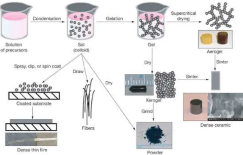
4.3.2 Sol-gel methodology
The sol-gel methodology is based on the hydrolysis and condensation of the glass former precursors (e.g. silicon and aluminium alkoxides, etc.) solubilised in specific solvents (e.g. water, ethanol, etc.). It also involves the addition of nitrates or chlorides of the glass modifiers (e.g. calcium nitrate, etc.) to the processing solution and the use of specific catalytic and/or initiator conditions (e.g. ammonium hydroxide, etc.). This approach presents a series of
21
advantages compared to the melt-quenching approach, namely: a better compositional control; morphological control (e.g. particles, fibers, membranes, etc.); lower processing temperatures; among others [54].
The main reactions occurring during the sol-gel methodology are the hydrolysis of the glass precursors and their subsequent condensation. For the first step to occur it is essential the presence of an hydrolyzing medium (e.g. water) in order to produce the hydroxides/acids of the glass formers (e.g. silicic acid, aluminium hydroxide, etc.). The condensation reaction (second step) occurs between the different hydroxides/acids with the release of water molecules. This step is usually catalysed by different compounds, e.g. ammonium hydroxide, nitric acid, etc. [55]. Depending on the targeted morphology, the methodology can include an additional step usually designated as gelation. In this step the colloidal solution of particles is aged allowing their condensation into a macroscopic system (Figure 5) [14].
Figure 5: Sol-gel process (https://www.llnl.gov/str/May05/Satcher.html).
The whole sol-gel methodology is performed at lower temperatures than the melt-quenching approach. This characteristic imparts the valuable advantage of enabling the preparation of glasses incorporating bioactive compounds (e.g. proteins, drugs, etc.) sensitive to high temperatures [56, 57]. Moreover, glasses produced by this methodology usually present higher specific surface area and bioactivity [58].
22
4.4 The polymeric component
The GICs curing reactions only occurs if the polymeric component present significant acidity. Throughout the development and optimization of GIC different polymers have been tested in the formulations. As examples, can be pointed out PAA, poly (acrylic acid-co-itaconic acid) and poly (acrylic-co-maleic acid) with structural formulas presented in Figure 6 [59].
Poly(acrylic acid)
Poly(acrylic acid-co-itaconic acid) Poly(acrylic-co-maleic acid)
Figure 6: Polymers already tested in the development of GIC formulations.
From all the polymeric components tested until today PAA continues to be the most used in GIC formulations. This is essentially due to its high acidity, availability and lower price when compared to the other tested polymers [59].
The PAA molecular weight (Mw) directly influences cement mechanical
behaviour and the cement paste initial viscosity. GIC mechanical performance is enhanced with PAA of high Mw, although, their high viscosity limits the
homogeneity and mixability of the cement pastes. In this perspective, it is important to obtain a compromise between these two properties. The optimal
formulations are prepared with polymeric components that have the highest Mw
without significantly affecting the viscosity of the cement paste [60-63].
4.5 GIC curing reactions
GICs are formed when an acid-base reaction occurs between the glass powder and an aqueous solution of PAA, creating an inorganic-organic interlinked structure. The curing mechanism is composed by two general steps, gelation (Figure 7) and maturation.
23
Figure 7: GICs curing reactions during the gelation stage [64].
During gelation, hydrated protons from the PAA/H2O penetrate the surface of
the glass particles and attack its basic sites (i.e. cations such as Na+, Ca2+, octahedral Al3+, etc.). The cations from the surface layer are leached to the cement matrix, promoting the ionic cross-linking of the PAA chains, through their carboxylate anions. This step usually occurs under a timeframe of approximately, 30 min. During the maturation step the tetrahedral aluminium present in the glass particle surface layer is leached under the same type of PAA acid attack. The longer timeframe of this maturation step (approximately, 24h) is attributed to the higher stability of the tetrahedral aluminium that is covalently bounded within the glass structure [34, 41, 50, 65-70].
4.6 Properties, applications and importance of GICs and its
constituents
4.6.1 Properties of GICs
There are a series of advantages on using GIC as cementation agent of different dental restorations, namely: chemical adhesion to tooth; anti-cariogenic effect; mechanical behaviour under compression loading; among others. The GIC chemical adhesion to the tooth is due to the dentin chemical structure. This is one of the main advantages of the GICs and is based on formation of ionic
cross-links between the PAA carboxylate anions and the Ca2+ from the dentin’s
hydroxyapatite [71]. Their anti-cariogenic effect is mainly related with the use of fluorine in the glass compositions. Fluorine is gradually leached from GIC to the
24
surrounding dentin strengthening the tooth resistance to bacterial proliferation [44, 72]. Moreover, it has been proven that GICs are able to act as a fluorine reservoir. In this sense GICs are not only leaching fluorine to the surrounding tissue but is also able to uptake additional fluorine from the surroundings (e.g. toothpastes) [73]. In relation to the mechanical behaviour, Kenny et al reported that it depends on the molecular weight (Mw) of PAA [74]. Additionally, the
powder to liquid ratio [75], concentration of PAA and the use of chelating agents also influences the final performance of the GICs. In general, the available formulations achieve compressive strengths up to 200 MPa and biaxial flexural strengths up to 50 MPa. The ratio of “bound” and “unbounded” water is a determinant factor affecting the GIC properties. In fact, the highest is the proportion of “unbounded” water the lower is the mechanical performance and the GIC general stability. Finally, the glass particle size also influences significantly the mechanical behaviour of the GICs and the cement curing reactions. The lower the glass particle size, the higher is its surface area and lower is the GIC crack deflection [76]. The main drawback of the reduction of the particle size is related with the faster setting kinetics. This variation reduces the time available to the medical doctor to manipulate the cement paste and to apply it at the intervention site. In this sense, it is always desirable to obtain a compromise between glass particle size, mechanical properties and GIC setting kinetics to develop optimized formulations.
A number of modifications can be made in the formulations to enhance the GIC properties, namely: (1) alternative polymers, as the polyacid component; (2) dried polymers blended with the glass and activation by the addition of water; (3) ceramic-metal hybrid cements; (4) metal-reinforced cement for enhanced mechanical properties; (5) resin-modified GICs (RMGICs) using initiators and resins capable of undergoing photochemical polymerization [35, 77].
GICs have demonstrated to possess biocompatibility and bioactivity. These cements are able to release osteoconductive ions such as calcium and fluoride to the surrounding tissue [65]. The incorporation of strontium in the glass formulations has been done for several years due to its characteristic properties, namely: radiopacifier, antibacterial properties and can help in the regeneration of healthy bone. In fact, it has been proven that low doses of
25
strontium (300 mg Kg-1 day-1 of Sr2+ for 9 weeks) can stimulate bone formation and inhibit bone resorption in both animal and humans. Strontium has an affinity for bone being incorporated by surface exchange and ionic substitution [78].
4.6.2 Applications of GICs
GICs have been tested in the cementation of different biomaterials dedicated to the repair or substitution of tooth and bone. Its main application area continues to be the dentistry field, where conventional GICs are used to cement different types of composites used to repair teeth or as temporary filler before the application of a definitive dental restoration. Non-conventional GICs, such as the RMGICs have been additionally proposed as definitive dental restorations [44, 79-81].
Hurrell-Gillingham and co-workers suggest that the main properties of GICs (including adhesion to mineralized tissues, minimal exotherm during setting and good biocompatibility) enables them to be exploited in the otology field [82]. Although, the most relevant and underexploited application of GIC remains to be in the orthopaedic field as bone cements [74].
4.6.3 Biocompatibility of GICs
GICs are not bioinert but should be classified as bioactive. Hatton and co-workers tested a set of GICs under in vitro cell culture and they demonstrated that some GICs are biocompatibility. Although, other studies proved that the ions present in GICs namely aluminium and fluoride are responsible for the toxicicity of some formulations. These studies also report a relation between the toxicity of the formulations and the ionic concentrations and culture conditions [83].
4.6.4 The importance of GIC cations in the regeneration of tissue
The GICs have been tested in vivo with encouraging results. The formation of bone was observed in some formulations after 6 weeks of implantation and being stable during 1 year. In addition, in the short term there is an inflammatory response observed in soft tissues adjacent to the GIC. The tissue reaction was caused by one or both of the following factors:26
(i) Reduction of tissue pH due to the acidic PAA. This is the most probable cause of tissue necrosis (a damage of the surrounding tissues).
(ii) Release of free glass particles from the cement to the surrounding tissues. This is probably induced by the excess of water originated on the surrounding tissue that migrates to the GIC affecting its curing reactions and promoting the release of glass particles. Glass particles are known to cause inflammatory response in adjacent soft tissues. In surgery (e.g. dentistry), it is important to avoid excess of the moisture contamination during placement of the GICs. A limited dose of ion release is the significant factor that determines tissue response to regeneration. In contrast, as previously mentioned, high levels of ionic leaching from the GIC can produce inflammatory response. Upon GIC application it is expectable an initial time period where inflammation occurs in the surrounding tissue due to higher levels of ionic and PAA leaching from the GIC. After this initial timeframe the inflammatory response is suppressed and the GIC appears with a layer of relatively mature bone tissue. At this stage the osteoconductive potential of the GIC is evident (a property not common in other types of bone cements) [83].
a) Aluminium and its toxicity
Aluminium in the ionic form (Al3+) has several implications in human health and its toxicity has been studied by several authors. This cation is responsible for many degenerative diseases including Alzheimer’s and Parkinson’s. Boyce and co-workers reported that encephalopathy and osteomalacia was detected in patients being subject to dialysis. It was observed that the water used in the dialysis contained high levels of aluminium. Bone biopsies detected the presence of high levels of aluminium in the bone, confirming that the origin of the detected pathologies was the aluminium present in the water used for the dialysis [84].
Another case study (a bone reconstruction of a woman with 52 years old) was reported by Reusche et al. This reconstruction was performed with a GIC prepared by mixing a calcium aluminium fluorosilicate glass and an aqueous solution of polycarboxylic acid. This GIC (Ionocem) contained high levels of
27
aluminium and six weeks after implantation the patient presented a fatal aluminium encephalopathy (see Figure 8) [85].
Figure 8: Aluminium inflammatory response of Al-based GICs resulting in an irregular
density at the specific positions (arrowheads) [85].
In general, it has been accepted that the release of aluminium present in the GICs formulations, used as bone cements, produces a deleterious effect in the health of the intervened patients [85]. Although, this has been considered a critical component of the glass compositions used to prepare GICs. In fact, it has been proposed that Al3+ has a critical role in the formation of the PAA carboxylate cross-links within the GIC cement matrix [86]. Under this perspective, Boyd and co-workers tested several aluminium-free glass compositions and their cement forming ability. Under these studies the same authors found that glass compositions where aluminium was substituted by zinc are able to produce GIC. The developed formulations presented properties that are consistent with their application in orthopaedic procedures, according to ISO5833 [78, 87].
b) Other relevant cations
There are a series of other cations that possess a relevant role in the setting chemistry and final properties of the GICs. As stated before, zinc has demonstrated that it is able to substitute aluminium in the glass compositions used to prepare GICs. Although not completely proven, it is reported that zinc imparted other relevant properties to the final GICs, namely: bactericidal and bioactivity. Zinc is also an essential trace element which presents effects on in
28
vitro and in vivo bone formation and bone protein synthesis. It promotes the
proliferation of osteoblasts and many biological functions. It promotes new bone formation in the surroundings of the implants [88]. Additionally, zinc deficiency may be a risk factor in the pathogenesis of osteoporosis [89].
Their bivalency makes the cations derived from the elements of the second group of the periodic table, namely, magnesium, calcium and strontium, suitable to act as PAA ionic cross-linkers. In fact, calcium has been described as the second most relevant cations in the GIC setting chemistry [90]. It has been reported in the glass composition since the initial development of the GICs. Its role is similar to aluminium in the cross-linking of the PAA network through calcium-carboxylate ionic linkages. Due to their similar characteristic, magnesium and strontium have an equivalent potential. In contrast, beryllium and barium are usually not included due to their toxicity.
The nature of strontium and its biological function has not been investigated as extensively as the role of calcium and magnesium of the same chemical family. Strontium is believed to have a participation in dental tissue mineralization due to their properties similar to those presented by calcium. Its antibacterial effect has been claimed but remains to be clarified: previous in vitro work on commercial RMGIC (Fuji II LC) showed that strontium was not cytotoxic when in contact with human osteoblastic cells. Strontium, as a substitute of calcium in hydroxyapatite, has been studied recently and claimed to prevent bone fractures [90]. In addition, strontium may inhibit osteoclastic turnover and promote the osteoblastic one. Moreover, bone strength increases and the risk of fractures decrease [91, 92]. It has been also reported to contribute to the increase in bone mass and volume when given at low doses, re-mineralizing skeletal lesions. It seems that strontium directly suppresses bone resorption and has no deleterious effect on bone mineralization. Strontium has also reported to have beneficial effects on bone formation in rodents and humans which results in increased trabecular volume. It has been also confirmed that strontium increases the bone strength and in bone density due to its higher atomic weight in comparison with calcium. Strontium is currently used as dopant of the crystalline structures of calcium salts to improve their biological properties. The successful incorporation of strontium ions in the composition of
29
hydroxyapatite, calcium phosphate cements and calcium silicates help on the proliferation and activity of osteoblastic cells [58]. Finally, Johnson et al. studied the effect of the incorporation of strontium in GICs. They reported a relation between the presence of strontium in the GIC and a marked reduction of bone ash; increase in the amount of magnesium and potassium in the bone; and by depressed calcium contents as compared to normal bone [93].
Drouet et al. studied the effect of strontium and magnesium in the formation of apatites. They observed that the magnesium taken up was found to be reversibly fixed to the apatite structure independently of their maturation stage. In contrast, the amount of reversibly fixed strontium decreased noticeably with maturation in a strontium-containing solution. These findings suggested that magnesium remained mostly on the surface of nanocrystalline apatites whereas strontium was progressively incorporated into the growing apatite domains [94]. Magnesium is a cation that presents high physiological interests in the biomedical field. It is essential to human metabolism and is naturally present in the bone. It may actually have stimulatory effects on the growth of new bone and it is classified as an essential element for the human body [88].

![Table 1: Mechanical properties of cortical and cancellous bone [10].](https://thumb-eu.123doks.com/thumbv2/123dok_br/17943169.853223/26.892.125.762.505.705/table-mechanical-properties-cortical-cancellous-bone.webp)
![Table 2: Fields of knowledge relevant for the development of biomaterials [13].](https://thumb-eu.123doks.com/thumbv2/123dok_br/17943169.853223/29.892.128.751.133.622/table-fields-knowledge-relevant-development-biomaterials.webp)
![Figure 7: GICs curing reactions during the gelation stage [64].](https://thumb-eu.123doks.com/thumbv2/123dok_br/17943169.853223/42.892.131.764.110.341/figure-gics-curing-reactions-gelation-stage.webp)
![Figure 8: Aluminium inflammatory response of Al-based GICs resulting in an irregular density at the specific positions (arrowheads) [85]](https://thumb-eu.123doks.com/thumbv2/123dok_br/17943169.853223/46.892.312.581.185.435/aluminium-inflammatory-response-resulting-irregular-specific-positions-arrowheads.webp)