Article
Antimicrobial Effects of Violacein against Planktonic
Cells and Biofilms of Staphylococcus aureus
Andressa H. M. Batista1,* ID, Anne C. D. Moreira1, Rafael M. de Carvalho1, Gleilton W. P. Sales1 ID, Patrícia C. N. Nogueira2, Thalles B. Grangeiro3 ID, Suelen C. Medeiros3, Edilberto R. Silveira2and Nádia A. P. Nogueira1
1 Clinical Department of Toxicological Analysis, Faculty of Pharmacy, Federal University of Ceará, Fortaleza 60356000, Brazil; [email protected] (A.C.D.M.); [email protected] (R.M.d.C.); [email protected] (G.W.P.S.); [email protected] (N.A.P.N.)
2 Organic Chemistry Department, Federal University of Ceará, Fortaleza 60356000, Brazil; [email protected] (P.C.N.N.); [email protected] (E.R.S.)
3 Biology Department, Federal University of Ceará, Fortaleza 60356000, Brazil; [email protected] (T.B.G.); [email protected] (S.C.M.)
* Correspondence: [email protected]; Tel.: +55-85-3366-8267 Received: 7 August 2017; Accepted: 9 September 2017; Published: 25 September 2017
Abstract:Violacein is an indole compound, produced byChromobacterium violaceum, a bacteria present in tropical and subtropical areas. Among its numerous biological activities, its antimicrobial potential stands out. This study aims to determine the antimicrobial activity of VIO onS. aureusin planktonic culture and biofilms. VIO showed excellent antimicrobial activity in inhibiting and killingS. aureusin planktonic cultures and biofilm formation. The minimum bactericidal concentration (5µg/mL) of VIO caused the death ofS. aureusafter 3–4 h of exposure and the minimum inhibitory concentration (1.25µg/mL) of VIO inhibited bacterial growth within the first 8 h of contact. Biofilm formation was also strongly inhibited by VIO (1.25µg/mL), in contrast to the higher resistance verified for S. aureusin mature biofilm (40µg/mL). The high bacterial metabolic activity favored VIO activity; however, the good activity observed during phases of reduced metabolism indicates that VIO action involves more than one mechanism. Thus, VIO is a promising molecule for the development of an antimicrobial drug for the eradication ofS. aureusinfections.
Keywords:violacein; antimicrobial activity; biofilms;Staphylococcus aureus
1. Introduction
Staphylococcus aureus, one of the main etiological agents of acquired infections in the community and the environment [1], has a remarkable ability to adapt and an enormous ability to rapidly develop resistance to countless antibiotics [2]. Its ability to produce numerous virulence factors contributes to its pathogenicity and ability to cause a variety of infectious diseases [3].
Biofilms are heterogeneous bacterial communities, irreversibly bound to a complex matrix consisting of DNA, proteins, and polysaccharides [3], which have an altered phenotype regarding growth rate and gene transcription [4]. Microbial growth in biofilm plays an important role during infection by providing different defense mechanisms to the microorganism. The biofilm matrix can prevent the access of immune cells, such as macrophages [5], as well as promote increased tolerance of microorganisms to antimicrobial agents [6].
The ability ofS. aureusto form biofilms on medical devices, such as catheters and prostheses, increases its virulence and contributes to treatment failure [4,7]. Compared to its planktonic state, S. aureusin biofilms shows a significant difference in gene expression and in its physiology [8].
Thus, due to the difficulty for antimicrobial agents to promote inhibition and eradication of S. aureusbiofilm, the search for new therapies is extremely important for the successful treatment of infectious diseases.
Violacein (VIO) is a purple bisindole metabolite, natural violet pigment produced by several Gram-negative bacteria, including the species Chromobacterium violaceum. VIO is a bioactive secondary metabolite, formed by the condensation of two tryptophan molecules through the action of proteins [9,10] (Figure1).
N
H
HO
N
O
H
N
H
O
1 2
3 4
5 6 7
8 9
10 11
12 13 14
15 16 17 18
19 20
21 22 23
δ δ
δ δ
δ
δ δ
Figure 1.Structure of violacein from the NMR.
VIO has attracted much attention in the scientific community due to its several pharmacological properties, such as antitumor, antibacterial, antiviral, antiparasitic, antioxidant action, as well as fungicide and leishmanicidal properties [10–13].
An excellent antibacterial activity of VIO has been demonstrated in vitro assays [13], as well as its synergistic potential in association with gentamicin, cefadroxil, azithromycin, and kanamycin, suggesting the possibility of using it as an alternative therapy [14], when administered in combination with other antibiotics, VIO is more effective than the use of antibiotics alone [10]. Nanoparticles loaded with VIO were two to five times more effective than VIO alone against strains of methicillin-resistant S. aureus(MRSA) [15].
Given the concern about the emergence of antibiotic-resistant strains, there is a great need for searching and investigating new molecules, as well as understanding their mechanism of action. In this context, VIO with its excellent antimicrobial activity especially onS. aureus, has emerged as a promising molecule. However, it is necessary to elucidate its antimicrobial mechanism of action so that VIO can become a new drug for the treatment of microbial infections [13,15]. The aim of this study was to determine the antistaphylococcal activity of VIO in planktonic cultures and biofilms.
2. Results
2.1. Structure Characterization
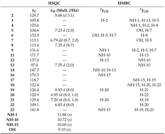
The1H-NMR spectrum showed 13 proton signals, four singlets atδ11.88, 10.72, 10.60, and 9.33; six doublets atδ8.93 (J= 8.0 Hz, H-19), 8.06 (J= 3.1 Hz, 2 H), 7.55 (J= 2.0 Hz, H-13), 7.35 (J= 8.7 Hz, H-8), 7.23 (J= 2.0 Hz, H-5), and 6.83 (J= 8.0 Hz, H-22); two triplets of doublet atδ7.20 (J= 8.0; 1.0 Hz, 21-H) and 6.95 (J= 8.0, 1.0 Hz, H-20); and, finally, a double doublet atδ6.79 (J= 8.7; 2.0 Hz, H-7).
Table 1.1H- and13C-NMR spectrals data of VIO.
HSQC HMBC
δC δH(Mult,J/Hz) 2JCH 3JCH
2 129.7 8.06 (d3.1)
3 105.8 — H-2 NH-1, H-13, H-5
4 125.6 — NH-1, H-2, H-8
5 104.6 7.23d(2.0) OH, H-7
6 152.9 — OH, H-5, H-7 H-8
7 113.1 6.79dd(8.7, 2.0) OH, H-5
8 113.4 7.35d(8.7)
9 131.6 — NH-1 H-2, H-5, H-7
11 171.7 — NH-10 H-13
12 137.0 — H-13 NH-10
13 97.0 7.55d(2.0) NH-10
14 147.7 — NH-10, H-13
16 170.3 — NH-15
17 118.7 — NH-15, H-19
18 122.4 — NH-15, H-20, H-22
19 126.4 8.93d(8.0) H-20 H-21
20 120.9 6.95td(8.0, 1.0) H-22
21 129.4 7.20td(8.0, 1.0) H-20 H-19
22 109.1 6.83d(8.0) H-20
23 141.8 — NH-15 H-19, H-21
NH-1 11.88 (s)
NH-10 10.72 (s)
NH-15 10.60 (s)
OH 9.33 (s)
DMSO-d6, 500 e 75 MHz.
All these data, together with the comparison with the NMR data in the literature [16], allowed the identification of the compound as (3E)-[3-(1,2-dihydro-5-(5-hydroxy-1H -indol-3-yl)-2-oxo-3H-pyrrol-3-ylidene)-1,3-dihydro]-2H-indol-2-one, known as VIO. The Supplementary Materials are available online. NMR are available as Supporting Information.
2.2. Antimicrobial Activity of VIO
The values of MIC and MBC ranged from 1.25 to 20µg/mL and 5 to 40µg/mL, respectively. Interestingly, VIO showed excellent activity inhibiting biofilm growth, with MBIC equal to MIC, indicating that its action occurs during biofilm formation, when the cells show intense metabolism (Table2).
Table 2.VIO antimicrobial activity on planktonic cultures and biofilms ofS. aureus.
Microbial Strain VIOµg/mL
MIC MBC MBIC
S. aureusATCC 6538P (OSSA) 1.25 5 1.25
S. aureusATCC 14458 (OSSA) 2.5 10 2.5
S. aureusATCC 3359 (ORSA) 20 40 20
S. aureusCCBH 5330 (ORSA) 5 20 5
MIC: minimum inhibitory concentration; MBC: minimum bactericidal concentration; MBIC: minimum biofilm inhibitory concentration; ATCC: American type culture collection; CCBH: culture collection of hospital-acquired bacteria. OSSA: oxacillin-susceptible strain, ORSA: oxacillin-resistant strain. Assays were performed in triplicate.
depending on concentration, against cells ofS. aureusATCC 6538P, completely preventing bacterial metabolism after 3–4 h of exposure to 5µμg/mL (Figure2).
0 4 8 12 16 20 24
2 4 6 8 10 12 14 MIC
*
p<0.05*
4xMIC 2xMIC Control*
*
*
**
*
*
*
*
*
*
*
*
*
* *
*
*
*
*
*
Time (h) lo g10 CF U /m LFigure 2. Effects of VIO on the viability of S. aureus ATCC 6538P. VIO (MIC = 1.25 μg/mL
μ
Figure 2.Effects of VIO on the viability ofS. aureusATCC 6538P. VIO (MIC = 1.25µg/mL); Control: without VIO. Detection limit was 102colony forming units per milliliter (CFU/mL) and the assays were performed in triplicate. Data are expressed as mean±SEM of three experiments. *p< 0.05 compared with the control.
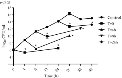
In the beginning of the exponential growth phase (4 h), when cells are young and have an intense metabolic activity, an initial reduction of 3 log10 CFU/mL was observed in the microbial population (Figure3). At the end of the exponential growth phase (8 h) and stationary growth phase, VIO inhibitory action can also be observed, but to a lower intensity (1–2 log10CFU/mL).
μ
μ
0 4 8 12 24 28 32 48
4 6 8 10 12 14 16 18 Control T=0 T=4h T=8h T=24h p<0.05
*
*
*
*
*
*
*
*
Time (h) lo g10 CF U /m L μ2.3. Antibiofilm Activity
VIO effect on mature biofilm viability was the concentration of 40µg/mL. Our results show total inhibition of bacterial metabolism and loss ofS. aureuscell viability in mature biofilm after 150 min of exposure to MBEC (Figure4).
μ
Cont
rol 160 80 40 20 10
OXA
0 2 4 6 8 10
MBEC
*
*
*
p<0.05
**
* *
* *
Concentration(µg/ml)
lo
g10
CF
U
/m
l
** *
**
*
μ
μ
Figure 4.Effect of different concentrations of VIO on the viability of biofilm formed byS. aureusATCC 6538P following a 24 h exposure. MBEC: minimum biofilm eradication concentration; Control: without VIO; OXA: oxacillin 20µg/mL. Detection limit was 102colony forming units per milliliter (CFU/mL) and the assays were performed in triplicate. Data are expressed as mean±SEM of three experiments.
***p< 0.05 compared with the control.
A better effect was obtained when VIO concentration was 2×MBEC, with a significantly greater reduction of cell viability (3 log10CFU/mL) being observed when compared to the MBEC activity (<1 log10CFU/mL) after 30 min of exposure and total cell inactivity between 90 and 120 min. The VIO subinhibitory concentration (0.5×MBEC) inhibited cell growth in biofilm when compared to the control (p< 0.05), but was not able to reduce the number of viable cells (Figure5).
μ
μ
0 30 60 90 120 150
2 4 6 8 10
Control
2 x MBEC 0.5 x MBEC MBEC
*
*
*
*
*
*
*
*
*
*
*
p<0.05
Time (min)
lo
g10
CF
U
/m
L
μ
3. Discussion
Many biological activities have been identified for VIO and among them we can highlight the anti-inflammatory, anti-diarrheal, anti-ulcer, anti-tumor, antimalarial, antimicrobial, and antimycobacterial activities [10,11,13,17–19].
Our results show VIO antimicrobial activity onS. aureusstrains, both resistant and susceptible to oxacillin, in suspension and in biofilm. All tested strains were susceptible to VIO and the OSSA strains showed greater susceptibility than ORSA strains.
S. aureusstrains are often responsible for hospital infections, increasing the costs to the health system and increasing mortality and morbidity rates. Furthermore, the resistance ofS. aureusto antimicrobial agents used in the treatment of infectious diseases hinders or even prevents treatment success, which is enhanced by its biofilm growth [20].
In order to consider VIO a molecule with potential for the development of a new chemotherapy drug for the treatment of staphylococcal infections, it is important to establish the kinetics of its antimicrobial action on cells in planktonic cultures and in biofilm. For that purpose, we performed the time–kill assay, which determines the time of action of a compound antimicrobial activity. The used strain wasS.aureusATCC 6538P, the most sensitive among the tested strains and the only one that had its growth in mature biofilm eradicated by the action of VIO at the tested concentration range (MBEC = 40µg/mL). TheS. aureusATCC 6538P strain was also used in other assays of this study.
The VIO MIC (1.25µg/mL) was able to inhibit the growth ofS. aureusfor up to 8 h, a period of intense metabolic activity. After this period, exponential growth was observed until 24 h of incubation, when growth was once again inhibited. The recovery of microbial growth after 8 h may be related to possible decomposition of VIO, as well as the reduction in cell activity levels, suggesting that VIO mechanism of action involves macromolecule synthesis.
The lower sensitivity ofS. aureusin mature biofilm, verified in our study, confirms the importance of the active microbial metabolism for VIO action, as the bacteria inside the biofilm have reduced metabolic activity [21]. The influence of cell metabolism on VIO activity is also confirmed by the VIO ability to inhibit biofilm formation.
Considering the occurrence of possible VIO decomposition after 8 h of incubation, as well as the possibility that VIO inhibitory action be favored by the high cell metabolism rate, one can infer that cell growth inhibition observed after 24 h, when cell division is minimal, may be associated with the high nutritional depletion and metabolite concentration.
VIO showed inhibitory activity in all stages ofS. aureusgrowth. In these assays, the substance is added at different stages of growth and its activity is assessed after 4 and 24 h. The fact that VIO was added to each of the different stages of bacterial growth might have prevented its decomposition and favored its action, even at the low bacterial metabolism stages. Thus, an initial decrease in the microbial population (up to 4 h) was demonstrated for all phases. This was not observed when VIO was added only at the beginning of incubation, in the time–kill assay, which reinforces the idea that VIO may have degraded after 4–8 h, in microbial culture, under the experimental conditions.
The ability of S. aureus to form biofilms is a major virulence factor and contributes to the success of the infectious process [20]. The greater resistance ofS. aureusin the mature biofilm matrix, when compared to that of its cells in planktonic culture, can be attributed to the ability of the bacterial biofilm to limit the access and dissemination of antimicrobial agents in its interior. In the biofilm, the cells are protected from antimicrobial agents, environmental conditions, and from the host’s immune response, and can display an increase of up to 1000 times the antimicrobial resistance rate [22].
4. Materials and Methods
4.1. Violacein
VIO extracted from the strain Chromobacterium violaceum ATCC 12472 was purified and characterized by nuclear magnetic resonance (NMR) [23].
4.2. Bacterial Strains
This study used four reference strains: S. aureus ATCC 6538P and S. aureus ATCC 14458 (oxacillin-susceptible strains-OSSA),S. aureusATCC 33591 andS. aureusCCBH 5330 (oxacillin-resistant strains-ORSA).
4.3. Determination of Minimum Inhibitory Concentration (MIC) and Minimum Bactericidal Concentration (MBC)
The MIC was determined using the microdilution method in culture broth [24]. Aliquots of the microbial suspensions (106 CFU/mL) in Brain–Heart Infusion Broth (BHI) (Merck Millipore Corporation) were added to the wells containing 95µL of BHI and 100µL of VIO (0.019–40µg/mL), in 0.1% Dimethyl Sulfoxide (DMSO). The microplates were incubated for 24 h/37◦C. The MIC was considered as the lowest VIO concentration capable of completely inhibiting microbial growth. As experiment controls, sterile BHI broth, amikacin, and 0.1% DMSO were used.
The MBC was determined by counting viable cells [25]. Briefly, 5µL of inoculum were collected and plated. The MBC was regarded as the lowest VIO concentration capable of determining the death of 99.9% ofS. aureuscells of the initial inoculum. The assays were performed in triplicate.
4.4. Time–Kill Assay
Aliquots of 20µL of VIO (MIC, 2×MIC, 4×MIC) were added to the microplate wells containing 100µL of BHI broth and 80µL of bacterial suspension (106CFU/mL). The microplates were incubated at 37◦C and 10µL aliquots were removed, diluted in 0.85% sterile saline solution and plated for colony counting, at predetermined time intervals (0, 2, 4, 6, 8, 10, 12, and 24 h) [26]. Microbial cultures with no exposure to VIO were used as experimental control. The assays were performed in triplicate and the results were expressed in log10CFU/mL.
4.5. Determination of VIO Effect on S. aureus at Different Stages of Microbial Metabolism
A microbial suspension was exposed to VIO MIC. VIO was added at the start of the incubation period (t= 0) and after 4, 8, and 24 h and microbial growth was determined after 4 and 24 h of exposure. Cultures ofS. aureus, without VIO addition and submitted to the same experimental conditions were used as control [27]. The experiment was performed in triplicate and the results expressed as log10CFU/mL.
4.6. Determination of Minimum Biofilm Inhibitory Concentration (MBIC)
4.7. Determination of Minimum Biofilm Eradication Concentration (MBEC)
MBEC was determined to evaluate VIO effect on mature biofilm viability [30]. TheS. aureusATCC 6538P strains were grown in TSB supplemented with 1% (p/v) of glucose (24 h/37◦C). Aliquots of 100µL (106CFU/mL) were transferred into microplates and incubated for 72 h/37◦C. 50µL of TSB and 50µL of VIO (10–160µg/mL) were added to the wells containing biofilm. The microplates were incubated for 24 h/37◦C and the viability of the remaining biofilms was measured by counting the colonies. Biofilms not treated with VIO were used as controls. MBEC was considered the lowest VIO concentration able to prevent microbial growth. The experiments were performed in triplicate and the results were expressed in log10CFU/mL.
4.8. Biofilm Time–Kill Assay
VIO aliquots of 20µL (0.5, 1, and 2×MBEC) were added to microplates containing 100µL of TSB and mature biofilm ofS. aureusATCC 6538P, formed in 72 h as previously described. The microplates were incubated at 37◦C, and after 24 h the remaining biofilm viability was measured by counting the colonies at the time of VIO addition and at predetermined time intervals (30, 60, 90, 120, and 150 min) after VIO addition. Aliquots of the biofilm serial dilutions were plated and the counting of colonies was performed after 24 h/37◦C. Biofilms not treated with VIO were used as controls. The experiments were performed in triplicate and the results were expressed in log10CFU/mL [30].
4.9. Statistical Analysis
Statistical analyses were performed using one-way ANOVA with post hoc Bonferroni test. The data were performed in triplicate. The charts are shown as mean±standard deviation. Data were considered significant whenp< 0.05.
5. Conclusions
VIO is a promising molecule for the development of an antimicrobial drug for the eradication of S. aureusbiofilm infections. However, while its action is enhanced by the active cell metabolism, it is also present in cells with reduced metabolism.
Supplementary Materials:The following are available online.1H- and13C-NMR of VIO.
Acknowledgments:We thank Coordination for Enhancement of Higher Education Personnel (CAPES) for their
financial support.
Author Contributions: A.H.M.B. assisted of growingC. violaceum, in the production of violacein and in the implementation of microbiological experiments and wrote the article; A.C.D.M. and G.W.P.S. assisted in the implementation of microbiological experiments; R.M.d.C. assisted in growingC. violaceumand in the production violacein; T.B.G. and S.C.M. provided the strainChromobacterium violaceumATCC 12472, assisted of growing C. violaceumand in the production violacein; E.R.S. and P.C.N.N. assisted in the purification and characterization of VIO; N.A.P.N. assisted in the implementation of microbiological experiments and contributed to the coordination of the study and wrote the article.
Conflicts of Interest:The authors declare no conflict of interest.
References
1. Lister, J.L.; HorswilL, A.R. Staphylococcus aureus biofilms: Recent developments in biofilm dispersal. Front. Cell. Infect. Microbiol.2014,4, 178. [CrossRef] [PubMed]
2. Mccallum, N.; Berger-Bächi, B.; Senn, M.M. Regulation of antibiotic resistance inStaphylococcus aureus.Int. J. Med. Microbiol.2010,300, 18–29. [CrossRef] [PubMed]
3. Shi, C.; Zhao, X.; Li, W.; Meng, R.; Liu, Z.; Liu, M.; Guo, N.; Yu, L. Inhibitory effect of totarol on exotoxin proteins hemolysin and enterotoxins secreted byStaphylococcus aureus.World J. Microbiol. Biotechnol.2015,31, 1565–1573. [CrossRef] [PubMed]
5. Scherr, T.D.; Hanke, M.L.; Huang, O.; James, D.B.; Horswill, A.R.; Bayles, K.W.; Fey, P.D.; Torres, V.J.; Kielian, T. Staphylococcus aureus biofilms induce macrophage dysfunction through leukocidin ab and alpha-toxin.mBio2015,6, e01021-15. [CrossRef] [PubMed]
6. De la Fuente-Núñez, C.; Reffuveille, F.; Fairfull-Smith, K.E.; Hancock, R.E. Effect of nitroxides on swarming motility and biofilm formation, multicellular behaviors in Pseudomonas aeruginosa. Antimicrob. Agents Chemother.2013,57, 4877–4881. [CrossRef] [PubMed]
7. Otto, M.Staphylococcalbiofilms.Curr. Top. Microbiol. Immunol.2008,322, 207–228, PMC2777538. [PubMed] 8. Beenken, K.E.; Dunman, P.M.; Mcaleese, F.; Macapagal, D.; Murphy, E.; Projan, S.J.; Blevins, J.S.; Smeltzer, M.S.
Global gene expression inStaphylococcus aureusbiofilms. J. Bacteriol. 2004,186, 4665–4684. [CrossRef] [PubMed]
9. Hoshino, T. Violacein and related tryptophan metabolites produced by Chromobacterium violaceum: Biosynthetic mechanism and pathway for construction of violacein core.Appl. Microbiol. Biotechnol.2011,91, 1463–1475. [CrossRef] [PubMed]
10. Choi, S.Y.; Yoon, K.H.; Lee, J.I.; Mitchell, R.J. Violacein: Properties and Production of a Versatile Bacterial Pigment.BioMed Res. Int.2015,2015, 1–8. [CrossRef] [PubMed]
11. Durán, N.; Justo, G.Z.; Ferreira, C.V.; Melo, P.S.; Cordi, L.; Martins, D. Violacein: Properties and biological activities.Biotechnol. Appl. Biochem.2007,48, 127–133. [CrossRef] [PubMed]
12. Anju, S.; Kumar, N.S.; Krishnakumar, B.; Kumar, B.S.D. Synergistic combination of violacein and azoles that leads to enhanced killing of major human pathogenic dermatophytic fungiTrichophyton rubrum.Front. Cell. Infect. Microbiol.2015,5, 57. [CrossRef] [PubMed]
13. Cazoto, L.L.; Martins, D.; Ribeiro, M.G.; Duran, N.; Nakazato, G. Antibacterial activity of violacein against Staphylococcus aureusisolated from bovine mastitis.J. Antibiot.2011,64, 395–397. [CrossRef] [PubMed] 14. Subramaniam, S.; Ravi, V.; Sivasubramanian, A. Synergistic antimicrobial profiling of violacein with
commercial antibiotics against pathogenic micro-organisms. Pharm. Biol. 2014, 52, 86–90. [CrossRef] [PubMed]
15. Martins, D.; Costa, F.; Brocchi, M.; Duran, N. Evaluation of the antibacterial activity of poly-(d, l-lactide-co-glycolide) nanoparticles containing violacein.J. Nanopart. Res.2011,13, 355–363. [CrossRef] 16. Ahmad, W.A.; Yusof, N.Z.; Nordin, N.; Zakaria, Z.A.; Rezali, M.F. Production and Characterization
of Violacein by Locally Isolated Chromobacterium violaceum Grown in Agricultural Wastes. Appl. Biochem. Biotechnol.2012,167, 1220–1234. [CrossRef] [PubMed]
17. Duran, N.; Menck, C.F.Chromobacterium violaceum: A review of pharmacological and industiral perspectives. Crit. Rev. Microbiol.2001,27, 201–222. [CrossRef] [PubMed]
18. Antonisamy, P.; Kannan, P.; Aravinthan, A.; Duraipandiyan, V.; Valan Arasu, M.; Ignacimuthu, S.; Abdullah AL-Dhabi, N.; Kim, J.H. Gastroprotective activity of violacein isolated fromChromobacterium violaceumon indomethacin-induced gastric lesions in rats: Investigation of potential of mechanisms of action.Sci. World J.
2014,2014, 1–10. [CrossRef] [PubMed]
19. Verinaud, L.; Lopes, S.C.P.; Prado, I.C.N.; Zanucoli, F.; Da Costa, T.A.; Di Gangi, R.; Issayama, L.K.; Carvalho, A.C.; Bonfanti, A.P.; Niederauer, G.F. Violacein treatment modulates acute and chronic inflammation through the suppression of cytokine production and induction of regulatory t cells.PLoS ONE
2015,10, e0125409. [CrossRef] [PubMed]
20. Savage, V.J.; Chopra, I.; O’neill, A.J.Staphylococcus aureusbiofilms promote horizontal transfer of antibiotic resistance.Antimicrob. Agents Chemother.2013,57, 1968–1970. [CrossRef] [PubMed]
21. Hoiby, N.; Bjarnsholt, T.; Givskov, M.; Molin, S.; Ciofu, O. Antibiotic resistance of bacterial biofilms.Int. J. Antimicrob. Agents2010,35, 322–332. [CrossRef] [PubMed]
22. Haussler, S.; Fuqua, C. Biofilms 2012: New discoveries and significant wrinkles in a dynamic field.J. Bacteriol.
2013,195, 2947–2958. [CrossRef] [PubMed]
23. Rettori, D.; Durán, N. Production, extraction and purificationof violacein: An antibiotic pigment producedby Chromobacterium violaceum.World J. Microbiol. Biotechnol.1988,14, 685–688. [CrossRef]
24. CLSI.Performance Standards for Antimicrobial Susceptibility Testing; Twenty-Fifth Informational Supplement; CLSI Document M100-S25; Clinical and Laboratory Standards Institute: Wayne, PA, USA, 2015.
26. Olajuyigbe, O.O.; Afolayan, A.J. In vitro antibacterial and time–kill assessment of crude methanolic stem bark extract of acacia mearnsii de wild against bacteria in shigellosis.Molecules2012,17, 2103–2118. [CrossRef] [PubMed]
27. Muroi, H.; Kubo, I. Antibacterial activity of anacardic acid and totarol, alone and in combination with methicillin, against methicillin resistantStaphylococcus aureus.J. Appl. Bacteriol.1996,80, 387–394. [CrossRef] [PubMed]
28. Nostro, A.; Roccaro, A.S.; Bisignano, G.; Marino, A.; Cannatelli, M.A.; Pizzimenti, F.C.; Cioni, P.L.; Procopio, F.; Blanco, A.R. Effects of oregano, carvacrol and thymol onStaphylococcus aureusandStaphylococcus epidermidisbiofilms.J. Med. Microbiol.2007,56, 519–523. [CrossRef] [PubMed]
29. Stepanovi´c, S.; Vukovi´c, D.; Daki´c, I.; Savi´c, B.; Švabi´c-Vlahovi´c, M. A modified microtiter-plate test for quantification ofStaphylococcalbiofilm formation.J. Microbiol. Methods2000,40, 175–179. [CrossRef] 30. Kwieci ´nski, J.; Eick, S.; Wójcik, K. Effects of tea tree (Melaleuca alternifolia) oil onStaphylococcus aureusin
biofilms and stationary growth phase.Int. J. Antimicrob. Agents2009,33, 343–347. [CrossRef] [PubMed]
Sample Availability:Samples of the compounds are not available from the authors.