UNIVERSIDADE FEDERAL DO CEARÁ
FACULDADE DE FARMÁCIA, ODONTOLOGIA E ENFERMAGEM
PROGRAMA DE PÓS-GRADUAÇÃO EM ODONTOLOGIA
BEATRIZ GONÇALVES NEVES
IDENTIFICAÇÃO MOLECULAR DE BACTÉRIAS EM LESÕES CARIOSAS
DENTINÁRIAS E EM BIOFILME DE CRIANÇAS COM DIFERENTES ESTÁGIOS
DA CÁRIE PRECOCE DA INFÂNCIA
FORTALEZA
BEATRIZ GONÇALVES NEVES
IDENTIFICAÇÃO MOLECULAR DE BACTÉRIAS EM LESÕES CARIOSAS DENTINÁRIAS E EM BIOFILME DE CRIANÇAS COM DIFERENTES ESTÁGIOS
DA CÁRIE PRECOCE DA INFÂNCIA
Tese apresentada ao Programa de Pós-Graduação em Odontologia da Faculdade de Farmácia, Odontologia e Enfermagem da Universidade Federal do Ceará como um dos requisitos para a obtenção do Título de Doutor em Odontologia.
Área de concentração: Clínica Odontológica
Orientadora: Profa. Dra.. Lidiany Karla Azevedo Rodrigues
Co-orientador: Prof. Dr. Rafael Nóbrega Stipp
Dados Internacionais de Catalogação na Publicação Universidade Federal do Ceará
Biblioteca de Ciências da Saúde
N422i Neves, Beatriz Gonçalves.
Identificação molecular de bactérias em lesões cariosas dentinárias e em biofilme de crianças com diferentes estágios da cárie precoce da infância. / Beatriz Gonçalves Neves. – 2014.
79 f. : il. color., enc. ; 30 cm.
Tese (doutorado) – Universidade Federal do Ceará; Centro de Ciências da Saúde; Faculdade de Farmácia, Odontologia e Enfermagem; Departamento de Odontologia; Programa de Pós-Graduação em Odontologia; Doutorado em Odontologia, Fortaleza, 2014.
Área de Concentração: Clínica Odontológica.
Orientação: Profa. Dra. Lidiany Karla Azevedo Rodrigues. Co-Orientação: Prof. Dr. Rafael Nóbrega Stipp.
1. Cárie Dentária. 2. Dente Decíduo. 3. Placa Dentária. 4. Dentina. 5. Reação em Cadeia da Polimerase em Tempo Real. I. Título.
A Deus, em primeiro lugar, por toda a força e coragem que me concedeu, durante essa trajetória, imprescindíveis para realização deste projeto, tão importante na minha vida e da minha família.
Aos meus queridos pais, Gilberto e Olivete, pelo exemplo de vida, incentivo e confiança, sem os quais não seria possível a realização deste trabalho. Obrigada por todo carinho e apoio ! Amo vocês !
Ao meu irmão, Bruno, e minha cunhada, Roberta, pelo carinho, amizade, respeito e pelas palavras de apoio, tão importantes nesta caminhada. Peço desculpas por não ter sido tão presente, como gostaria, durante os preparativos para a chegada do nosso querido Pedro, mas o meu coração estava sempre com vocês.
AGRADECIMENTOS ESPECIAIS
À minha orientadora, Profa. Dra. Lidiany Karla Azevedo Rodrigues, pelo exemplo de profissional e pesquisadora que não mediu esforços na colaboração para o desenvolvimento desta pesquisa. Agradeço por ter acreditado neste projeto. Obrigada pelo incentivo constante e por contribuir para o meu crescimento profissional.
Ao meu co-orientador, Prof. Dr. Rafael Nóbrega Stipp, agradeço por ter me acolhido em um momento tão difícil e especial desta tese. Agradeço pelas orientações na área de Microbiologia e Biologia Molecular e pela contribuição ímpar para o desenvolvimento deste trabalho.
À equipe maravilhosa, Daniela da Silva Bezerra e Sarah Florindo de Figueiredo Guedes, os desafios e as dificuldades de cada momento contribuíram para que nos tornássemos amigas e companheiras até o final. Agradeço pela amizade e companheirismo fortalecidos durante a saudável convivência.
AGRADECIMENTOS
À Universidade Federal do Ceará (UFC), por meio do seu Magnífico Reitor Prof. Dr. Jesualdo Pereira Farias.
À Faculdade de Farmácia, Odontologia e Enfermagem da Universidade Federal do Ceará, na pessoa de sua diretora, Profa. Maria Goretti Rodrigues de Queiroz e vice-diretor Prof. Sérgio Lima Santiago.
Ao Coordenador do curso de Odontologia da Universidade Federal do Ceará, Prof. Dr. Fabrício Bitu Sousa.
À coordenação do Programa de Pós-Graduação em Odontologia da UFC, por meio da Coordenadora Profa. Dra. Lidiany Azevedo Rodrigues e da vice-coordenadora Profa. Dra. Cristiane Sá Roriz Fonteles.
Ao Programa de Pós-Graduação de Odontologia (PPGO) da Universidade Federal do Ceará (UFC) e a todos os professores envolvidos, pela generosidade, bem como pelo estímulo, apoio e ensinamentos.
À Faculdade de Odontologia da Universidade de Campinas (UNICAMP) – Piracicaba e Laboratório de Imunologia e Biologia Molecular, na pessoa do Prof. Dr. Rafael Nóbrega Stipp, pela acolhida, disponibilidade e estrutura oferecidas para realização dos experimentos desta tese.
À Coordenação do Curso de Odontologia da Universidade Federal do Ceará (UFC) – Campus Sobral, em especial aos professores Mario Áureo Gomes Moreira e Rodrigo Citó Rego.
Aos funcionários do PPGO da UFC, Lúcia Ribeiro Marques Lustosa e Janaine Marques Leal, pela constante colaboração e disponibilidade.
Ao Davi Queiroz, técnico do Laboratório do PPGO da UFC, pelo apoio, auxílio e disponibilidade sempre presentes.
Aos funcionários do Curso de Odontologia sempre dispostos a ajudar e solucionar os problemas surgidos.
Às colegas do Curso de Odontologia da UFC – Campus Sobral, Alrieta Henrique Teixeira, Ana Karine Macedo, Celiane Mary Carneiro Tapety, Hellíada Vasconcelos
Chaves, Iriana Carla Junqueira Zanin, Karuza Maria Alves Pereira e Mariana
Ramalho de Farias, pelo apoio e compreensão.
A Cristiane Tomaz Rocha e Patricia Leal Dantas Lobo, amigas e colegas de disciplina da Odontopediatria, pela compreensão, incentivo, apoio, ajuda e disponibilidade, imprescindíveis para que eu conseguisse concluir esta tese.
Às amigas Denise Sá Maia Casseli e Virgínia Régia Souza da Silveira, pela força, estímulo e incentivo constantes, bem como pela amizade desenvolvida ao longo de nossa convivência. Obrigada pelo carinho de vocês.
À Juliana Nunes Botelho, por toda a ajuda, companhia e carinho para comigo em vários momentos durante a minha estada em Piracicaba.
Aos colegas pós-graduandos da Faculdade de Odontologia da Unicamp – Piracicaba, Erika Harth, Marcelle Buso, Natalia Vizoto, Tarsila Camargo, Lívia Alves, Felipe Joia,
Ítalo Sarto e Thais Oliveira, pela ajuda e atenção nos momentos em que precisei, nas
minhas várias temporadas em Piracicaba. Meu agradecimento.
Aos colegas de doutorado da UFC, Ana Patrícia Souza de Lima, Denise Lins de Sousa, Fabianni Apolônio, Ramille Araújo Lima, Juliana Marques Paiva, Mary Anne
Sampaio de Mello, Vanara Passos Florêncio e Patrícia Maria Costa de Oliveira, pela
À Profa. Dra. Maria Izabel Florindo Guedes, pela disponibilidade em ajudar quando precisei.
À amiga Patrícia Maria Costa de Oliveira, pela ajuda em vários momentos desta pesquisa. Muito obrigada!
Aos alunos de iniciação científica, especialmente Edyr Pereira e Caroline Salema, que contribuíram para o bom andamento deste trabalho.
Ao colega Paulo Goberlânio de Barros Silva, pela colaboração para a intepretação dos resultados. Obrigada pela paciência!
A todos os meus familiares, Vovó Alzirinha, meus avós que já partiram, “in memoriam” Boanerges, Cazuza e Joanila, tios, primos e primas, presentes em todos os momentos da minha vida. Em especial, minhas primas-irmãs, Clarissa Lima de Oliveira, Mariana Lima de Oliveira e Marília de Oliveira Araújo, que, apesar da distância física, estão sempre ao meu lado.
Aos meus padrinhos Júlio e Lusenir Timbó, pelo incentivo e apoio de sempre.
A minha sempre amiga Fernanda Campos Machado, companheira do Mestrado e, hoje, mesmo distante, fisicamente, continua presente me apoiando em todos os momentos da minha vida.
À Maria Elena Evangelista, muito querida, por toda a ajuda e apoio em casa, minimizando o meu cansaço. Obrigada!
Aos pequenos pacientes e seus responsáveis, pela compreensão, contribuição e participação na execução deste trabalho. O atendimento dessas crianças resultou em grande aprendizado.
Aos meus amigos, que aproveito para me desculpar pela ausência em muitos momentos importantes na vida de vocês, quando não foi possível estar presente para me dedicar a esta tese. Sei que me compreenderam, pela importância da causa.
Ao Conselho Nacional de Desenvolvimento Científico e Tecnológico (CNPq), pelo apoio financeiro para esta pesquisa, por meio do Edital Universal.
À Coordenação de Aperfeiçoamento de Pessoal de Nível Superior (Capes), pela concessão de bolsa em determinado período deste Doutorado.
E a todos que, direta ou indiretamente, contribuíram para a realização deste trabalho, o meu sincero agradecimento.
RESUMO
progressão da doença cárie. A presença de Bifidobacterium spp. e S. mutans apresentou forte associação com os estágios mais avançados da CPI. Em relação às lesões dentinárias, o aumento da concentração de bactérias Bifidobacterium spp. e do grupo L. casei evidenciou um papel importante destas bactérias na atividade de lesões dentinárias.
ABSTRACT
Bifidobacterium spp. and S. mutans presented a strong association with the development of the more advanced stages of ECC. Regarding the activity of dentine lesions, higher detection levels of the group L. casei and Bifidobacterium spp. showed an important role of these bacteria in the dentine caries activity.
SUMÁRIO
RESUMO
ABSTRACT
1 INTRODUÇÃO GERAL...17
2 PROPOSIÇÃO...22
3 CAPÍTULOS...23
3.1 CAPÍTULO 1...24
Molecular analysis of biofilm bacteria associated to different stages of early childhood caries 3.1.1 Introduction...27
3.1.2 Methods...28
3.1.3 Results...31
3.1.4 Discussion...32
3.1.5 References...36
3.2 CAPÍTULO 2 ...46
Molecular detection of bacteria associated to caries activity in dentinal lesions 3.2.1 Introduction...49
3.2.2 Methods...50
3.2.3 Results ...54
3.2.4 Discussion...55
3.2.5 References...60
4 CONCLUSÃO GERAL...70
REFERÊNCIAS GERAIS...71
APÊNDICES...75
1 INTRODUÇÃO GERAL
A cárie dentária é uma doença biofilme-açúcar-dependente, uma vez que é necessário que as bactérias bucais se organizem sobre os dentes na forma de biofilme e que haja ingestão frequente de açúcar para que ocorra desmineralização dos tecidos duros dentais (FEJERSKOV, 2004; LOESCHE, 1986). Dentre os fatores etiológicos, destacam-se o acúmulo de biofilme na superfície do dente, o frequente consumo de carboidratos fermentáveis e a susceptibilidade do indivíduo, após um período de tempo (MARSH, 1994). Esta desordem constitui uma das mais comuns doenças causadas por microrganismos em humanos e ainda é considerada como um significante problema de saúde pública em vários países (RAMOS-GOMEZ et al., 2002; PETERSEN et al., 2005), inclusive no Brasil. Em crianças, apresenta-se como a doença crônica mais prevalente da infância (de SILVA-SANIGORSKI et al., 2010).
Os últimos levantamentos epidemiológicos de saúde bucal (BRASIL, 2012) apontam desigualdades entre grupos populacionais (COSTA et al., 2013), sendo que no relatório de saúde bucal de 2004, (BRASIL, 2004), o Brasil não atingiu a meta estabelecida pela Organização Mundial de Saúde (OMS), a qual preconizava que 50% das crianças de zero a cinco anos deveriam estar livres de cárie. Este levantamento verificou uma prevalência de quase 27% na experiência de cárie em crianças entre 18 e 36 meses. Em 2010, o levantamento em saúde bucal SB Brasil não incluiu nos exames o grupo da faixa etária de 18-36 meses, no entanto observou-se que aos 5 anos de idade, uma criança brasileira possui, em média, o índice de 2,43 dentes com experiência de cárie, com predomínio do componente cariado, sendo responsável por mais de 80% do índice (BRASIL, 2012).
A cárie precoce da infância (CPI), anteriormente conhecida como “cárie de mamadeira” e aceita mundialmente pelo termo “Early Childhood Caries’’ (ECC), é definida como a presença de uma ou mais superfícies cariadas (lesões cavitadas ou não-cavitadas), perdidas (devido à cárie) ou restauradas em qualquer dente decíduo em uma criança com menos de seis anos de idade (AAPD, 2014). O termo na língua inglesa Early Childhood Caries” foi traduzida para a língua portuguesa como “cárie de estabelecimento precoce”, “cárie de primeira infância”, ou com maior aceitação “cárie precoce da infância” (FELDENS , 2013). A presença de padrões atípicos, progressivos, agudos ou rampantes desta doença é designada cárie precoce da infância severa. Em crianças em idade pré-escolar, a grande quantidade de
sócio-econômica (SCHWENDICKE et al., 2014) têm sido identificados como fatores predisponentes ao desenvolvimento de cárie.
A CPI é normalmente iniciada com lesões de mancha branca nos incisivos maxilares decíduos ao longo da margem gengival, onde o biofilme dentário normalmente acumula. Esta doença apresenta uma evolução rápida e progressiva, podendo ser dolorosa e causar sérias consequências para a criança (BERKOWITZ, 2003). Dentre as sequelas iniciais da doença não tratada pode-se citar dor, infecção e abscessos. Ainda pode acarretar várias consequências na saúde e desenvolvimento da criança, incluindo atraso no crescimento, problemas nutricionais e de sono, baixa auto-estima, além de prejuízo no rendimento escolar (RAMOS-GOMEZ et al., 2002). Além disso, crianças acometidas por esta desordem podem apresentar um maior risco para o desenvolvimento de novas lesões de cárie em ambas dentições decídua e permanente (TINANOFF & O´SULLIVAN, 1997). Inicialmente, acreditava-se que a CPI estava associada ao uso prolongado de mamadeira (ISMAIL & SOHN, 1999), no entanto, somente o fator etiológico dieta não é capaz de causar a severidade característica desta doença (CLEATON-JONES et al., 2000), apesar de exercer um papel imprescindível na manifestação clínica desta infecção (BERKOWITZ, 2003).
Há evidências consideráveis de que micro-organismos Gram-positivos acidogênicos, acidúricos e com capacidade de aderência às superfícies dentárias, pertencentes a um grupo heterogêneo denominado estreptococos do grupo mutans (EGM), são especialmente envolvidos na dinâmica do processo carioso (LOESCHE, 1986; PARISOTTO et al., 2010). Estas bactérias são fortemente associadas com cárie dentária em virtude dos seus atributos metabólicos, ecológicos e epidemiológicos (LOESCHE, 1986). Dentre os EGM, Streptococcus mutans constitui uma espécie bacteriana predominante na microbiota de crianças pré-escolares com cárie de estabelecimento precoce (BECKER et al., 2002; BERKOWITZ, 2003; SAXENA et al., 2008). Apesar de que a associação entre S. mutans e CPI esteja estabelecida na literatura (BERKOWITZ, 2003; KOHLER et al., 1988; VAN HOUTE et al., 1982), tem sido demonstrado que nem todos os indivíduos que são colonizados por EGM apresentam a doença (CARLSSON, OLSSON & BRATTHALL, 1985; MATTOS-GRANER et al., 2001) e que também a cárie pode ocorrer em sua ausência (AAS et al., 2008; GROSS et al., 2010; TANNER et al., 2011).
cariogênicos mais pesquisados, estudos têm sugerido que a etiologia da cárie dentária pode envolver comunidades mais complexas de espécies bacterianas (BECKER et al., 2002; VAN HOUTE, 1994).
Dessa forma, análises quantitativas e qualitativas de ecossistemas polimicrobianos
como biofilme dentário e dentina cariada são desafiadoras, uma vez que as comunidades
microbianas podem consistir em centenas de diferentes espécies bacterianas. É relatado na
literatura existir mais de 700 espécies de bactérias na cavidade oral, sendo espécies
cultiváveis apenas 50-60% (DEWHIRST et al , 2010). O advento de novos métodos
moleculares através de identificação e quantificação bacteriana tem tornado possível reavaliar
a patogênese de infecções orais de forma mais precisa. Em pesquisas utilizando identificação molecular de bactérias, estudos têm relatado que diversas comunidades bacterianas, incluindo novas espécies, estão associadas com a cárie dentária (BECKER et al., 2002; MUNSON et al.; 2004; AAS et al., 2008; TANNER et al., 2011; BELDA-FERRE et al., 2012; GROSS et al., 2012; JIANG et al., 2014; PETERSON et al. 2014). Além do S. mutans, espécies acidúricas e acidogênicas do grupo estreptococos não-mutans e algumas espécies dos gêneros
Veillonella, Lactobacillus, Bifidobacterium, Propionibacterium, Actinomyces, e Atopobium
podem também exercer uma importante função na progressão da cárie (BECKER et al., 2002; MUNSON et al., 2004; CHHOUR et al., 2005; AAS et al., 2008; BELDA-FERRE et al., 2012; GROSS et al., 2012). Estes achados apoiam a hipótese da placa ecológica, que propõe que o S. mutans é apenas um dos muitos micro-organismos envolvidos com a patogênese da cárie (MARSH, 2003; AAS et al., 2008; TAKAHASHI & NYVAD, 2011).
No estudo de AAS et al. (2008), foi observado que em 10 a 20% dos pacientes que apresentavam cárie severa não foram identificados níveis detectáveis de S. mutans, mas apresentavam outras espécies produtoras de ácidos. Além disso, em algumas lesões cariosas
S. mutans pode ser um componente bacteriano minoritário do biofilme dental.
Em um estudo com cárie, BECKER et al. (2002) identificaram 10 novos filotipos na microbiota associada com lesões de cárie em um único indivíduo com cárie de
estabelecimento precoce. Nesta mesma pesquisa, espécies bacterianas encontradas em
crianças com cárie de estabelecimento precoce foram comparadas com aquelas identificadas
em crianças livres de cárie. Algumas espécies como Streptococcus sanguinis foram associadas com saúde bucal, enquanto outras como S. mutans, Lactobacillus fermentum e algumas espécies dos gêneros Streptococcus spp., Veillonella spp., Actinomyces spp.,
Corby et al. (2005), através de métodos moleculares de identificação como checkerboard hybridization, verificaram bactérias associadas com cárie dentária e saúde em um grupo de 204 gêmeos de idade entre 1,5 e 7 anos. As bactérias Actinomyces spp., S. mutans, e Lactobacillus spp. foram detectadas em abundância no grupo cárie-ativo. Em contraste, espécies bacterianas, incluindo Streptococcus parasanguinis, Abiotrophia defectiva, Streptococcus mitis, Streptococcus oralis, e S. sanguinis predominaram na microbiota indígena bacteriana de indivíduos livres de cárie.
Estudos têm demonstrado que espécies consideradas como colonizadores iniciais da placa dentária, como estreptococos do grupo mitis, podem apresentar a capacidade de produzir álcali, gerando um impacto no equilíbrio do pH do biofilme (NASCIMENTO et al., 2009). No entanto, em situações de acidúria e acidogenicidade, tais bactérias, comumente associadas à saúde dental, podem apresentar relação com atividade de cárie dentária (MARCHANT et al., 2001; AAS et al., 2008 GROSS et al., 2012).
Com o advento de métodos e sistemas de diagnóstico ao longo dos últimos anos, houve a inserção do ICDAS II (International Caries Detection & Assessment System), que é um sistema de detecção de cárie baseado na inspeção visual e inclui avaliação de lesões cariosas cavitadas e não-cavitadas. Esse sistema foi desenvolvido para uso na prática clínica, bem como em pesquisas clínicas e epidemiológicas (PITTS, 2004). O método provou ser bem aceito e prático, baseado na sua padronização de critérios e possibilidade de comparação com outros índices (SHOAIB et al., 2009).
Com as mudanças no perfil de cárie nas últimas décadas (redução na prevalência e na progressão das lesões em uma parcela da população), a avaliação da atividade de cárie também se faz necessária juntamente com o diagnóstico, pois as lesões podem ter sido ou vir a ser inativadas por meio de medidas preventivas e mudanças comportamentais, tornando a atividade um fator importante na escolha do tratamento. Porém, pouco se tem pesquisado sobre a atividade das lesões em estudos de cárie em pré-escolares nos últimos anos (PIOSEVAN et al., 2013), embora possa ser determinada pelo critério de Nyvad, um dos principais índices descritos e validados, o qual é baseado no diagnostico táctil e visual da lesão (NYVAD et al., 1999; MACHIULSKIENE et al., 1998).
2 PROPOSIÇÃO
Esta tese de doutorado será apresentada em capítulos, tendo como objetivo:
Detectar a presença e quantificar através da técnica de reação em cadeia da polimerase
quantitativa (qPCR) as espécies/grupos de bactérias Actinomyces naeslundii, Bifidobacterium spp., Lactobacillus acidophilus, grupo Lactobacillus casei, grupo Mitis, Streptococcus gordonii e Streptococcus mutans em biofilme de crianças pré-escolares com diferentes estágios de progressão de cárie precoce da infância e em lesões
cariosas dentinárias ativas e inativas bem como verificar a associação destes
3 CAPÍTULOS
Esta tese está baseada no Artigo 46 do Regimento Interno do Programa de Pós-
Graduação em Odontologia da Universidade Federal do Ceará, que regulamenta o formato
alternativo para dissertações de Mestrado e teses de Doutorado e permite a inserção de artigos
científicos de autoria e co-autoria do candidato. Por se tratarem de pesquisas envolvendo seres
humanos, ou parte deles, o projeto de pesquisa referente a este trabalho foi submetido à
apreciação do Comitê de Ética em Pesquisa da Faculdade de Medicina da Universidade
Federal do Ceará, tendo sido aprovado sob protocolo nº 158/2011 (Anexo). Assim sendo, esta
tese de Doutorado é composta por dois capítulos que contém artigos que serão submetidos
para publicação em periódicos científicos, conforme descrito abaixo:
CAPÍTULO 1
“Molecular analysis of biofilm bacteria associated to different stages of early childhood caries”
B.G. Neves, R.N. Stipp, D.S. Bezerra, S.F.F. Guedes, L.K.A. Rodrigues
Este artigo será submetido à publicação no periódico “Caries Research”.
CAPÍTULO 2
“Molecular detection of bacteria associated to caries activity in dentinal lesions”
B.G. Neves, R.N. Stipp, D.S. Bezerra, S.F.F. Guedes, L.K.A. Rodrigues
CAPÍTULO 1
Molecular analysis of biofilm bacteria associated to different stages of early childhood caries
B.G. Nevesa, R.N. Stippb, D.S. Bezerraa , S.F.F. Guedesa, L.K.A. Rodriguesa
a
Post-graduation Program, Faculty of Pharmacy, Dentistry and Nursing, Federal University
of Ceará, Fortaleza, Ceará, Brazil; bDepartment of Microbiology and Immunology, Piracicaba Dental School, State University of Campinas, Piracicaba, SP, Brazil
Short title: Bacteria detection in stages of early childhood caries
Key words: Dental Plaque, Bacteria, Early Childhood Caries, Quantitative Polymerase Chain Reaction, Preschoolers
Address all correspondence to:
Lidiany K. A. Rodrigues, DDS, MSc, PhD, Associate Professor
Postgraduate Program in Dentistry, Faculty of Pharmacy, Dentistry and Nursing
Federal University of Ceará, Fortaleza, Ceará, Brazil.
Rua Monsenhor Furtado, 1273 - Zip Code: 60430-355
Phone- #+558533668410/ Fax- #+558533668232
Email: lidianykarla@yahoo.com
Este artigo está escrito de acordo com as normas de publicação do periódico Caries Research.
Conflict of interests
ABSTRACT
Several bacterial species have been associated to early childhood caries (ECC), a prevalent public health problem among preschool children. The aim of this study was to quantify
INTRODUCTION
Dental caries is a progressive disorder associated with a complex etiology, including genetic, microbial, environmental and behavioral factors [Peterson et al., 2013] and remains as a major public health problem, being considered pandemic worldwide [Jiang et al., 2014; Peterson et al., 2014]. Early childhood caries (ECC) may cause pain and infection in preschool children, reduce quality of life and compromise primary dentition soon after eruption [AAPD, 2014; Gross et al., 2012]. Frequent sugared diet causes a growing acidification of dental biofilm (i.e., dental plaque), subsequently leading to an increase of the proportion of acidogenic/aciduric bacteria with the rapid progression of caries lesions [Takahashi and Nyvad, 2011; Burne et al., 2012].
The oral biofilm is composed by a structurally organized microbial community, being considered as a dynamic and extremely complex ecosystem [Marsh, 2006] and its role in the onset and progression of dental caries is unquestionable [Dige et al., 2014]. The human oral microbiome contains a diverse polymicrobial population, consisting of over 600 species [Dewhirst et al., 2010]. Efforts have been made to determine the bacterial species associated to oral health and disease [Dige et al., 2014]. However, researches of biofilms are particularly challenging and relevant, due to their dynamic changes in response to the constant exposure to complex diet-host-microbial interactions occurring in the oral cavity [Klein et al., 2011].
Streptococcus mutans (MS) has been identified as a major microbial pathogen in the development of early childhood caries [Koehler et al., 1988; Berkowitz, 2003; Parisotto et al., 2010a], however not all children with ECC harbor this bacteria [Becker et al., 2002; Tanner et al., 2011; Gross et al., 2012]. Thus, it is accepted that this association is not absolute, since dental caries-associated microbiota is highly complex and multiple members of the community can be implicated in the caries development [McLean et al, 2012; Obata et al, 2014]. ECC studies have detected other pathogens involved in dental caries such as Bifidobacterium [Becker et al., 2002; Aas et al., 2008; Mantzourani et al., 2009; Palmer et al., 2010], Lactobacillus [Becker et al., 2002; Aas et al., 2008] and Actinomyces [Marchant et al., 2001]. All these microorganisms are potential acid producers and able to survive in acid environment [Takahashi & Nyvad, 2011].
2009]. In addition, studies indicate that microorganisms present in dental plaque that have been frequently related with oral health [Li et al., 2004; Corby et al., 2005; Takahashi and Nyvad, 2008], have now been also associated with caries, then being considered as alternative pathogens [Gross et al., 2012] such as Streptococcus oralis, Actinomyces naeslundii [Marchant et al., 2001; Aas et al., 2008] and Streptococcus parasanguinis [Gross et al., 2012].
Considering that relatively few molecular studies have been performed using qPCR for studying specific bacteria associated with early childhood caries or oral health, the aim of the present study was to quantify Actinomyces naeslundii, Bifidobacterium spp., Lactobacillus acidophilus, Lactobacillus casei group, Mitis group, Streptococcus gordonii and Streptococcus mutans in dental biofilm from children with different stages of early childhood caries.
METHODS
Ethics statement
The study design and informed consent were approved by the Ethics Committee of the Federal University of Ceará (COMEPE/UFC) (Protocol Number 158/2011). Signed informed consents were obtained from the parents or guardians of all subjects. Samples were collected only after getting the approval from the children and their parents.
Study population
The study sample consisted of 75 children aged 2 to 5 years from public nurseries and preeschools from a suburban area in Fortaleza, Ceará, Brazil. Inclusion criteria were: the child was under 71 months of age; child had not be treated with antibiotics in the 3 months prior to the study and subjects had no salivary gland disorders or systemic diseases. Children, who did not cooperate with the dental exams, were excluded from the study.
(seen only when the tooth is dry); 2 = visible non-cavitated lesion seen when wet and dry; 3 = cavitation in enamel; 4 = non-cavitated lesion extending into dentine seen as an undermining shadow; 5 = cavitated lesion with visible dentine: < 50% of surface; and 6 = extensive cavitated lesions with visible dentine in more than 50% of the surface.
Screening of 420 children was performed in the current study. After meeting inclusion and exclusion criteria, 75 children were included in the final sample size. The distribution of the groups were defined by the different degrees of dental caries status, according to ICDAS coding:
• CF: caries-free children (n=20): code 0. Children who presented no caries lesions.
• ECL: children with the most severe score classified as enamel carious lesions (n=17):
dmft ≥ 1 (including surfaces with ICDAS codes 2 and 3)
• DCL: children with the most severe score classified as dentine carious lesions (n=38): dmft ≥ 1 (including surfaces with ICDAS codes 5 and 6)
All children involved in this study were enrolled in a dental care program that included preventive counseling and dental treatment at the dental clinic of the Faculty of Dentistry, Federal University of Ceará, Fortaleza, Ceará.
Biofilm collection
Dental biofilm samples were collected at children’s schools. Pooled supragengival
plaque sample from each child was taken with autoclaved spoon excavators from vestibular and lingual surfaces of teeth, including anterior and posterior primary teeth. In children with dentin lesions, plaque was not collected from cavities. The participants were asked not to brush their teeth 24 h before sampling.
Plaque samples were transferred into a sterile 1.5 mL microcentrifuge tube that
contained 150 µL of TE (10 mM Tris-HCl, 1 mM EDTA pH 7.6). All of the samples were immediately transported on ice to the laboratory, not exceeding one hour after collection. Samples were stored and kept frozen at -20°C until DNA extraction.
Laboratory methods
Bacterial strains and culture conditions
Streptococcus gordonii and Streptococcus mutans was investigated using specific forward and reverse primers as listed in Table 1.
The bacteria strains used as positive controls to test the specificity of the primers and for
qPCR standard curve preparations included Actinomyces naeslundii (ATCC 12104), Bifidobacterium animalis subsp. lactis BB-12® (Chr. Hansen), Lactobacillus acidophilus (ATCC 4356), Lactobacillus paracasei subsp. paracasei (ATCC 335), Streptococcus gordonii (ATCC 35105), Streptococcus mitis (ATCC 49456 – NTCC 12261) and Streptococcus mutans (UA159).
Isolated bacteria were cultured in Brain-Heart Infusion broth as recommended by
Bergey’s Manual of Determinative Bacteriology [Holt et al. 1994]. The cells were centrifuged and washed in sterile saline solution (NaCl 0.9%). The quality and purity of bacterial cultures
were checked by Gram staining.
DNA extraction
Extraction and purification of DNA from dental plaque samples and bacterial
cultures
All samples were transferred into a fresh 2 mL screw cap tube and the cells were
mechanically lysed with 0.16g of 0.1mm diameter zirconia beads on a Mini-Bead Beater
homogenizer (Biospec Products, Bartlesville, OK, USA) and subject to beating for 1 min at
maximun power. DNA was recovered from all samples using an organic extraction protocol
based on phenol/chloroform purification and alcohol precipitation [Wilson, 2001]. The DNA
concentration (A
260nm
) and purity (A
260nm
/A
280nm) of the samples were evaluated by a
NanoDrop 2000 spectrophotometer (Thermo Fisher Scientific, Wilmington, DE, USA).
Electrophoresis of the extracted DNA was performed on a 1.2% agarose gel in
Tris/borate/EDTA buffer and stained with 0.1 µg/ ml ethidium bromide.
Preparation of PCR standards and quantification of target bacterial DNA in
plaque samples by quantitative PCR (qPCR)
Quantitative real-time assays
Serial dilutions starting from 300 ng to 0.0003 ng (10-fold) of reference bacterial DNA
targeted bacteria. A standard DNA amplification curve and a melting-point product curve
were obtained for each primer set/run. SYBR Green real-time PCR amplifications were
performed using MicroAmpFast Optical 48-Well Reaction Plate (Applied Biosystems, Foster
City, CA, USA) in StepOne Real-Time PCR System (Applied Biosystems) covered with
Optical Adhesive Film (Applied Biosystems). Each reaction mixture (10 µL) contained 5 µL
of 2x Power SYBR Green Mastermix (Applied Biosystems), 0.3 µL of each appropriate
forward/reverse primer, 2 µL of DNA sample and 2.4 µL of water free of nucleases. Negative
control included reactions without template to rule out primers dimers formation or presence
of contaminating DNA. All samples were analyzed in duplicates. The final analyses were
based on the mean of the two reactions. The standard curves were used to transform the cycle
threshold (Ct) values to the mass of DNA and the results of the concentrations of bacteria in
plaque samples were normalized relative as a percentage of the total bacterial load estimated
by the primer Bacteria 16S rDNA [Nadkarni et a., 2002].
Statistical analysis
Data were tabulated in Microsoft Excel and exported to statistical software Statistical
Package for Social Sciences (SPSS) version 17.0, on which all analyzes were performed
considering a confidence level of 95%.
After examining the pattern of sample distribution (normality test Kolmogorov-Smirnov
test) data were expressed as mean and standard deviation and compared between groups using
Kruskal-Wallis test. Data were dichotomized according to the presence or absence of bacteria
in the different groups and as the number of samples with higher or lower values and equal to
its median assessment for bivariate (chi-square test) and multivariate (multinomial logistic
regression).
RESULTS
In a cross-sectional design, bacterial species were detected and compared to different
stages of caries. The mean d1-6 mfs for the ECL group was 3.58 and for DCL group was
15.55. Levels of each species were calculated as a percent of total bacteria for each sample.
Table 2 shows mean and median proportions of oral bacteria in plaque oral samples. S.
mutans displayed higher concentration in biofilm of children with dentine carious lesions. There was a statistically significant difference in median S. mutans and Bifidobacterium spp.
acidophilus and Casei group were almost absent in oral plaque samples from all groups
(Median=0.00%). Bifidobacterium spp. concentrations were augmented significantly in the
DCL group compared to the others (p < 0.001). No differences in relative proportions were
observed for any stage of caries for any of the following species S. gordonii, Mitis group and
A. naeslundii.
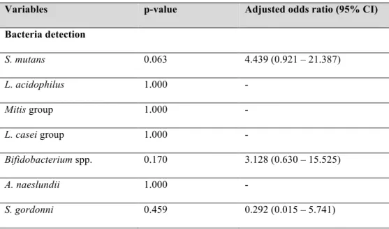
Table 3 shows the association between ECC stages and bacterial composition. After
bivariate analysis, the variable that showed statistical significant association with ECL and CF
groups was Bifidobacterium spp. (odds ratio =4.5). Considering CF versus DCL group, the
bivariate analysis also revealed strong association with the presence of S. mutans
(odds ratio=34) and Bifidobacterium spp (odds ratio =11.2). These variables were submitted
to a multiple logistic regression analysis in order to obtain the best model possibility. The
multivariate modeling indicated that Streptococcus mutans was strongly associated to the
progressive stages of early childhood caries, with an odds ratio of 21.5. Moreover,
Bifidobacterium was strongly associated with biofilm of children with dentine caries lesions
(odds ratio=5.9) (Table 4 and 5).
DISCUSSION
A detailed investigation of the composition of oral biofilm microbiota is essential for a
better understanding of the etiology of early childhood caries. Analysis of the data presented
in this study demonstrates considerable differences in bacterial diversity and biofilm
composition at different stages of ECC progression, as described from previous studies
[Becker et al., 2002; Corby et al., 2005; Kanasi et al., 2010a; Gross et al., 2012; Jiang et al.,
2014].
In the current study, the presence of S. mutans in dental plaque was strongly associated
with biofilm in children with cavitated dentin lesions (odds ratio=21) in multivariate analysis,
which is consistent with previous literature reports [Becker et al., 2002; Kanasi et al., 2010a;
Parisotto et al., 2010b; Gross et al., 2012; Jiang et al., 2014]. Streptococcus mutans is highly
acidogenic and aciduric with great capacity to adhere to enamel. It is considered the chief
pathogen associated with caries initiation [Loesche, 1986; Tanzer et al., 2001] and also
identified as a candidate risk factor for caries progression [Gross et al., 2012]. Moreover, we
observed low levels of S. mutans in dental plaque samples with higher concentration in the
biofilm of the DCL group (p<0.05) (Table 2), when compared to CF and ECL groups, making
concentrations, this can be explained because S. mutans functions in caries modulation seem not to be entirely dependent on bacterial levels, since virulence factors expressed by this species are shown to alter the biofilm structure and to promote ecological shifts leading to an acidogenic and acid-tolerant microbiota [Xiao et al., 2012; Mattos-Graner et al., 2014]. This study has not found association with the presence of S. mutans and biofilm of children with enamel caries lesions. However, it is important to emphasize that patients from this study with advanced stage of caries also presented enamel lesions. Therefore, in this study, S. mutans is associated with the progression of the disease and not with individual caries lesions, considering that the sampling method was not site-specific.
Interestingly, members of the L. casei group and L. acidophilus were rarely identified or absent in most dental plaque samples even in DCL group, showing no association of these bacteria in biofilm with caries, which corroborates previous investigations [Marchant et al., 2001; Gross et al., 2012; Dige et al., 2014; Simón-Soro et al., 2014]. In contrast, some species of Lactobacilli have been found in biofilm in earlier studies [Parisotto et al., 2010b; Kanasi et al., 2010b] and also identified as metabolically active species in pH 4.5 [McLean et al., 2012]. Nevertheless, Lactobacillus are normally associated with the more advanced caries, being frequently detected in carious dentine [Byun et al., 2004; Chhour et al., 2005]. These conflicting results may be due to high heterogeneity between individuals [Gross et al., 2010] and also to variations in evaluation methods being molecular methods more accurate than culture-based techniques [Nyvad et al., 2013]. Besides, as long as no biofilm was collected from over dentinal lesions and considering that Lactobacilli are weakly adherent to smooth surfaces, they are more present in retentive sites such as cavities [van Houte, 1996; Gross et al., 2010], therefore it was not found in the biofilm samples of the current study.
[Takahashi and Nyvad, 2011].
Most dental plaque samples from the current study presented among their composition Actinomyces naeslundii, Mitis group and S. gordonii. Nevertheless, no significantly differences in the relative levels of these species were observed for any stage of caries. Non-mutans bacteria (mainly non-Non-mutans streptococci and Actinomyces) are considered common members of health-associated microbiota [Corby et al., 2005; Gross et al., 2012] and also initial colonizers of tooth surfaces [Li et al., 2004; Takahashi and Nyvad, 2008; Dige et al., 2009]. Some metabolic activities of these bacteria may modulate dynamic caries processes, such as the use of lactate by Actinomyces naeslundii as a carbon source for growth converting it in weaker acids [Takahashi and Yamada, 1996] and the capacity of non-mutans streptococci
to induce the arginine deiminase system producing alkalis [Nascimento et al., 2009], which
can culminate in an increased pH in biofilms.
On the other hand, in certain circumstances, such as an acidic environment, non-MS may enhance their acidogenicity and acidurance adaptively [Takahashi and Yamada, 1999] playing a critical role for destabilizing the plaque homeostasis by facilitating a shift of the
demineralization/remineralization balance [Takahashi and Nyvad, 2008]. Thus, these
microorganisms were associated with dental caries in numerous studies and considered as
alternative pathogens contributing to the disease process [Marchant et al., 2001; Becker et al., 2002; Aas et al., 2008; Belda-Ferre et al., 2012; Gross et al., 2012; Jiang et al., 2013; Peterson et al., 2013].
The discrepancies findings regarding the association of these species with health or disease might have been modulated by local conditions, since Actinomyces are as versatile to adjust oneself to different conditions in dental biofilm environment as are the non-MS [Takahashi and Nyvad, 2008]. This way, the molecular mechanisms through which non-MS bacteria participates in caries initiation remain unclear and further studies are needed to identify the ecological shifts leading to cariogenic biofilms [Mattos-Graner et al., 2014].
Despite all these factors, MS, lactobacilli and Bifidobacterium are more acidogenic, aciduric and competitive than non-mutans streptococci under severely acidic conditions [Takahashi, and Nyvad, 2011].
associated with localized healthy tooth surfaces and caries lesions are similar within the same oral cavity [Corby et al., 2005]. However, there are limitations to this procedure. According to Nyvad et al. [2013], individual site-specific sampling is more appropriate when trying to compare bacterial profiles with each stage of dental caries. Therefore, the bacterial composition observed on this study could have varied if specific sites were included.
Molecular techniques are currently available to explore the bacterial microbiota, since some pathogens are not routinely cultivable [Paster et al., 2001]. Quantitative PCR represents an approach highly specific and considered as the gold standard for quantitative accuracy, however it has been underutilized [Peterson et al., 2011]. Its main limitation consists in not differentiate between live and dead cells [Alvarez et al., 2013], however a recent technique using propidium monoazide have made possible to assess cell viability with qPCR [Yasunaga et al., 2013]. While informative, this study does not provide data about the metabolic activity of the biofilm community. It seems essential to know what bacteria are doing [Takahashi and Nyvad, 2008]; since describing the identity of an organism does not necessary mean that the functional feature is apparent [Nyvad et al., 2013].
The presence and higher concentration of Bifidobacterium spp. and Streptococcus mutans in the biofilm of children with dentine caries lesions were strongly associated to the progression of early childhood caries, being considered as potentially key pathogens for ECC. Besides, it seems important to identify the metabolically active species present within these complex multi-species communities at the onset and progression of early childhood caries and also better understand how the bacterial population changes from healthy to diseased states in this dynamic/bacterial process.
Acknowledgements
This study was supported by CNPq - National Counsel of Technological and Scientific Development (Process # 475346/2011-4 MCT/CNPq 14/2011). We kindly thank the researcher Karina Maria Olbrich dos Santos (EMBRAPA) for providing Bifidobacterium
REFERENCES
American Academy of Pediatric Dentistry: Policy on Early Childhood Caries (ECC): classifications, consequences and preventive strategies. Reference Manual. Pediatric Dentistry 2014;36:50-52.
Aas JA, Griffen AL, Dardis SR, Lee AM, Olsen I, Dewhirst FE, Leys EJ, Paster BJ: Bacteria of dental caries in primary and permanent teeth in children and young adults. J Clin Microbiol 2008;46:1407–1417.
Becker MR, Paster BJ, Leys EJ, Moeschberger ML, Kenyon SG, Galvin JL, Boches SK, Dewhirst FE, Griffen AL: Molecular analysis of bacterial species associated with childhood caries. J Clin Microbiol 2002;40:1001–1009.
Belda-Ferre P, Alcaraz LD, Cabrera-Rubio R, Romero H, Simón-Soro A, Pignatelli M, Mira A: The oral metagenome in health and disease. ISME J 2012;6:46–56.
Berkowitz RJ: Acquisition and transmission of mutans streptococci. J Calif Dent Assoc 2003;31:135–138.
Byun R, Nadkarni MA, Chhour KL, Martin FE, Jacques NA, Hunter N: Quantitative analysis of diverse Lactobacillus species present in advanced dental caries. J Clin Microbiol. 2004;42:3128-3136.
Burne RA, Zeng L, Ahn SJ, Palmer SR, Liu Y, Lefebure T, Stanhope MJ, Nascimento MM: Progress dissecting the oral microbiome in caries and health. Adv Dent Res 2012;24:77–80.
Chhour K, Nadkarni MA, Byun R, Martin FE, Jacques NA, Hunter N: Molecular analysis of microbial diversity in advanced caries. J Clin Microbiol 2005;43:843–849.
Corby PM, Lyons-Weiler J, Bretz WA, Hart TC, Aas JA, Boumenna T, Goss J, Corby AL, Junior HM, Weyant RJ, Paster BJ: Microbial risk indicators of early childhood caries. J Clin Microbiol 2005;43:5753–5759.
Dewhirst FE, Chen T, Izard J, Paster BJ, Tanner AC, Yu WH, Lakshmanan A, Wade WG. The human oral microbiome. J Bacteriol 2010;192:5002-5017.
Dige I, Raarup MK, Nyengaard JR, Kilian M, Nyvad B. Actinomyces naeslundii in initial dental biofilm formation. Microbiology. 2009;155:2116-26.
Dige I, Grønkjær L, Nyvad B: Molecular studies of the structural ecology of natural occlusal caries. Caries Res 2014;48(5):451-60.
Furet J, Quéneé P, Tailliez P. Molecular quantification of lactic acid bacteria in fermented milk products using real-time quantitative PCR. International Journal of Food Microbiology 2004;97:197–207.
Gross EL, Leys EJ, Gasparovich SR, Firestone ND, Schwartzbaum JA, Janies DA, Asnani K, Griffen AL: Bacterial 16S sequence analysis of severe caries in young permanent teeth. J Clin Microbiol 2010;48:4121–4128.
mutans: dental caries onset linked to multiple species by 16S rRNA community analysis. PLoS One 2012; 7:e47722.
Holt JG,Krieg NR, Sneath PHA, Staley JT,Williams ST: Bergey’s Manual of Determinative Bacteriology 1994: 9 Edition. Williams & Wilkins. Baltimore.
Ismail AI, Sohn W, Tellez M, Amaya A, Sen A, Hasson H, Pitts NB: The International Caries Detection and Assessment System (ICDAS): an integrated system for measuring dental caries. Community Dent Oral Epidemiol 2007; 35:170–178.
Jiang W, Ling Z, Lin X, Chen Y, Zhang J, Yu J, Xiang C, Chen H. Pyrosequencing analysis of oral microbiota shifting in various caries states in childhood. Microb Ecol 2014;67:962– 969.
Li J, Helmerhorst, Leone CW, Troxler RF, Yaskell T, Haffajee AD, Socrasnsky SS, Oppenheim FG: Identification of early microbial coloniz- ers in human dental biofilm. J Appl Microbiol 2004;97:1311–1318.
Loesche WJ. Role of Streptococcus mutans in human dental decay. Microbiol Rev 1986; 50:353-380
Kanasi E, Dewhirst FE, Chalmers NI, Kent R, Moore A: Clonal analysis of the microbiota of severe early childhood caries. Caries Res 2010a; 44:485–497.
Kanasi E, Johansson I, Lu SC, Kressin NR, Nunn ME, Kent R Jr, Tanner AC.Microbial risk markers for childhood caries in pediatricians' offices. J Dent Res 2010b; 89:378-383.
Klein MI, Xiao J, Heydorn A, Koo H:An analytical tool-box for comprehensive biochemical, structural and transcriptome evaluation of oral biofilms mediated by mutans streptococci. J Vis Exp 2011; 25: pii: 2512. doi: 10.3791/2512.
Koehler B, Andreen I, Jonsson B: The earlier the colonization by mutans streptococci, the higher the caries prevalence at 4 years of age. Oral Microbiol Immunol 1988;3:14-17.
Mantzourani M, Gilbert SC, Sulong HNH, Sheehy EC, Tank S, Fenlon M, et al.: The isolation of bifidobacteria from occlusal carious lesions in children and adults. Caries Res 2009;43:308–313.
Marsh PD:Dental plaque as a biofilm and a microbial community - implications for health and disease. BMC Oral Health. 2006;15: Suppl 1:S14.
Mattos-Graner, RO, Klein MI, Smith DJ. Lessons learned from clinical studies: roles of mutans streptococci in the pathogenesis of dental caries. Curr Oral Health Rep 2014; 1:70–78.
McLean JS, Fansler SJ, Majors PD, McAteer K, Allen LZ, Shirtliff ME, Lux R, Shi W: Identifying low pH active and lactate-utilizing taxa within oral microbiome communities from healthy children using stable isotope probing techniques.PLoS One 2012;7:e32219.
Nascimento MM, Burne RA: Caries prevention by arginine metabolism in oral biofilms: translating science into clinical success. Curr Oral Heal Reports 2014;1:79–85.
Nascimento MM, Gordan VV, Garvan CW, Browngardt CM, Burne RA. Correlations of oral bacterial arginine and urea catabolism with caries experience.Oral Microbiol Immunol 2009; 24:89-95.
Nyvad B, Machiulskiene V, Baelum V: Reliability of a new caries diagnostic system differ- entiating between active and inactive caries lesions. Caries Res 1999;33:252–260.
Nyvad B, Machiulskiene V, Baelum V: Construct and predictive validity of clinical caries di- agnostic criteria assessing lesion activity. J Dent Res 2003;82:117–122.
Nyvad B, Crielaard, Mira A, Takahashi N, Beighton D: Dental caries in a molecular microbiological perspective. Caries Res 2013; 47: 89– 102.
Obata J, Takeshita T, Shibata Y, Yamanaka W, Unemori M, Akamine A, Yamashita Y: Identification of the microbiota in carious dentin lesions using 16S rRNA gene sequencing. PLoS One 2014;9:e103712.
Palmer CA, Kent R, Jr, Loo CY, Hughes CV, Stutius E, Pradhan N, Dahlan M, Kanasi E, Arevalo Vasquez SS, Tanner AC: Diet and caries-associated bacteria in severe early childhood caries. J Dent Res 2010; 89:1224–1229.
Park S, Kim YK, Kook J: Development of quantitative real-time PCR primers for detecting 42 bacterial species. Arch Microbiol 2013;195:473–482.
Parisotto TM, Steiner-Oliveira C, Silva CM, Rodrigues LK, Nobre-dos-Santos M: Early childhood caries and mutans streptococci: a systematic review. Oral Health Prev Dent. 2010a;8:59-70.
Parisotto TM, Steiner-Oliveira C, Duque C, Peres RCR, Rodrigues LKA, Nobre-dos-Santos M: Relationship among microbiological composition and presence of dental plaque, sugar exposure, social factors and different stages of early childhood caries. Arch Oral Biol 2010b;55:365–73.
Paster BJ, Boches SK, Galvin JL, Ericson RE, Lau CN, Levanos VA, Sahasrabudhe A, Dewhirst FE: Bacterial diversity in human subgingival plaque. J. Bacteriol 2001;183:3770– 3783.
Peterson SN, Snesrud E, Schork NJ, Bretz WA: Dental caries pathogenicity: a genomic and metagenomic perspective. Int Dent J 2011;61:11–22.
Peterson SN, Snesrud E, Liu J, Ong AC, Kilian M, Schork NJ, Bretz W.: The dental plaque microbiome in health and disease. PLoS One 2013;8:e58487.
Peterson SN, Meissner T, Su AI, Snesrud E, Ong AC, Schork NJ, Bretz WA: Functional expression of dental plaque microbiota. Front Cell Infect Microbiol 2014;4:108.
Rintillä T, Kassinen A, Malinen E, Krogius L, Palva A: Development of an extensive set of 16S rDNA-targeted primers for quantification of pathogenic and indigenous bacteria in faecal samples by real-time PCR. Journal of Applied Microbiology 2004;97:1166–1177.
Simón-Soro A, Guillen-Navarro M, Mira A: Metatranscriptomics reveals overall active bacterial composition in caries lesions. Journal of Oral Microbiology 2014;6,25443
Tanner AC, Mathney JM, Kent RL, Chalmers NI, Hughes CV, Loo CY, Pradhan N, Kanasi E, Hwang J, Dahlan MA, Papadopolou E, Dewhirst FE: Cultivable anaerobic microbiota of severe early childhood caries. J Clin Microbiol 2011;49:1464-1474.
Takahashi N, Nyvad B: Caries ecology revisited: microbial dynamics and the caries process. Caries Res. 2008;42:409-18.
Takahashi N, Nyvad B: The role of bacteria in the caries process: ecological perspectives. J Dent Res 2011;90:294–303.
Takahashi, N. & Yamada, T: Catabolic pathway for aerobic degradation of lactate by Actinomyces naeslundii. Oral Microbiol Immunol 1996;11:193–198.
Takahashi N, Yamada T: Acid-induced acidogenicity and acid tolerance of non-mutans streptococci. Oral Microbiol Immunol 1999;14:43–48.
Tanzer JM, Livingston J, Thompson a M: The microbiology of primary dental caries in humans. J Dent Educ 2001;65:1028–1037.
van Houte J, Lopman J, Kent R: The final pH of bacteria comprising the predominant f lora on sound and carious human root and enamel surfaces. J Dent Res 1996;75:1008–1014.
Wilson K: Preparation of genomic DNA from bacteria. Curr Protoc Mol Biol 2001;Chapter 2:Unit 2.4.
Wolff, D, Freese C, Maier-Kraus T, Krueger T, Wolff B: Bacterial biofilm composition in caries and caries-free subjects. Caries Res 2013;47:69–77.
Xiao J, Klein MI, Falsetta ML, Lu B, Delahunty CM, Yates JR 3rd, Heydorn A, Koo H: The exopolysaccharide matrix modulates the interaction between 3D architecture and virulence of a mixed-species oral biofilm. PLoS Pathog 2012;8:e1002623.
Table 1. Primers that were used for qPCR assays.
Target species Sequence (5’ 3’)
Annealing temperature
(°C)
Amplicon length
(bp)
References
Bacteria 16S rDNA F: TCCTACGGGAGGCAGCAGT
R:GGACTACCAGGGTATCTAATCCTGTT 57 466 Nadkarni et al., 2002
Actinomyces naeslundii F:CTGCTGCTGACATCGCCGCTCGTA
R:TCCGCTCGCGCCACCTCTCGTTA 62 144 Park et al., 2013
Bifidobacterium spp.1 F: TCGCGTC(C/T)GGTGTGAAAG
R: CCACATCCAGC(A/G)TCCAC 58 243 Rintilla et al., 2004
Lactobacillus acidophilus F: GATCGCATGATCAGCTTATA
R: AGTCTCTCAACTCGGCTATG 60 124 Furet et al., 2004
L. casei group2 F: GCGGACGGGTGAGTAACACG
R: GCTTACGCCATCTTTCAGCCAA 60 121 Furet et al., 2004
Mitis group3 F:TAGAACGCTGAAGGAAGGAGC
R: GCAACATCTACTGTTATGCGG 60 133 Wolff et al., 2013
Streptococcus gordonii F: CAGGAAGGGATGTTGGTGTT
R: GACTCTCTTGGCGACGAATC 60 136 Wolff et al., 2013
Streptococcus mutans F: AGCCATGCGCAATCAACAGGTT
R: CGCAACGCGAACATCTTGATCAG 64 415 Yano et al., 2002
!
1. Bifidobacterium longum, B. minimum, B. angulatum, B. catenulatum, B. pseudocatenulatum, B. dentium, B. ruminantium,! B. thermophilum, B. subtile, B. bifidum, B. boum, B. lactis, B. animalis, B. choerinum, B. gallicum, B. pseudolongum! subsp. globosum, B. pseudolongum subsp. pseudolongum, B. magnum, B. infantis, B. indicum, B. gallinarum, B. pullorum,! B. saeculare, B. suis!
2. L . casei group: L. casei, L. paracasei, L. rhamnosus and L. zeae.!
Table 2. Dental plaque bacteria of the caries-free, caries-active with enamel lesions and caries-active with dentine lesion groups determined by qPCR as a percentage of the total bacteria load.
Oral bacteria Groups Mean (%) ±SD Median (%) p-value
S. mutans CF 0.190 0.680 0.000 <0.001
ECL 0.052 0.118 0.001
DCL 1.191 2.068 0.250*
L. acidophilus CF 0.000 0.000 0.000 0.373
ECL 0.000 0.000 0.000
DCL 0.000 0.001 0.000
Mitis group CF 1.052 0.884 0.876 0.580
ECL 1.573 1.453 1.178
DCL 3.361 6.123 0.892
L. casei group CF 0.000 0.000 0.000 0.132
ECL 0.000 0.000 0.000
DCL 0.001 0.002 0.000
Bifidobacterium spp. CF 0.000 0.000 0.000 <0.001
ECL 0.000 0.001 0.000
DCL 0.088 0.213 0.002*
A. naeslundii CF 18.062 17.021 15.053 0.092
ECL 14.239 14.904 9.620
DCL 20.942 15.034 16.419
S. gordonii CF 0.513 0.777 0.294 0.075
ECL 0.811 0.948 0.541
DCL 2.821 5.518 0.570
SD= standard deviation; CF=caries-free; ECL= enamel caries lesions; DCL=dentine caries lesions
The data are mean ± standard deviation and median values of concentrations of bacterial species as a percent of total bacteria load.
Table 3. Bivariate analysis of the relationships between ECC status and related factors.
Odds ratio (95% CI)
Variables CF n (%)
ECL n (%)
DCL
n (%) p-value CF X ECL CF X DCL ECL X DCL Bifidobacterium <0.001 4.5 (1.1 -19.2) 11.2 (3.0 -41.6) 2.4 (0.7 - 8.2)
positive 4 (20) 9 (52.9) 28 (73.7)
negative 16 (80) 8 (47.1) 10 (26.3)
S mutans < 0.001 4.0 (0.9 -17.4) 34.0 (7.5 - 153.6) 9.5 (2.3 - 39.1) positive 4 (20) 9 (52.9) 34 (89.5)
negative 16 (80) 8 (47.1) 4 (10.5)
L. acidophilus 0.218
positive ND ND 3 (7.9)
negative 20 (100) 17 (100) 35 (92.1)
L. casei group 0.177
positive ND 1 (5.9) ND
negative 20 (100) 16 (94.1) 38 (100)
Mitis group 1.000
positive 20 (100) 17 (100) 38 (100)
negative ND ND ND
S. gordonii 0.343 0.8 (0.0 -14.6) 5.9 (0.2 - 152.4 ) 7.0 (0.3 - 181.1)
positive 19 (95) 16 (94.1) 38 (100)
negative 1 (5) 1 (5.9) ND
A. naeslundii 1.000
positive 20 (100) 17 (100) 38 (100)
negative ND ND ND
CF=caries-free; ECL= enamel caries lesions; DCL=dentine caries lesions; ND= not detected
Table 4. Multivariate modeling for enamel caries lesions
Variables p-value Adjusted odds ratio (95% CI)
Bacteria detection
S. mutans 0.063 4.439 (0.921 – 21.387)
L. acidophilus 1.000 -
Mitis group 1.000 -
L. casei group 1.000 -
Bifidobacterium spp. 0.170 3.128 (0.630 – 15.525)
A. naeslundii 1.000 -
Table 5. Multivariate modeling for dentine caries lesions
Variables p-value Adjusted odds ratio (95% CI)
Bacteria detection
S. mutans <0.001* 21.501 (4.299 – 107.544)
L. acidophilus 1.000 -
Mitis group 1.000 -
L. casei group 1.000 -
Bifidobacterium 0.033* 5.903 (1.153 -30.221)
A. naeslundii 1.000 -
S. gordonni 0.999 -
CAPÍTULO 2
Molecular detection of bacteria associated to caries activity in dentinal lesions
B.G. Nevesa, R.N. Stippb, D.S. Bezerraa , S.F.F. Guedesa, L.K.A. Rodriguesa
a
Post-graduation Program, Faculty of Pharmacy, Dentistry and Nursing, Federal University of Ceará, Fortaleza, Ceará, Brazil; bDepartment of Microbiology and Immunology, Piracicaba Dental School, State University of Campinas, Piracicaba, SP, Brazil
Short title: Dentine caries-associated bacteria in ECC-lesions
Key words: Early Childhood Caries, Quantitative Polymerase Chain Reaction, Bacteria, Dentin Caries, Preschoolers
Address all correspondence to:
Lidiany K. A. Rodrigues, DDS, MSc, PhD, Associate Professor
Postgraduate Program in Dentistry, Faculty of Pharmacy, Dentistry and Nursing
Federal University of Ceará, Fortaleza, Ceará, Brazil.
Rua Monsenhor Furtado, 1273 - Zip Code: 60430-355
Phone- #+558533668410/ Fax- #+558533668232
Email: lidianykarla@yahoo.com
Este artigo está escrito de acordo com as normas de publicação do periódico Caries Research.
Conflict of interests
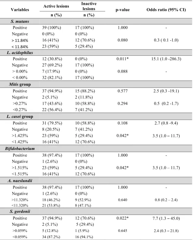
ABSTRACT
Early childhood caries (ECC) is a microbial infection that severely compromises the dentition
of young children. Few studies have focused on oral bacterial community changes within
carious lesion activity. The aim of this study was to quantify Actinomyces naeslundii,
Bifidobacterium spp., Mitis group, Lactobacillus acidophilus, Lactobacillus casei group, Streptococcus gordonii and Streptococcus mutans in active and inactive carious dentine lesions in ECC- children by using quantitative polymerase chain reaction (qPCR). Fifty-six dentine lesions samples, classified as active (n=39) or inactive (n=17), were collected from children aged 2 to 5 years. Relative quantification revealed that Bifidobacterium spp. and L.
casei group were significantly more abundant in active dentine lesions (p<0.05).
Concentrations of Actinomyces naeslundii, Mitis group and Streptococcus gordonni were not significantly different when comparing dentin lesion activity. The relative proportion of S. mutans was significantly greater in inactive than in active lesions (p<0.05). Bifidobacterium
spp and L. casei group demonstrated a positive correlation (p=0.001) in active lesions. The
positive detection of L. acidophilus (OR=15.1) and S. gordonii (OR=7.7) was significantly associated to the active lesions. The data indicate that higher detection levels of
Bifidobacterium spp. and L. casei group may be linked to dentin lesion activity. Also, the
INTRODUCTION
Dental caries is one of the most prevalent diseases of childhood worldwide, especially
in socially disadvantaged populations [Berkowitz, 2003]. Several factors, including
microbial, genetic, immunological, behavioral and environmental, are involved and
contribute to its development [Peterson et al., 2013]. In particular, in preschool children, this
condition defined as early childhood caries (ECC), can devastate the primary teeth, affect
child’s self-esteem, impact general health and lead to nutritional deficiency [Ramos-Goméz
et al., 2002]. Because of the aggressive pattern of ECC, areas of demineralization can
rapidly progress, develop cavitation and involve dental pulp tissues, causing serious
consequences such as pain and pulp infection [AAPD, 2014; Obata et al., 2014].
Cavitated dentine carious lesions are considered as the last stage of dental caries and
also diverse ecosystems with high variability [Belda-Ferre et al., 2012]. Dentine provides a
different environment for bacteria involved in caries progression, where only specialized
bacteria are able to colonize and exploit [Simon-Sóro et al., 2013]. The bacterial profile in
enamel and dentinal caries are significantly different [Simón-Soro et al., 2013; Obata et al., 2014] , since the microbiota in dentine is constantly submitted to changes, such as nutrient availability, oxygen concentration and pH [Lima et al., 2011; Takahashi and Nyvad, 2011].
Moreover, this tissue contains a higher proportion of organic matrix and lower inorganic component than enamel. Thus, the critical pH for dentine dissolution is higher compared to
enamel, which allow colonization of bacteria that are not as acidogenic and aciduric as those
required for initial enamel demineralization [Kinaoush et al., 2013], but also proteolytic
bacteria are involved [Simón-Soro et al., 2013].
The advent of molecular researches to characterize the oral microbiota in health and
disease is revealing the diversity of oral biofilms and dentinal caries and finding new
candidates for disease-associated bacterial species [Becker et al., 2002; Aas et al., 2008;
Corby et al., 2005; Gross et al., 2012] . Considering that the microbiota involved in dental
caries are known to be highly diverse and variable [Martin et al., 2002; Takahashi and
Nyvad, 2011], understanding the microbial etiology of caries and how environmental
conditions in the oral cavity impact the disease process continues to change as technology
advances [Kianoush et al. 2014]. Although the strong association of mutans streptococci and
ECC is established in the literature [Kohler et al, 1988; Berkowitz, 2003; Kanasi et al.,
2010; Parisotto et al., 2010], it seems that these bacteria are not present in all children with
caries [Mattos-Graner et al., 2001; Aas et al., 2008]. Beyond S. mutans, molecular