with Different Levels of Ginsenoside Rb1 Degradation to
Compound K
Kyung-Ah Kim1, IL-Hoon Jung1, Se-Hoon Park2, Young-Tae Ahn2, Chul-Sung Huh2, Dong-Hyun Kim1* 1Department of Life and Nanopharmaceutical Sciences and Department of Pharmacy, Kyung Hee University, Seoul, Korea,2R &B D Center, Korea Yakult Co., Ltd., Yongin-si, Kyunggi-do, Korea
Abstract
Panax ginseng (family Araliaceae) which contains ginsenoside Rb1 as a main constituent is traditionally used as a remedy for cancer, inflammation, stress, and ageing. The ginsenoside Rb1 in orally administered ginseng is metabolized to bioactive compounds by gut microbiota before their absorptions to the blood. However, its metabolizing activities in individuals are significantly different as we previously demonstrated. Here, we selected 5 samples with fecal activity potently metabolizing ginsenoside Rb1 to compound K (FPG; metabolic activity, 0.05860.029 pmol/min/mg) and 5 samples with fecal activity non-metabolizing ginsenoside Rb1 to compound K (FNG) from a pool of 100 subjects investigated in a previous study and analyzed fecal microbiota by 16S rRNA gene pyrosequencing. Taxonomy-based analysis showed that the population levels of Firmicutesand Proteobacteriain FPG were lower than in FNG, but those of Bacteroidetes and Tenericutes in FPG were higher than in FNG. At the genus level, the population levels of
Clostridiales_uc_g, Oscillibacter, Ruminococcus, Holdemania,and Sutterella in FPG were significantly higher than in FNG, but that ofLeuconostocin FPG was lower than in FNG. The population levels ofBacteroides and Bifidobacterium,which potently metabolizes ginsenoside Rb1 to compound K were dramatically increased in FPG. The gut microbiota compositions of FPG and FNG were segregated on PCO2 by Principal Coordinate Analysis. Intestinal bacterial metabolism of ginseng, particularly ginsenoside Rb1, may be dependent on the composition of gut microbiota, such as
Ruminococcus spp., Bacteroides spp. and Bifidobacterium spp.
Citation:Kim K-A, Jung I-H, Park S-H, Ahn Y-T, Huh C-S, et al. (2013) Comparative Analysis of the Gut Microbiota in People with Different Levels of Ginsenoside Rb1 Degradation to Compound K. PLoS ONE 8(4): e62409. doi:10.1371/journal.pone.0062409
Editor:Manlio Vinciguerra, University College London, United Kingdom
ReceivedJanuary 3, 2013;AcceptedMarch 21, 2013;PublishedApril 29, 2013
Copyright:ß2013 Kim et al. This is an open-access article distributed under the terms of the Creative Commons Attribution License, which permits unrestricted use, distribution, and reproduction in any medium, provided the original author and source are credited.
Funding:This study was financially supported by the research fund of Korean Food and Drug Administration (09172KFDA996). The funders had no role in study design, data collection and analysis, decision to publish, or preparation of the manuscript.
Competing Interests:The authors confirm here again that the authors including Se-Hoon Park Young-Tae Ahn, and Chul-Sung Huh who work for R &B D Center, Korea Yakult Co., Ltd. (Yongin-si, Kyunggi-do, 446–901, Korea) have declared that no competing interests exist. This does not alter the authors’ adherence to all the PLOS ONE policies on sharing data and materials.
* E-mail: [email protected]
Introduction
Most of traditional Chinese medicines (TCM) are orally administered to humans. The components of orally administered TCM are therefore inevitably brought into contact with intestinal microbiota in the alimentary tract [1,2]. In human, gut is trillions of individual microbes residing [3,4]. The exact membership of this highly complex gut ecosystem, known as the microbiome, varies between individuals. Integral to this picture is the interplay between gut bacteria and diet as well as between gut microbes and health [4,5,6]. The global rise of diets, such as fat and chronic diseases, such as obesity and bowel disease, is increasingly being linked with perturbations in gut flora. The gut microbiota has the ability to metabolize drugs and other xenobiotics more extensively than any other part of the body [7,8,9]. Thus, gut microbiota may transform the constituents of orally administered TCM to bioactive compounds before they get absorbed from the gastro-intestinal tract [2,10,11].
Ginseng (the root of Panax ginseng C.A. Meyer, Araliaceae), which contains ginsenosides as major constituents, is frequently used as a traditional medicine in Asian countries [2]. The
compound K (FNG) from a pool of 100 subjects investigated in a previous study [22] and analyzed fecal microbiota by 16S rRNA gene pyrosequencing.
Materials and Methods
Materials
p-Nitrophenyl-b-D-glucopyranoside was purchased from Sigm-Aldrich (St. Louis, MO). Ginsenoside Rb1 (purity, .92%) and compound K (purity,.95%) were isolated using the previously published method of Baeet al. [16,20].
Subjects
From a pool of 100 subjects analyzed in a previous study [22], we selected 5 samples with FPG (sample No. 3, 10, 29, 47, and 95; 38.0613.3 years) and 5 samples with FNG (sample No. 20, 23, 31, 75, and 88; 38.4612.7 years). Exclusion criteria included smoking and current medication, especially regular or current use of antibiotics. The recruitment of subjects and the consent procedure as well as the collection of their stools were approved by the ethics committee for the Care and Use of Clinical Study in the Medical School, Kyung Hee University (KMC IRB 0922-08-A1). The participants provided their written informed consent to participate in the study.
Sample Preparation
The human fecal samples (about 1 g) were prepared according to the previous method [22], were collected in plastic cups 9 h after fasting, and then carefully mixed with a spatula and suspended with cold 9 ml saline. The fecal suspension was centrifuged at 5006g for 5 min. The supernatant was then centrifuged at 10,0006g for 20 min. The resulting precipitates were used as a metabolic enzyme source for the assay of enzyme activity. The preparation and assay of the enzyme source were performed within 24 h under anaerobic conditions.
Assay ofb-D-glucosidase,b-D-glucuronidase,b -D-galactosidase anda-L-rhamnosidase Activities
For the assay of b-D-glucosidase, b-D-glucuronidase, b -D-galactosidase, and a-L-rhamonosidase activities, the reaction mixture (total volume of 0.5 ml) was composed of 0.2 ml of 1 mM p-nitrophenyl-b-D-glucopyranoside, p-nitrophenyl-b -D-glucuronide, p-nitrophenyl-b-D-galactopyranoside, or p-nitrophe-nyl-a-L-rhamnopyranoside as substrate respectively, 0.2 ml of 0.1 M phosphate buffer (pH 7.0), and 0.1 ml of the fecal enzyme fraction. The reaction mixture was incubated at 37uC for 20 min. The reaction was stopped by the addition of 0.5 ml of 0.5 N NaOH, centrifuged at 3,0006g for 10 min and measured the absorbance at 405 nm (vis spectrophotometer, Shimadzu UV-1201). The activities were expressed in pmol per minute per milligram protein and the protein content was assayed by Bradford method [23].
Assay of Intestinal Bacterial Enzyme Activity Metabolizing Ginsenoside Rb1, and Ginseng Extract to Compound K
For the fecal enzyme activity for ginsenoside Rb1 or ginseng extract, the reaction mixture (0.5 ml) containing 0.125 ml of the human fecal suspension and 0.1 mM ginsenoside Rb1 (or 0.5 mg ginseng extract) was incubated at 37uC for 4 h, and 1.5 ml of MeOH was added to stop the reaction. The reaction mixture was centrifuged at 3,0006g for 10 min and the level of ginsenoside Rb1 in the resulting supernatant was analyzed by HPLC.
HPLC Analysis
The reaction mixture was analyzed by Hewlett Packard Series 1050 HPLC system. The instrument was controlled and the data were processed by a HP Chemstation (Rev. A. 09.03). The analytical column was an Agilent Hypersil ODS (10064.6 mm i.d., 5mm; Agilent Technologies, USA) protected by a C18
Security Guard Cartridge (Phenomenex, Torrance, CA). The elution solvent was acentonitrile (ACN) and distilled and deionized water (DDW). Ginsenoside Rb1 was analyzed using a linear gradient 0,70% ACN in DDW including 0.05% formic acid for Table 1.Number of sequence analyzed, observed diversity richness (OTUs), estimated OTU richness (ACE and Chao1), and coverage.
Phylotype
Group Number Total reads OTUs ACE Chao1 Goods Coverage
FPG 3 10320 1262 2183.3 1912.8 0.95
10 4959 713 1374.7 1168.0 0.94
29 9271 1039 2083.4 1624.8 0.95
47 7108 271 330.7 336.0 0.99
95 9536 1015 1933.3 1574.3 0.95
Mean6SD 823862186 8606382 15816765 13236612 0.9660.02
FNG 20 12807 1293 1874.7 1913.3 0.96
23 11353 436 740.8 631.0 0.99
31 3851 419 706.1 596.2 0.96
75 2453 249 492.6 414.1 0.96
88 5077 300 484.5 413.2 0.98
Mean6SD 710864661 5396428 7936634 8596579 0.9760.01
The cutoff value of phylotype is equal to or greater than 97% similarity. OTUs, operational taxonomic units; FPG, fecal activity potently metabolizing ginsenoside Rb1 to compound K; FNG, fecal activity non-metabolizing ginsenoside Rb1 to compound K.
doi:10.1371/journal.pone.0062409.t001
15 min and an isocratic elution for 5 min in 70% ACN at a flow rate of 1.0 ml/min and detected at 203 nm. A sample volume of 20ml was used for injection. The retention times of Rb1, and
compound K were 10.5, and 15.6 min, respectively.
DNA Extraction, Pyrosequencing, and Data Analysis Genomic DNA was extracted from fecal sample using a commercial DNA isolation kit (QIAamp DNA stool mini kit, Qiagen, Hilden, Germany) by following the manufacturer’s protocol. For pyrosequencing, amplification of genomic DNA was performed using barcoded primers, which targeted the V1 to V3 region of the bacterial 16S rRNA gene. The amplification and Figure 1. Fecal metabolic activities for glucopyranoside, glucuronide, p-nitrophenyl-b-D-galactopyranoside, p-nitrophenyl-a-L-rhamnopyranoside and ginsenoside Rb1 in 10 Koreans.(A) Hydrolytic activity of p-nitrophenyl-b -D-glucopyranoside (PNG). (B) Hydrolytic activitiy of ginsenoside Rb1 to compound K. The relationships between PNG hyfrolyzing and ginsenoside Rb1 degrading activities (C), between PNG hydrolyzing and compound K forming activities (D), b-D-glucuronide hydrolyzing and compound K forming activities (E),b-D-galactopyranoside hydrolyzing and compound K forming activities (F),a-L-rhamnopyranoside hydrolyzing and compound K forming activities (G), and between ginsenoside Rb1 degrading and compound K forming activities (H). We selected 5 samples with FPG (fecal activity potently metabolizing ginsenoside Rb1 to compound K) and 5 samples with FNG (fecal activity non-metabolizing ginsenoside Rb1 to compound K) from a pool of 100 subjects investigated in a previous study [22]. FPG is black bars (in A and B) and closed circles (in C, D and E). FNG is white bars (in A and B) and open circles (in C, D and E). Grayish bars (in A and B) are average values of 10 samples. All values indicate mean6SD. **p,0.01.
sequencing were performed according to the methods described by Chun et al. [24] and completed by Chunlab Inc. (Seoul, Korea) using a 454 GS FLX Titanium Sequencing System (Roche, Branford, CT). Sequence reads were identified using EzTaxon-e database (http://eztaxon-e.ezbiocloud.net/; [25]) on the basis of 16S rRNA sequence data. Number of sequence analyzed, observed diversity richness (operational taxonomic units, OTUs), estimated OTU richness (ACE and Chao1), and coverage in the present pyrosequencing were indicated in Table 1.
Statistics
The data are expressed as the means6standard deviation. Statistical analysis of the data was performed with Student’st-test. Differences with a p,0.05 were considered to be statistically significant.
Results and Discussion
The pharmacological activities of orally administrated herbal medical components such as ginsenoside Rb1 are enhanced by gut microbiota [16]. Furthermore, the capacities of transformation of bioactive compounds are variable between individuals. Therefore, to understand the difference of gut microbiota related to the fecal metabolism of ginsenoside Rb1 to compound K between individuals, we selected 10 samples from a pool of 100 subjects analyzed in a previous study [22]; 5 samples with FPG and 5 samples with FNG. The activity of the former group (FPG) potently metabolizing ginsenoside Rb1 to compound K was 0.05860.029 pmol/min/mg whereas the latter group (FNG) did not metabolize ginsenoside Rb1 to compound K (Fig. 1B). However, using p-nitrophenyl-b-D-glucopyranoside as a substrate,
b-glucosidase activity between FPG and FPG was not significantly different (Fig. 1A). Furthermore, their p-nitrophenyl-b -D-glucopyranoside-hydrolyzing b-glucosidase activities were not proportional to their ginsenoside Rb1 degrading activities or compound K-forming activities (Fig. 1C and D). In addition, the activities of b-glucuronidase hydrolyzing p-nitrophenyl-b
-D-glucuronide, b-galactosidase hydrolyzing p-nitrophenyl-b -D-galactopyranoside, and a-rhamnosidase hydrolyzing p-nitrophe-nyl-a-L-rhamnopyranoside were not proportional to compound K-forming activities (Fig. 1E, 1F and 1G). However, ginsenoside Rb1-degrading activities were proportional to their compound K-forming activities (Fig. 1H).
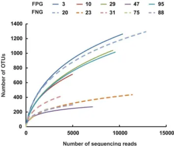
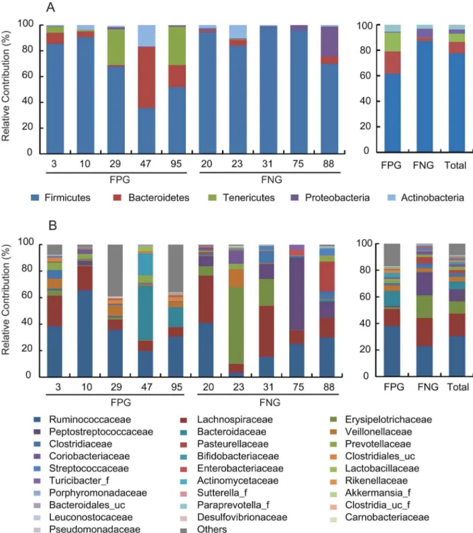
Next, we analyzed the gut microbiota compositions of FPG and FNG by pyrosequencing. As demonstrated by the rarefaction curves (Fig. 2) and the number of sequences analyzed and estimated OTU richness (Table 1), bacterial richness and diversity in FPG showed a tendency to be higher than in FNG with no significant difference. Furthermore, taxonomy-based analysis showed a modulation of the populations of the dominant intestinal microbiota. The distributions of the major phyla (Firmicutes, Bacteroidetes, Tenericutes, Proteobacteria and Actinobacteria) are con-sistent with previous human gut studies [26,27,28]. However, the main dominants in FPG were Firmicutes, Bacteriodetes, and Tenericutes, while those in FNG were Firmicutes and Proteobacteria (Figure 3A). Of them, the population levels of Firmicutes and Proteobacteria in FPG were lower than in FNG, but those of BacteroidetesandTenericutesin FPG were higher than in FNG. At the family level, among the relative abundance of 16 major family groups, an average of 74.3% of all sequences belonged to the 8 families comprising Firmicutes: Ruminococcaceae, Lachnospiraceae, Erysipelotrichaceae, Peptostreptococcaceae, Veillonellaceae, Clostridiaceae, Clostridiales_uc_g, and Streptococcaceae(Fig. 3B). The two families of Bacteroidetes (Bacteroidaceae and Prevotellaceae) accounted for an average of 8.4% of sequences while other families (Pasteurellaceae, phylum Proteobacteria; Coriobacteriaceae and Bifidobacteriaceae,phylum Figure 2. Rarefaction curves. Rarefaction analysis of V1–V3
pyrosequencing tags of the 16S rRNA gene in fecal microbiota from FPG (fecal activity potently metabolizing ginsenoside Rb1 to compound K) or FNG (fecal activity non-metabolizing ginsenoside Rb1 to compound K).
doi:10.1371/journal.pone.0062409.g002
Table 2.The difference between FPG and FNG in the
composition (percent of total sequences) of fecal bacterial genera.
Genus Compositiona)(%)
Total FPG FNG P value
Faecalibacterium 15.7614.75 14.58618.24 16.84612.39 0.960
Clostridium_g4 8.27615.39 0.7160.96 15.83619.72 0.175
Bacteroides 6.50613.66 14.17618.87 0.3760.63 0.077
Catenibacterium 6.06616.04 0.3360.70 11.80622.27 0.331
Roseburia 4.5263.96 4.8164.86 4.2763.40 0.401
Ruminococcaceae_uc 2.9262.83 4.1963.46 1.6561.42 0.222
Eubacterium_g9 2.7564.68 0.7361.02 4.7866.17 0.204
Haemophilus 2.6667.37 0.0160.02 5.96610.89 0.316
Prevotella 2.4362.37 3.0262.71 1.8462.11 0.744
Clostridium 2.3562.92 1.5262.44 3.1763.39 0.157
Dorea 2.0461.66 1.1961.22 2.8961.70 0.066
Bifidobacterium 1.7265.20 3.3167.37 0.0160.01 0.291
Clostridiales_uc_g 1.2761.14 2.0861.12 0.4660.08 0.030
Ruminococcus 0.8260.98 1.3461.19 0.3060.19 0.046
Oscillibacter 0.1160.15 0.2260.14 0.00660.01 0.022
Leuconostoc 0.0760.10 0.00460.005 0.13360.104 0.043
Holdemania 0.0360.04 0.05260.047 ndb) 0.021
Sutterella 0.0160.02 0.02660.019 0.00160.003 0.006
a)Mean
6SD (n = 5).
b)not detected. FPG, fecal activity potently metabolizing ginsenoside Rb1 to compound K; FNG, fecal activity non-metabolizing ginsenoside Rb1 to compound K.
doi:10.1371/journal.pone.0062409.t002
Actinobacteria) accounted for an average of 5.9% of sequences. Interestingly, the population levels ofRuminococcaceae, Bacteroidaceae, Sutterella_ f, Clostridiales_uc,Bifidobacteriaceae,andRikenellaceaein FPG were higher than in FNG, while those of Lachnospiraceae, Erysipelotrichaceae, Peptostreptococcaceae, Streptococcaceae and Leuconosto-caceae were enriched in FNG rather than in FPG. At the genus level, the three most abundant genera were Faecalibacterium, Clostridium_g4,and Bacteroides, which accounted for an average of 30.5% of sequences (Table 2). The population levels of
Clostridiales_uc_g, Oscillibacter, Ruminococcus, Holdemania,andSutterella in FPG were significantly higher than in FNG, but that of Leuconostoc in FPG was lower than in FNG. Furthermore, the population levels of Bacteroides and Bifidobacterium, were dramati-cally increased in FPG whereas those ofClostridium_g4, Catenibacter-ium, Eubacterium_g9,andHaemophiluswere dramatically increased in FNG. Interestingly, in our previous work, we have found that BacteroidesandBifidobacteriumcould metabolize ginsenoside Rb1 to compound K [16].
Figure 3. The composition of fecal microbiota in 10 Koreans.The relative contribution of dominant phyla (A) and families (B) identified from pyrosequencing data is shown (individual samples are on the left panels and pooled samples are on the right panels). FPG, fecal activity potently metabolizing ginsenoside Rb1 to compound K; FNG, fecal activity non-metabolizing ginsenoside Rb1 to compound K.
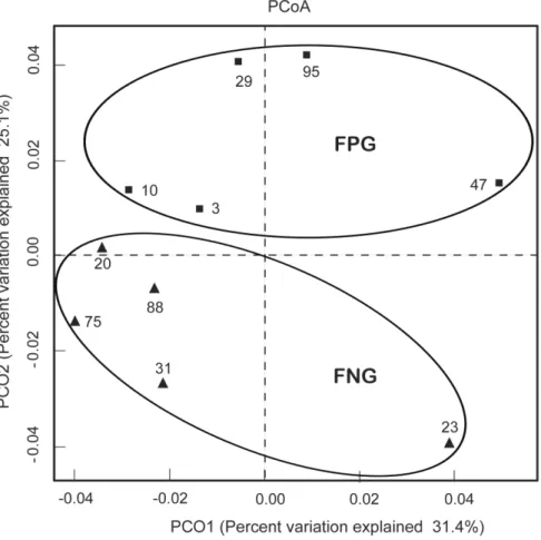
We also processed all these sequences at the same length and position to match the length and position of the gut microbiota 16S rRNA gene sequences, computed all pair-wise distances between FPG and FNG and performed Principal Coordinate Analysis (PCoA) to cluster these communities along axes of maximal variance (Fig. 4D). Gut microbial community of each group member was clustered and the maximum variations were 31.4% (PCO1) and 25.1% (PCO2). Furthermore, the gut microbiota compositions of FPG and FNG were segregated on PCO2 by Principal Coordinate Analysis. The difference may be related to gut bacteria metabolizing ginsenoside Rb1, suggesting that diet style, as well as host genetics may affect in molding gut microbiota.
Based on these findings, the difference of the pharmacological effects of orally administrated TCM and its constituents between individuals may be dependent on the composition of gut microbiota.
Author Contributions
Conceived and designed the experiments: KAK DHK. Performed the experiments: KAK IHJ. Analyzed the data: KAK SHP YTA CSH DHK. Contributed reagents/materials/analysis tools: SHP YTA CSH. Wrote the paper: KAK DHK.
References
1. Crow JM (2011) Microbiome: That healthy gut feeling. Nature 480: S88–89. 2. Kim D (2012) Chemical Diversity of Panax ginseng, Panax quinquifolium and
Panax notoginseng. J Ginseng Res 36: 1–15.
3. Simon GL, Gorbach SL (1986) The human intestinal microflora. Dig Dis Sci 31: 147S–162S.
4. De Filippo C, Cavalieri D, Di Paola M, Ramazzotti M, Poullet JB, et al. (2010) Impact of diet in shaping gut microbiota revealed by a comparative study in children from Europe and rural Africa. Proc Natl Acad Sci U S A 107: 14691– 14696.
5. O’Keefe SJ (2008) Nutrition and colonic health: the critical role of the microbiota. Curr Opin Gastroenterol 24: 51–58.
6. Zoetendal EG AA, Akkermans-van Vliet WM, De Visser JAGM, De Vos WM (2001) The host genotype affects the bacterial community in the human gastrointestinal tract. Microb Ecol Health and Dis 13: 129–134.
7. Scheline RR (1973) Metabolism of foreign compounds by gastrointestinal microorganisms. Pharmacol Rev 25: 451–523.
8. Mikov M (1994) The metabolism of drugs by the gut flora. Eur J Drug Metab Pharmacokinet 19: 201–207.
9. Sousa T, Paterson R, Moore V, Carlsson A, Abrahamsson B, et al. (2008) The gastrointestinal microbiota as a site for the biotransformation of drugs. Int J Pharm 363: 1–25.
10. Akao T, Kanaoka M, Kobashi K (1998) Appearance of compound K, a major metabolite of ginsenoside Rb1 by intestinal bacteria, in rat plasma after oral administration–measurement of compound K by enzyme immunoassay. Biol Pharm Bull 21: 245–249.
11. Akao T, Kida H, Kanaoka M, Hattori M, Kobashi K (1998) Intestinal bacterial hydrolysis is required for the appearance of compound K in rat plasma after oral administration of ginsenoside Rb1 from Panax ginseng. J Pharm Pharmacol 50: 1155–1160.
12. Joh EH, Lee IA, Jung IH, Kim DH (2011) Ginsenoside Rb1 and its metabolite compound K inhibit IRAK-1 activation–the key step of inflammation. Biochem Pharmacol 82: 278–286.
Figure 4. Principal coordinate analysis (PCoA) plot.The plot showed the clustering pattern between FPG and FNG based on weighted pairwise Fast UniFrac analysis. FPG, fecal activity potently metabolizing ginsenoside Rb1 to compound K; FNG, fecal activity non-metabolizing ginsenoside Rb1 to compound K. doi:10.1371/journal.pone.0062409.g004
13. Choo MK, Sakurai H, Kim DH, Saiki I (2008) A ginseng saponin metabolite suppresses tumor necrosis factor-alpha-promoted metastasis by suppressing nuclear factor-kappaB signaling in murine colon cancer cells. Oncol Rep 19: 595–600.
14. Wakabayashi C, Hasegawa H, Murata J, Saiki I (1997) In vivo antimetastatic action of ginseng protopanaxadiol saponins is based on their intestinal bacterial metabolites after oral administration. Oncol Res 9: 411–417.
15. Shin YW, Kim DH (2005) Antipruritic effect of ginsenoside rb1 and compound k in scratching behavior mouse models. J Pharmacol Sci 99: 83–88. 16. Bae EA, Park SY, Kim DH (2000) Constitutive beta-glucosidases hydrolyzing
ginsenoside Rb1 and Rb2 from human intestinal bacteria. Biol Pharm Bull 23: 1481–1485.
17. Lee J, Lee E, Kim D, Yoo J, Koh B (2009) Studies on absorption, distribution and metabolism of ginseng in humans after oral administration. J Ethnopharmacol 122: 143–148.
18. Tawab MA, Bahr U, Karas M, Wurglics M, Schubert-Zsilavecz M (2003) Degradation of ginsenosides in humans after oral administration. Drug Metab Dispos 31: 1065–1071.
19. Park EK, Choo MK, Han MJ, Kim DH (2004) Ginsenoside Rh1 possesses antiallergic and anti-inflammatory activities. Int Arch Allergy Immunol 133: 113–120.
20. Bae EA, Choo MK, Park EK, Park SY, Shin HY, et al. (2002) Metabolism of ginsenoside R(c) by human intestinal bacteria and its related antiallergic activity. Biol Pharm Bull 25: 743–747.
21. Kim YS, Kim JJ, Cho KH, Jung WS, Moon SK, et al. (2008) Biotransformation of ginsenoside Rb1, crocin, amygdalin, geniposide, puerarin, ginsenoside Re, hesperidin, poncirin, glycyrrhizin, and baicalin by human fecal microflora and its relation to cytotoxicity against tumor cells. J Microbiol Biotechnol 18: 1109– 1114.
22. Choi JY HS, Kim Y, Jang SE, Kim NJ, Han MJ, et al. (2011) Metabolic activities of ginseng and its constituents, ginsenoside Rb1 and Rg1, by human intestinal microflora. Journal of Ginseng Research 35: 301–307.
23. Bradford MM (1976) A rapid and sensitive method for the quantitation of microgram quantities of protein utilizing the principle of protein-dye binding. Anal Biochem 72: 248–254.
24. Chun J, Kim KY, Lee JH, Choi Y (2010) The analysis of oral microbial communities of wild-type and toll-like receptor 2-deficient mice using a 454 GS FLX Titanium pyrosequencer. BMC Microbiol 10: 101.
25. Kim OS, Cho YJ, Lee K, Yoon SH, Kim M, et al. (2012) Introducing EzTaxon-e: a prokaryotic 16S rRNA gene sequence database with phylotypes that represent uncultured species. Int J Syst Evol Microbiol 62: 716–721. 26. Turnbaugh PJ, Hamady M, Yatsunenko T, Cantarel BL, Duncan A, et al.
(2009) A core gut microbiome in obese and lean twins. Nature 457: 480–484. 27. Yildirim S, Yeoman CJ, Sipos M, Torralba M, Wilson BA, et al. (2010)
Characterization of the fecal microbiome from non-human wild primates reveals species specific microbial communities. PLoS One 5: e13963.