A Comparative Analysis of Gene Expression
Profiles during Skin Regeneration in
Mus
and
Acomys
Jason Orr Brant1,2*, Maria-Cecilia Lopez2,3, Henry V. Baker2,3, W. Brad Barbazuk1,2, Malcolm Maden1,2
1Department of Biology, University of Florida, Gainesville, Florida, United States of America,2UF Genetics Institute, University of Florida, Gainesville, Florida, United States of America,3Department of Molecular Genetics and Microbiology, University of Florida, Gainesville, Florida, United States of America
Abstract
The African spiny mouse (Acomysspp.) can heal full thickness excisional skin wounds in a scar-free manner with regeneration of all dermal components including hair and associated structures. ComparingAcomysscar-free healing fromMusscarring identifies gene expres-sion differences that discriminate these processes. We have performed an extensive com-parison of gene expression profiles in response to 8mm full-thickness excisional wounds at days 3, 5, 7 and 14 post-wounding betweenAcomysandMusto characterize differences in wound healing, and identify mechanisms involved in scar-free healing. We also identify sim-ilarities with scar-free healing observed in fetal wounds. While wounding inMuselicits a strong inflammatory response, wounding inAcomysproduces a moderated immune response and little to no increase in expression for most cytokines and chemokines assayed. We also identified differences in the ECM profiles of theAcomyswounds, which appear to have a collagen profile more similar to fetal wounds, with larger increases in expression of collagen types III and V. In contrast,Muswounds have very high levels of col-lagen XII. This data suggests that an overall lack of induction of cytokines and chemokines, coupled with an ECM profile more similar to fetal wounds, may underlie scar-free wound healing inAcomysskin. These data identify candidate genes for further testing in order to elucidate the causal mechanisms of scar-free healing.
Introduction
Wound healing is a dynamic and highly coordinated series of complex events, that has been described extensively [1]. In order to attain tissue integrity following wounding in adult mam-malian tissue, the healing process occurs in three overlapping phases: inflammation, tissue for-mation and tissue remodeling. Immediately following wounding, hemostasis occurs in the presence of aggregated platelets. During the inflammatory phase first neutrophils, and subse-quently monocytes, infiltrate the wound and eliminate tissue debris and contaminating OPEN ACCESS
Citation:Brant JO, Lopez M-C, Baker HV, Barbazuk WB, Maden M (2015) A Comparative Analysis of Gene Expression Profiles during Skin Regeneration inMusandAcomys. PLoS ONE 10(11): e0142931. doi:10.1371/journal.pone.0142931
Editor:Paul McNeil, Medical College of Georgia, UNITED STATES
Received:September 14, 2015
Accepted:October 28, 2015
Published:November 25, 2015
Copyright:© 2015 Brant et al. This is an open access article distributed under the terms of the Creative Commons Attribution License, which permits unrestricted use, distribution, and reproduction in any medium, provided the original author and source are credited.
Data Availability Statement:All relevant data are within the paper and its Supporting Information files. The microarray dataset can be found at the NCBI's Gene Expression Omnibus under the accession number GSE74387.
bacteria through phagocytosis. Granulation tissue is formed during the tissue formation phase of wound healing. This is characterized as a loose matrix of fibronectin and immature collagen fibers supporting migration of proliferative fibroblasts and vascularization of the wound bed. This newly formed tissue is then covered by a new wound epidermis formed by migration of cells from the wound edge, and results in the restoration of tissue continuity of the wound. In the final phase of wound healing the granulation tissue is remodeled, which results in an altered collagen profile and reduced vascularity. The end result of this series of events is scar tissue comprised of non-functional dermal tissue covered by a smooth, hairless epidermis.
In contrast, wounding of fetal mammalian tissue, up to the middle of the third trimester, results in scar free healing. This phenotypic difference in wound healing outcomes has lead to numerous studies comparing fetal and adult wound healing in order to determine what is responsible for the improved outcome (reviewed in [2]). These studies have been highly infor-mative and have shown differences in several processes involved in wound healing between adult and fetal tissue; fetal wounds show a blunted inflammatory response, reduced fibrosis and vascularization, and a different extracellular matrix (ECM) profile.
A consistently observed characteristic associated with fetal skin wounding is a substantially blunted inflammatory immune response relative to that initiated from adult wounding [3–5]. This is due in part to reduced levels ofPdgfa,Tgf-β1 andTgf-β2[6]. The number of immune cells present in fetal wounds is also decreased, with fetal wounds having fewer macrophages that are present for a shorter duration [7]. Fetal wounds also contain fewer neutrophils and they demonstrate reduced phagocytic activity [8,9]. The reduced presence of immune cells results in reduced levels of inflammatory cytokines and growth factors such as Tgf-β1 [10]. There are also differences in the ECM composition between fetal and adult wounds. The ECM has been shown to regulate cytokines and growth factors and to promote cell migration through the wound, and as such it is considered an important component of wound healing [11]. The fetal ECM has a higher ratio of collagen III:collagen I and higher levels of collagen V as well [12–14]. In addition, fetal wounds have increased levels of matrix metalloproteinases (MMPs) and lower levels of tissue inhibitors of matrix metalloproteinases (TIMPs), with these ratios reversed in adult wounds [15–17], suggesting that there is more active degradation and remodeling of wound tissue in fetal wounds.
Despite the extensive data comparing fetal scar-free and adult scarring mechanisms, very lit-tle has successfully been translated into improved outcomes following wounding in adult tis-sue. This is in part due to the inherent differences between fetal and adult tissues. Furthermore, our ability to compare scar-free healing vs scarring in two adult tissues has, until recently, been limited to contrasting wound repair between evolutionarily distant vertebrates. Although Uro-deles have a remarkable capacity for regeneration [18,19], it is difficult to translate findings from such disparate groups into improved clinical outcomes.
We have recently shown that the African spiny mouse, a member of theMuridae approxi-mately 20MY diverged fromMus[20], can heal full thickness excisional skin wounds in a scar-free manner with regeneration of all dermal components including regeneration of hair and associated structures [21]. This discovery enables comparison of scar-free healing vs scarring in two closely related adult mammalian species.
In order to examine differences in wound healing betweenAcomysandMusand to identify genes driving the mechanism of scar-free healing, we have performed an extensive comparison of gene expression profiles for 8mm full-thickness excisional wounds at days 3, 5, 7 and 14 post-wounding. Additionally, we asked whether scar-free healing inAcomysshares any similar-ities with scar-free healing described above for fetal tissues. Here we present evidence of expres-sion differences in genes participating in several aspects of wound healing betweenMusand Acomys, especially in the inflammatory pathway and the digestion and deposition of the ECM. Competing Interests:The authors have declared
While wounding inMuselicits a strong inflammatory response, the response inAcomys wounds is substantially muted and exhibits little to no increase in expression for most cyto-kines and chemocyto-kines assayed. We also show that the ECM profiles of theAcomyswounds indicate large increases in expression of collagen types III and V, and little collagen XII relative toMuswounds, indicating they are more similar to fetal rather than adult wounds. This data suggests that an overall lack of induction of cytokines and chemokines, coupled with an ECM profile more similar to fetal wounds, may be responsible for this remarkable scar-free wound healing observed inAcomysskin.
Methods
Animals
All experiments were performed following guidelines of theGuide for the Care and Use of Lab-oratory Animals of the National Institutes of Health. The protocol was approved by the Institu-tional Animal Care and Use Committee (IACUC) at the University of Florida (Protocol Numbers: 201203505 (Mus) and 201207707 (Acomys)), and all animals were housed under the care of the University of Florida’s Animal Care Services. All surgeries were performed under isoflurane inhalational anesthesia and all efforts were made to minimize suffering. CD-1 out-bred mice (Charles River Laboratories) were used asMuscontrols for all experiments. Animals were between 6 months to 1 year old at time of experiments; One to two 8-mm biopsy punches were excised from the dorsal skin of anaesthetized, shaved animals. Excised skin was immedi-ately placed in RNALater (Qiagen Cat. 76104) at room temperature for 24 hours and then stored at -80°C. Wounds were excised at appropriate time points (3, 5, 7 or 14 days post wounding), excluding surrounding normal skin, and stored as before for normal skin.
RNA Extraction
Tissue samples were removed from -80°C and thawed at 4°C for 24 hours before processing. Total RNA was extracted using RNeasy Fibrous Tissue Mini Kit (Qiagen Cat. 74704) following the manufactures recommended protocol, with tissue homogenization being performed using a rotor stator type tissue homogenizer (ProScientific Bio-Gen PRO200 Homogenizer; Multi-Gen 7XL Multi-Generator Probes) in RLT Buffer (Qiagen Cat. 74704). RNA quality was assayed using an Agilent 2200 TapeStation (Agilent, Andover, MA). All samples had a RIN score6.0.
RT-qPCR analysis
cDNA was generated from 500ng of RNA using iScriptTMcDNA Synthesis Kit (Bio-Rad cat. no. 170–8891) following manufacturer’s protocol. Real-time PCR was performed using Sso-FastTMEvaGreen1Supermix (Bio-Rad cat. no. 172–5200) following manufacturer’s protocol on a Bio-Rad CFX-96 Real-Time PCR Detection System. Fold change of expression was calcu-lated using theΔΔCt relative expression method [22]. Change in expression was expressed as normal skin versus wounded skin. Expression values for RT-PCR arrays (RT2Qiagen cat. no. PAMM-121Z) were calculated usingHsp90ab1(Heat shock protein 90 alpha, class B member 1) andGusb(Glucuronidase, beta) as reference genes.
Microarray analysis
100ng of total RNA was processed for microarray analysis using the GeneChip1WT PLUS Reagent Kit (Affymetrix, Santa Clara, CA) following manufacturer’s recommended
GeneChip1Mouse Gene 2.0 ST Array (MoGene 2.0) for 16 h at 45°C and washed following Affymetrix fluidics protocols FS450_001. Microarrays were normalized using the robust multiarray average (RMA) method as implemented in Partek Genomics Suite 6.6 (Partek Incorporated, St Louis MO). Only annotated probe sets were used in the subsequent analysis. BRB-ArrayTools (version 4.3.0 Stable Release, developed by Richard Simon & BRB-ArrayTools Development Team (http://linus.nci.nih.gov/BRB-ArrayTools.html)) was utilized to identify significant genes (p<0.001) using the class prediction tool. Leave-one-out-cross-validation
studies and Monte Carlo simulations were performed using BRB-Array Tools. Venn diagrams were generated using the R package VennDiagram (http://cran.r-project.org/web/packages/ VennDiagram/index.html).
Pathway analysis was performed using WEB-based Gene Set Analysis Toolkit (WebGestalt) [23,24]. Gene Ontology enrichment analysis summarization was performed using REViGO [25]. Semantic similarity-based scatterplots were generated using REViGO and modified in R using the REViGO generated R script.
Data availability
The microarray dataset discussed in this publication has been deposited in NCBI’s Gene Expression Omnibus and is accessible through GEO Series accession number GSE74387. (http://www.ncbi.nlm.nih.gov/geo/query/acc.cgi?acc=GSE74387)
Results
Pathway focused RT-PCR arrays
In order to elucidate the observed differences in wound healing betweenMusandAcomyswe examined the expression profile of full-thickness excisional wounds at 3 and 5 days post-wounding (compared to normal skin) using pathway-focused RT-PCR arrays with 84 genes targeted for involvement in wound healing (RT2Qiagen cat. no. PAMM-121Z). InMusthere were 21 genes up and 1 gene downregulated at day 3 post-wounding, and 24 up and 3 downre-gulated genes at day 5 post wounding, as compared to normal skin. InAcomys, there were 14 genes up and 1 gene downregulated at day 3, and 14 up and 2 downregulated at day 5 post wounding compared to normal skin (S1 Table) (p-values0.01). At both day 3 and 5 in the Mus, the top upregulated genes are those involved in the inflammatory pathway, as well as neu-trophil and macrophage activation and migration, with a substantial increase in c-x-c motif chemokines, interleukins and growth factors (Table 1). In comparison, the top upregulated genes in theAcomys, at both days 3 and 5, are those involved in tissue remodeling, including extracellular matrix degradation and deposition, with the inflammatory response being quite blunted (Table 1).
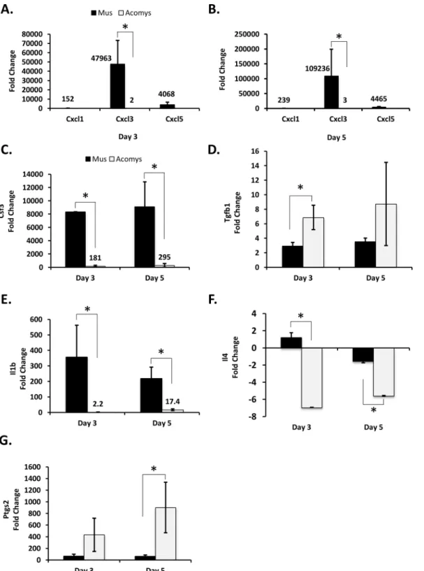
Examining expression differences in genes known to participate in the inflammation pathway and the extra-cellular matrix during cutaneous wound healing. The c-x-c motif family of inflammatory chemokines acts by attracting immune cells to the wound site. InMus woundsCxcl1,Cxcl3andCxcl5were all significantly upregulated to high degree (from>150
forCxcl1and>4000-fold forCxcl3andCxcl5). In contrast, the increase inCxcl3was relatively
modest inAcomys, with only a 2 to 3 fold increase in day 3 and 5 wounds compared to normal skin (Fig 1A and 1B). There were no detectable transcripts forCxcl1, and very low levels of transcripts detected forCxcl5inAcomys(Fig 1A and 1B).
wounds inMus. In contrast, the levels ofCsf3inAcomys, while substantially upregulated (~200 to 300 fold) represent approximately 2% of the levels observed inMus(Fig 1C).
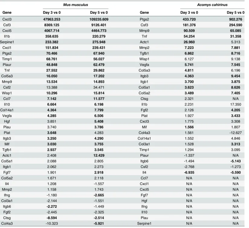
Tgf-β1(Transforming growth factor beta 1), thought to play a role in nearly all stages of wound healing, was ~3 fold upregulated inMusin day 3 and 5 wounds compared to normal skin. Expression levels ofTgf-β1inAcomyswere approximately twice that observed inMus, witha 7-fold increase in expression observed in wounds vs. unwounded skin (Fig 1D). Table 1. Differentially expressed genes in day 3 and 5 wounds. Differentially expressed genes in day 3 and 5 wounds, compared to normal skin, within each species. Bold entries are those with a p-value0.01.
Mus musculus Acomys cahirinus
Gene Day 3 vs 0 Day 5 vs 0 Gene Day 3 vs 0 Day 5 vs 0
Cxcl3 47963.253 109235.609 Ptgs2 433.720 902.276
Csf3 8369.125 9126.401 Csf3 181.376 294.590
Cxcl5 4067.714 4464.773 Mmp9 90.509 65.085
Il1b 358.635 220.279 Tnf 54.254 31.358
Serpine1 233.382 275.948 Actc1 26.960 5.313
Cxcl1 151.834 239.431 Mmp2 7.223 7.881
Ptgs2 70.466 67.940 Tgfb1 6.862 8.716
Timp1 68.761 56.027 Wisp1 6.127 9.138
Plaur 48.848 62.479 Vegfa 5.741 7.545
Tnf 27.552 29.862 Col5a3 4.811 6.196
Col5a3 16.050 17.202 Itgb3 4.363 9.454
Mmp9 13.534 14.893 Itgb1 3.700 3.875
Csf2 13.388 34.471 Col5a1 3.623 8.626
Wisp1 10.296 15.814 Col5a2 3.489 7.405
Ccl7 7.142 11.577 Ctsg 2.321 N/A
Il10 6.664 6.198 Il1b 2.231 17.350
Col14a1 4.364 7.799 Fgf2 2.126 4.205
Vegfa 4.285 6.506 Plat 1.927 3.433
Hgf 3.851 5.408 Cxcl3 1.775 3.308
Plau 3.740 3.786 Mif 1.566 1.807
Plat 3.648 4.283 Col4a3 1.561 -12.627
Itgb3 3.250 4.290 Col14a1 1.552 4.846
Mif 3.030 3.755 Col3a1 1.528 3.313
Tgfb1 2.937 3.545 Timp1 1.294 3.095
Actc1 2.408 12.429 Plaur -1.337 N/A
Col5a1 2.088 2.805 Itgb6 -1.494 -5.143
Itgb1 2.062 2.273 Csf2 -2.768 -1.273
Fgf7 1.901 2.918 Il4 -6.935 -5.590
Col5a2 1.671 2.118 Ccl7 N/A N/A
Il4 1.208 -1.557 Cxcl1 N/A N/A
Mmp2 1.158 1.743 Cxcl5 N/A N/A
Ifng -1.180 -2.665 Fgf7 N/A N/A
Col3a1 -2.144 -1.551 Hgf N/A N/A
Itgb6 -2.272 -1.449 Ifng N/A N/A
Fgf2 -2.445 -2.325 Il10 N/A N/A
Ctsg -8.594 -2.514 Plau N/A N/A
Col4a3 -10.323 -5.921 Serpine1 N/A N/A
The interleukin family of secreted cytokines facilitates intercellular communication between immune cells. The pro-inflammatory cytokineIl1β, can act pleiotropically to induce a diverse Fig 1. Immune response appears blunted inAcomysfollowing wounding.RT-qPCR analysis of day 3 and 5 wounds, compared to normal skin. Data are presented as mean±SEM.*p0.01; n = 3.Musis represented by black bars andAcomysis represented by white bars. The Cxcl cytokines,Cxcl1,Cxcl3
andCxcl5, were analyzed for day 3 (A) and day 5 (B) wounds. The remaining genes analyzed were: (C)Csf3, (D)Tgf-β1(E)Il1β, (F)Il4, and (G)Ptgs2.
range of effects, such as proliferation and differentiation, lymphocyte activation, angiogenesis and leukocyte attraction [29]. Interleukin 4 (Il4) is another pleiotropic cytokine that has been shown to stimulate production of components of the ECM [30–32]. The levels of Interleukins 1βand 4 mRNA were significantly higher inMusthan inAcomys(Fig 1E and 1F). The expres-sion ofIl1βwas expressed ~350 fold higher and ~220 fold higher inMusday 3 and day 5 wounds relative to unwounded skin, respectively. InAcomysthe expression ofIl1βis also ele-vated in wounded vs. non-wounded skin. However, the magnitude of the elevation in expres-sion ofIl1βis greatly reduced inAcomysrelative toMuswithAcomysday 3 and day 5 wounds exhibiting a 2.2 and 17.4 fold, increase in expression relative to unwounded skin, respectively. (Fig 1E.) There was essentially no change observed inIl4expression inMuswounds relative to unwounded skin (<2-fold), while inAcomys,Il4expression was reduced by 7 and 6.6 fold in
day 3 and 5 wounds relative to unwounded skin, respectively (Fig 1F.)
Prostaglandin-endoperoxide synthase 2 (Ptgs2, also known as cyclooxygenase-2 (Cox-2) is an immediate early response gene that is upregulated immediately after wounding [33], and has been shown to impair wound healing and to promote scar formation [34,35]. Interestingly, the levels ofPtgs2mRNA were significantly higher inAcomysthan those observed inMus(Fig 1G), with an increase of ~ 70 fold inMusand over 400 fold inAcomysobserved in both day 3 and 5 wounds.
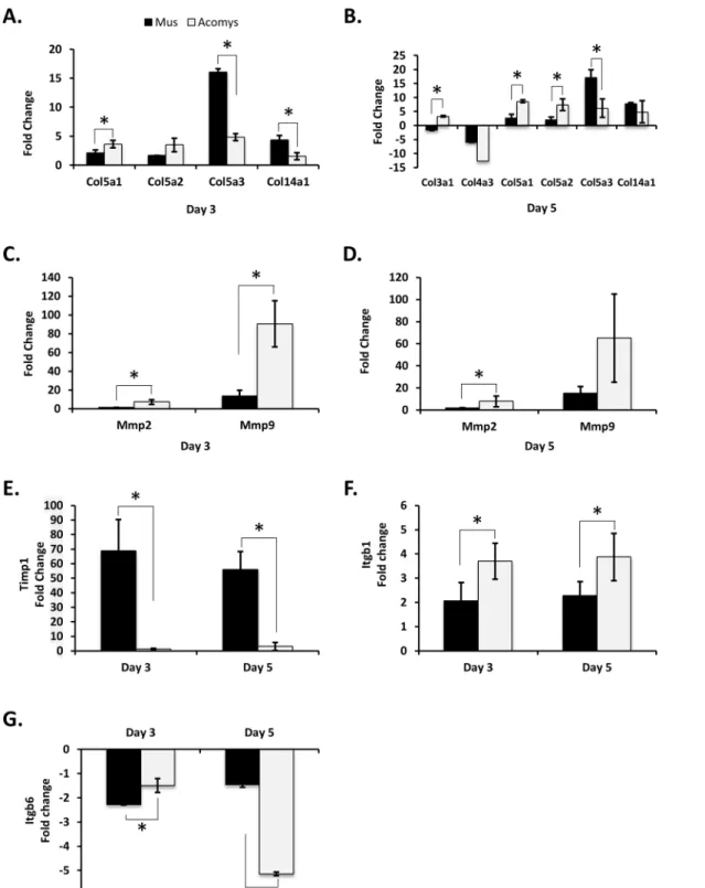
Extracellular matrix. Several genes associated with the organization of the extracellular matrix showed significant differences in mRNA levels betweenMusandAcomyswounds (Fig 2). At days 3 and 5 post wounding, there appears to be a different collagen expression profile betweenMusandAcomys(Fig 2A and 2B). In day 3 woundsCol5a2was expressed at signifi-cantly higher levels inAcomys, compared to unwounded skin, whileCol5a3andCol14a1were significantly higher inMus, compared to unwounded skin (Fig 2A). At 5 days post wounding, bothCol5a1andCol5a2showed significantly higher increases in expression, as compared to unwounded skin, inAcomys, whileCol5a3was higher inMus.Col4a3was downregulated in MusandAcomysin day 5 wounds, although the difference between the species was not signifi-cant (Fig 2B). In addition to components of the ECM, there were also genes involved in ECM degradation which showed differences in their gene expression profiles betweenMusand Acomys(Fig 2C–2E). The matrix metalloproteinasesMmp2andMmp9were both significantly upregulated at days 3 and 5 post wounding in theAcomys(Fig 2C and 2D).Mmp2was upregu-lated ~ 7.5 fold in both day 3 and 5 wounds inAcomys, with no significant changes observed in Mus.Mmp9was upregulated ~90 fold at day 3, and ~65 fold at day 5 post wounding inAcomys. InMus,Mm9was upregulated ~13 and 15 fold in day 3 and 5 wounds, respectively (Fig 2C and 2D). AlthoughMmp9was upregulated roughly 15 fold inMusat days 3 and 5, an inhibitor of metalloproteinases,Timp1, was upregulated greater than ~50 fold inMus, with no significant increase observed forTimp1inAcomys(Fig 2E). Taken together, this suggests that ECM degra-dation is more active inAcomyswound healing than inMus.
downregulated -1.5 fold and -5.1 fold in day 3 and 5 wounds, respectively (Fig 2G). These data suggest that the levels ofItgβ6are increasing inMusand decreasing inAcomysfrom days 3 to 5 Fig 2. Differences in ECM composition, matrix digestion and cell motility betweenAcomysandMus.RT-qPCR analysis was performed for the following genes: Collagens were analyzed for day 3 (A) and day 5 (B) wounds.Mmp2and9were analyzed for day 3 (C) and day 5 (D) wounds,Timp1(E),
Itgβ1(F), andItgβ6(G). All labels, symbols and calculations are as those described inFig 1.
post wounding, with no change in expression for either species in day 7 and 14 wounds (by microarray analysis).
Microarray analysis
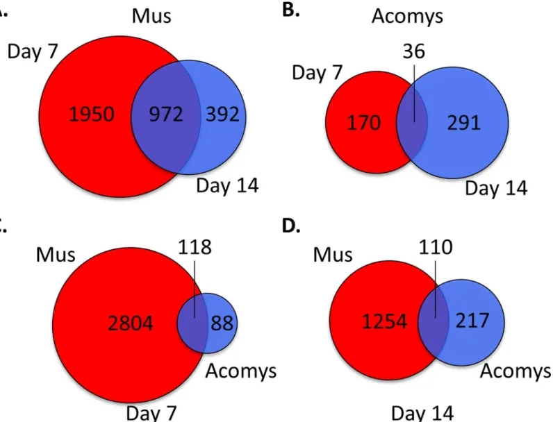
To examine the mechanisms involved in wound healing, genome wide gene expression analysis of full-depth excisional skin wounds at two additional time points, day 7 and day 14 after wounding, in bothMusandAcomyswere evaluated with Affymetrix GeneChip1microarrays. Changes in gene expression levels were considered significant if the fold change in expression was at least 1.4 fold, in either direction, and with a p-value of significance of0.001. At day 7 after wounding there were a total of 2922 genes exhibiting differential expression relative to normal skin inMus(S2 Table). There were 1872 genes that were upregulated and 1050 genes downregulated. At day 14 there were 1364 genes showing statistically significant changes in gene expression compared to normal skin. Of these, 795 were upregulated and 569 were down-regulated. Comparing genes with altered expression at both days 7 and 14 post wounding, we find that there were 1950 unique to day 7, 392 unique to day 14, and 972 common to both (Fig 3A).
InAcomysthere were a total of 206 genes showing differential expression at day 7 after wounding compared to normal skin (S2 Table). Of these, 176 were upregulated and 30 were downregulated. At day 14 there were 327 genes with altered expression compared to normal skin, with 159 genes upregulated and 168 downregulated. Comparing days 7 and 14, there were 170 genes whose change in expression was unique to day 7, 291 unique to day 14, and 30 common to both days 7 and 14 (Fig 3B).
At 7 days post wounding inMus, there was considerable upregulation of genes associated with the inflammatory response, in agreement with our observations by RT-PCR at days 3 and 5. There were a wide array of pro inflammatory genes, including most chemokines and cyto-kines, whose expression in the wound were increased from ~3 to 90-fold, as well as most inter-leukins, with interleukin 1 beta (Il1β) showing the largest increase at ~145-fold (S2 Table).
While most members of the collagen family were moderately upregulated, as would be expected in a healing wound, collagen 12a1 (Col12a1) was increased by nearly 30 fold inMus (this increased expression persisted in the day 14 post wounding samples as well). This was unexpected since there is a lack of evidence in the literature for the role ofCol12a1in dermal wound healing. We confirmed the high levels ofCol12a1inMusand lack ofCol12a1inAcomys by immunofluorescence using 2 different antibodies (Brant et al. manuscript under review).
In stark contrast to the high degree of response observed inMus, the overall response to wounding inAcomysappears to be reduced, not only in the number of genes affected, but in the magnitude of change as well (from +63 to -2.3 fold). The genes with the largest increase in expression were not those associated with the inflammatory pathways, as observed inMus, but were instead genes associated with the organization of the extracellular-matrix, such as colla-gens and platelet-derived growth factor binding proteins, as well as peptidases and matrix metalloproteinases (S2 Table).
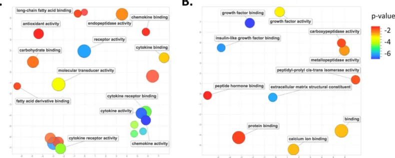
Those genes whose altered expression is unique to eitherMus(2804) orAcomys(88) could provide insight into the observed difference in wound healing, i.e. scaring inMusversus scar-free wound healing inAcomys. We performed an analysis of gene ontology (GO) terms associ-ated with these unique genes. At day 7 post wounding inMusthere was a marked enrichment for GO terms associated with the inflammatory pathway, including cytokine receptor activity, chemokine and cytokine activity, cytokine receptor binding, cytokine and chemokine binding, fatty acid and carbohydrate binding, as well as endopeptidase and antioxidant activity (Fig 4A). A pathway enrichment analysis identified the top enriched pathways for genes unique to day 7 Muswounds to be, as expected, those involved in the inflammatory pathways, including cyto-kine-cytokine receptor interaction, chemokine signaling pathway, Leishmanias, osteoclast dif-ferentiation, toll-like receptor signaling pathway and cell adhesion molecules (S3 Table). Fig 3. Venn diagram of differentially expressed genes in day 7 and 14 wounds, compared to normal skin.Visual representation of the number of unique differentially genes at days 7 and 14 and those common to both days for (A)Musand (B)Acomys. Unique and common differentially expressed genes betweenAcomysandMusfor (C) day 7 and (D) day 14. For (A) and (B), day 7 genes are in red, day 14 genes in blue and common genes are the overlap. For (C) and (D)Musgenes are in red andAcomysgenes are in blue and common genes are the overlap.
In contrast, the top enriched GO terms associated with those genes whose change in expres-sion is unique to day 7 wounds inAcomysare those associated with the extracellular matrix reorganization, growth factors and hormone binding, including extracellular matrix structural constituent, metalloproteinase activity, carboxypeptidase activity, growth factor activity and binding, and peptide hormone binding (Fig 4B). The top enriched pathways inAcomyswere those involved in protein digestion and absorption, extracellular matrix receptor interactions and focal adhesions (S4 Table).
A pathway analysis of the 1364 genes with significantly altered expression in day 14 wounds inMusrevealed an enrichment for genes associated with cell adhesion and migration, struc-tural morphogenesis, muscle contraction and contractile fibers and myofibril components, as well as a lingering upregulation of cytokines, chemokines and interleukins (S2 Table). The results of a pathway analysis of the 327 altered genes inAcomyswere more similar toMusfor day 14 than for day 7 wounds. Enriched pathways were observed for contractile fiber compo-nents and myofibril genes, as inMus, as well genes associated with tissue development and morphogenesis (S2 Table).
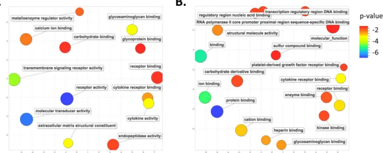
We again performed an analysis of GO terms for those genes whose altered expression is unique to eitherMus(1254) orAcomys(217) at day 14 post-wounding (Fig 3D). The 1254 genes inMusexhibit a persistence of enrichment for genes associated with the inflammation pathway, such as cytokine activity, cytokine receptor binding, and receptor binding and activ-ity. There was also an enrichment for genes involved in the extracellular matrix organization, including metalloenzyme regulator activity and extracellular matrix structural constituent, as well as transmembrane signaling receptor activity and carbohydrate, glycosaminoglycan, glyco-protein and calcium ion binding (Fig 5A). A pathway analysis of these same genes reveals a large number of genes involved in cellular respiration, oxidative phosphorylation, fructose and mannose metabolism, metabolic pathways, as well as the genes involved with the lysosome and focal adhesions (S5 Table).
Fig 4. GO term enrichment analysis for differentially expressed genes unique to each species at day 7 post-wounding.Scatter plot representing the summarized GO term analysis of differentially expressed genes at day 7 post wounding in (A)Musand (B)Acomys. Semantically similar GO terms are plotted near to each other on the (unit-less) X-Y axes, such that functionally similar terms are located nearby, and more unrelated terms are further apart in space. The size of the individual plots represents the frequency of the GO term in the database so that larger bubbles represent more general terms and smaller bubbles are more specific terms. The color indicates the p-value of enrichment of each GO term.
An enrichment analysis for the GO terms associated with the 217 genes unique toAcomys at day 14 wounds reveals genes involved in the regulation of transcription, such as regulatory and transcription regulatory region binding and RNA-polII DNA binding. There was also an enrichment for carbohydrate, protein and ion binding, as well genes involved with cytokine receptor and platelet-derived growth factor receptor binding (Fig 5B). A pathway analysis for these same genes shows enrichment for growth factor pathways, including insulin, VEGF, toll-like receptor, adipocytokine and chemokine signaling pathways (S6 Table).
Discussion
The recent discovery of the remarkable capacity ofAcomys cahirinusto regenerate full thick-ness excisional skin wounds in a scar free manner [21] provides a unique opportunity to com-pare scar free wound healing with scaring in two adult mammalian tissues. Previous studies were limited to comparing wound healing in adult tissues to fetal tissues, which have a limited ability for scar free healing. Although much has been learned from these studies, there are inherent differences between fetal and adult tissues which imposes limits on these comparative studies and as such, very little has translated into improved clinical outcomes. In this study, we have used RT-PCR and microarray analyses to generate gene expression profiles of full thick-ness excisional skin wounds in bothMusandAcomysto compare scar free wound healing to scaring in adult tissue and to determine if there are any similarities to scar free healing observed in fetal tissues. We have identified changes in gene expression patterns for several aspects of wound healing betweenMusandAcomys, particularly for those genes involved in the inflam-matory pathway and the deposition and digestion of the extracellular matrix, and have identi-fied similarities to fetal wounds.
There are several notable differences between fetal and adult wounds, which are predomi-nately in the inflammatory pathway and the collagen profile of the ECM and in expression lev-els of various growth factors. Previous studies have shown that the inflammatory response is quite blunted in fetal wounds [4,5]. Additionally, it has been shown that ectopically induced Fig 5. GO term enrichment analysis for differentially expressed genes unique to each species at day 14 post-wounding.Scatter plot representing the summarized GO term analysis of differentially expressed genes at day 14 post wounding in (A)Musand (B)Acomys. All labels, symbols and calculations are as those described inFig 4.
inflammation in fetal wounds increases fibrosis and results in scar formation [38,39]. Our data indicates that wounding initiates a strong inflammatory response inMus, typical of the adult mammalian response, while the inflammatory response inAcomysappears to be modest, simi-lar to fetal wounds.
Immediately after wounding platelets enter the wound bed and releasePdgfaandTgf-β1/2 [40–42]. Studies have shown thatPdgfaandTgf-β1/2 levels are lower in fetal compared to adult wounds [43]. In contrast to fetal wounds, our data indicate no difference inPdgfaexpression in day 3 and 5 wounds, and an increase inTgf-β1 expression inAcomys.
Shortly after platelets enter the wound, neutrophils are recruited by neutrophil attracting chemokines and activated by neutrophil activating cytokines (reviewed extensively in [44–46]). Studies have shown that neutrophil levels are higher in adult wounds compared to fetal wounds [9]. Counts of circulating leukocytes, from non-wounded animals, revealed that neutrophils are present inAcomysat only 20% the levels observed inMus(Brant et al. manuscript under review). The impact of reduced circulating neutrophil levels is unknown at this time but cer-tainly warrants further study. By day 2 after wounding, macrophages constitute the predomi-nant blood-derived cell type in the wound bed and initiate release of pro inflammatory cytokines and growth factors [47]. Our data show a striking difference in the expression levels of several genes thought to play important roles in inflammation. The expression of the chemo-kineCxcl3is only slighter higher inAcomyswounds, compared to normal skin, andCxcl1and Cxcl5are barely detectable by RT-PCR, in contrast to the high levels of induction observed in Mus. As the c-x-c like family of chemokines is expressed by macrophages and neutrophils, this observed difference in expression could in part be explained by the observed reduction in circu-lating neutrophils inAcomys. Additionally, the induction of granulocyte stimulating factor Csf3mRNA is much lower inAcomysas well, suggesting an overall diminished role of neutro-phils inAcomys. We have additional data that shows that while macrophages are present in Acomysin very early wounds, they are confined to the underlying fascial connective tissue and are not present in the wound bed at later time points (Brant et al. manuscript in review). The increased upregulation ofTgf-β1 is intriguing in light of the seemingly blunted immune response observed inAcomys, given its role in promoting inflammation, among others, during wound healing. It is also interesting to note that while differences have been observed for vari-ous growth factors between fetal and adult wounds [34,48–50], our data indicate that there were no statistically significant changes observed in expression levels of the growth factors Csf2,Ctgf,Egf,Fgf2,Fgf10andVegfabetweenMusandAcomyswounds (S1 Table).
also known to inducePtgs2expression [58], although the expression ofPtgs2is much higher in Acomyswounds compared to normal skin than inMuswounds. The high levels ofPtgs2in Acomyswounds is interesting, in that it also seems contrary to data suggestingPtgs2levels are correlated with scar formation in skin wounds. Wilgus et al. showed that thePtgs2inhibitor Celecoxib1decreased inflammation in incisional wounds and reduced scar formation [35] and have also shown that high levels ofPtgs2promote scarring and delay wound closure in fetal wounds [34].
The collagen composition of fetal skin has been shown to differ from adult skin [12–14], with fetal skin having higher levels of collagen type III and V than adult skin, among other dif-ferences. The expression of collagens inAcomyswounds suggests that they have a profile simi-lar to fetal skin, with higher levels of expression for collagens 3a1, 5a1, 5a2, and lower levels of expression of collagens 5a3 and 14a1. In addition to differences in the composition of the ECM, there are notable differences in the expression of matrix metalloproteinases involved in the degradation of the ECM. Fetal skin wounds have been shown to have a higher matrix metalloproteinase to tissue inhibitor of metalloproteinase ratio [16,17]. Similar to fetal wounds,Acomyswounds have significantly higher levels of expression ofMmp2andMmp9, and lower levels of expression ofTimp1in day 3 and 5 wounds compared toMus. This suggests that there is a higher turnover of ECM components and an increase in cell migration through the wound bed inAcomys. Fetal wounds have also been shown to have essentially no myofibro-blasts, which are thought to interact with the ECM and aid in wound closure through contrac-tion, while both scaring fetal and adult wounds have high levels of contractile myofibroblasts appearing 7 days post wounding [59]. AlthoughAcomyswounds heal in a scar-free manner, similar to fetal wounds, our data indicate there is no difference in the expression of smooth muscle actin, a marker of myofibroblasts, betweenMusandAcomyswounds from days 3, 5 (S1 Table), 7 and 14 (S2 Table).
We considered the possibility that some of the observed differences in expression could be the result of inefficient cross species hybridization of oligos used in the RT-PCR arrays. Although the primers used in the arrays were designed forMus, there was detectable amplifica-tion for 54/84 genes inAcomys, compared to 79/84 forMus. This could be due to the fact that not all genes from the array are expressed in skin or wounds in both species. Alternatively, some genes may have sufficiently diverged from the mouse, such thatMusspecific primers do not anneal. In either case, the majority of genes have detectable levels of amplification, suggest-ing a fairly high degree of conservation, at least for codsuggest-ing regions. In order to confirm that we are in fact amplifying the correct mRNA, we have cloned and sequenced theTgf-β1,Timp1and Mmp9RT-PCR amplicons fromAcomys. A BLAST using the resulting sequences against the Mustranscript database reveals high identity matches for all three sequences (S1A–S1C Fig). The percent identity forTgf-β1 andTimp1was 98% and 100%, respectively. Although the per-cent identity forMmp9was 74% forMus, there were higher identity matches in other rodent species, e.g.Peromyscus maniculatus(Deer mouse, 91%).
analyses using the whole dataset, i.e. using both unique and common sets of differentially expressed genes, suggesting that the observed enriched pathways are largely driven by those genes which are uniquely differentially expressed in each species and not necessarily by those whose change is common to both species.
In summary, the data presented here provide insights into scar free healing of full-thickness excisional wounds ofAcomys, and provides a starting point of potential gene candidates that may be further studied, in hopes of devising potential strategies for improved clinical outcomes in preventing scarring in humans.
Supporting Information
S1 Fig. BLAST search results ofAcomysRT-PCR amplicons againstMustranscript data-base.BLAST results of cloned and sequencedAcomysRT-PCR amplicons against theMus transcript database for (A)Tgf-β1, (B)Timp1, and (C)Mmp9.
(PDF)
S1 Table. Genes analyzed by RT-PCR in day 3 and 5 wounds.List of genes analyzed by Wound-Healing RT2Profiler Array in day 3 and 5 wounds, compared to normal skin, within each species. Bold entries are those with a p-value0.01.
(DOCX)
S2 Table. Results of microarray analysis.Tables of p-values and fold-change in expression with each analysis in its own worksheet.
(XLSX)
S3 Table. Pathway analysis ofMusday 7 wounds.Pathway analysis of differentially expressed genes between day 7 wounds and normal skin inMus.
(DOCX)
S4 Table. Pathway analysis ofAcomysday 7 wounds.Pathway analysis of differentially expressed genes between day 7 wounds and normal skin inAcomys.
(DOCX)
S5 Table. Pathway analysis ofMusday 14 wounds.Pathway analysis of differentially expressed genes between day 14 wounds and normal skin inMus.
(DOCX)
S6 Table. Pathway analysis ofAcomysday 14 wounds.Pathway analysis of differentially expressed genes between day 14 wounds and normal skin inAcomys.
(DOCX)
Acknowledgments
Author Contributions
Conceived and designed the experiments: JOB MM. Performed the experiments: JOB. Ana-lyzed the data: JOB MCL. Contributed reagents/materials/analysis tools: JOB MM BB HVB MCL. Wrote the paper: JOB.
References
1. Clark RAF, Henson PM. The Molecular and cellular biology of wound repair. New York: Plenum Press; 1988. xxii, 597 p. p.
2. Rolfe KJ, Grobbelaar AO. A review of fetal scarless healing. ISRN dermatology. 2012; 2012:698034. Epub 2012/06/08. doi:10.5402/2012/698034PMID:22675640; PubMed Central PMCID:
PMC3362931.
3. Satish L, Kathju S. Cellular and Molecular Characteristics of Scarless versus Fibrotic Wound Healing. Dermatology research and practice. 2010; 2010:790234. Epub 2011/01/22. doi:10.1155/2010/790234 PMID:21253544; PubMed Central PMCID: PMC3021858.
4. Liechty KW, Crombleholme TM, Cass DL, Martin B, Adzick NS. Diminished interleukin-8 (IL-8) produc-tion in the fetal wound healing response. The Journal of surgical research. 1998; 77(1):80–4. Epub 1998/08/12. doi:10.1006/jsre.1998.5345PMID:9698538.
5. Liechty KW, Adzick NS, Crombleholme TM. Diminished interleukin 6 (IL-6) production during scarless human fetal wound repair. Cytokine. 2000; 12(6):671–6. Epub 2000/06/14. doi:10.1006/cyto.1999. 0598PMID:10843743.
6. Olutoye OO, Barone EJ, Yager DR, Cohen IK, Diegelmann RF. Collagen induces cytokine release by fetal platelets: implications in scarless healing. Journal of pediatric surgery. 1997; 32(6):827–30. Epub 1997/06/01. PMID:9200079.
7. Cowin AJ, Brosnan MP, Holmes TM, Ferguson MW. Endogenous inflammatory response to dermal wound healing in the fetal and adult mouse. Developmental dynamics: an official publication of the American Association of Anatomists. 1998; 212(3):385–93. Epub 1998/07/22. doi:10.1002/(SICI) 1097-0177(199807)212:3<385::AID-AJA6>3.0.CO;2-DPMID:9671942.
8. Adzick NS, Harrison MR, Glick PL, Beckstead JH, Villa RL, Scheuenstuhl H, et al. Comparison of fetal, newborn, and adult wound healing by histologic, enzyme-histochemical, and hydroxyproline determina-tions. Journal of pediatric surgery. 1985; 20(4):315–9. Epub 1985/08/01. PMID:4045654.
9. Jennings RW, Adzick NS, Longaker MT, Duncan BW, Scheuenstuhl H, Hunt TK. Ontogeny of fetal sheep polymorphonuclear leukocyte phagocytosis. Journal of pediatric surgery. 1991; 26(7):853–5. Epub 1991/07/01. PMID:1895198.
10. Sullivan KM, Lorenz HP, Meuli M, Lin RY, Adzick NS. A model of scarless human fetal wound repair is deficient in transforming growth factor beta. Journal of pediatric surgery. 1995; 30(2):198–202; discus-sion -3. Epub 1995/02/01. PMID:7738738.
11. Schultz GS, Wysocki A. Interactions between extracellular matrix and growth factors in wound healing. Wound repair and regeneration: official publication of the Wound Healing Society [and] the European Tissue Repair Society. 2009; 17(2):153–62. Epub 2009/03/27. doi:10.1111/j.1524-475X.2009.00466.x PMID:19320882.
12. Smith LT, Holbrook KA, Madri JA. Collagen types I, III, and V in human embryonic and fetal skin. The American journal of anatomy. 1986; 175(4):507–21. Epub 1986/04/01. doi:10.1002/aja.1001750409 PMID:3521252.
13. Hallock GG, Rice DC, Merkel JR, DiPaolo BR. Analysis of collagen content in the fetal wound. Annals of plastic surgery. 1988; 21(4):310–5. Epub 1988/10/01. PMID:3232919.
14. Cuttle L, Nataatmadja M, Fraser JF, Kempf M, Kimble RM, Hayes MT. Collagen in the scarless fetal skin wound: detection with picrosirius-polarization. Wound repair and regeneration: official publication of the Wound Healing Society [and] the European Tissue Repair Society. 2005; 13(2):198–204. Epub 2005/04/15. doi:10.1111/j.1067-1927.2005.130211.xPMID:15828945.
15. Gill SE, Parks WC. Metalloproteinases and their inhibitors: regulators of wound healing. The interna-tional journal of biochemistry & cell biology. 2008; 40(6–7):1334–47. Epub 2007/12/18. doi:10.1016/j. biocel.2007.10.024PMID:18083622; PubMed Central PMCID: PMC2746915.
16. Peled ZM, Phelps ED, Updike DL, Chang J, Krummel TM, Howard EW, et al. Matrix metalloproteinases and the ontogeny of scarless repair: the other side of the wound healing balance. Plastic and recon-structive surgery. 2002; 110(3):801–11. Epub 2002/08/13. PMID:12172142.
reconstructive surgery. 2003; 111(7):2273–85. Epub 2003/06/10. doi:10.1097/01.PRS.0000060102. 57809.DAPMID:12794470.
18. Levesque M, Villiard E, Roy S. Skin wound healing in axolotls: a scarless process. Journal of experi-mental zoology Part B, Molecular and developexperi-mental evolution. 2010; 314(8):684–97. Epub 2010/08/ 19. doi:10.1002/jez.b.21371PMID:20718005.
19. Seifert AW, Monaghan JR, Voss SR, Maden M. Skin regeneration in adult axolotls: a blueprint for scar-free healing in vertebrates. PLoS ONE. 2012; 7(4):e32875. Epub 2012/04/10. doi:10.1371/journal. pone.0032875PMID:22485136; PubMed Central PMCID: PMC3317654.
20. Steppan S, Adkins R, Anderson J. Phylogeny and divergence-date estimates of rapid radiations in mur-oid rodents based on multiple nuclear genes. Systematic biology. 2004; 53(4):533–53. Epub 2004/09/ 17. doi:10.1080/10635150490468701PMID:15371245.
21. Seifert AW, Kiama SG, Seifert MG, Goheen JR, Palmer TM, Maden M. Skin shedding and tissue regen-eration in African spiny mice (Acomys). Nature. 2012; 489(7417):561–5. Epub 2012/09/29. doi:10. 1038/nature11499PMID:23018966; PubMed Central PMCID: PMC3480082.
22. Livak KJ, Schmittgen TD. Analysis of relative gene expression data using real-time quantitative PCR and the 2(-Delta Delta C(T)) Method. Methods. 2001; 25(4):402–8. Epub 2002/02/16. doi:10.1006/ meth.2001.1262PMID:11846609.
23. Zhang B, Kirov S, Snoddy J. WebGestalt: an integrated system for exploring gene sets in various bio-logical contexts. Nucleic Acids Res. 2005; 33(Web Server issue):W741–8. Epub 2005/06/28. doi:10. 1093/nar/gki475PMID:15980575; PubMed Central PMCID: PMC1160236.
24. Wang J, Duncan D, Shi Z, Zhang B. WEB-based GEne SeT AnaLysis Toolkit (WebGestalt): update 2013. Nucleic Acids Res. 2013; 41(Web Server issue):W77–83. Epub 2013/05/25. doi:10.1093/nar/ gkt439PMID:23703215; PubMed Central PMCID: PMC3692109.
25. Supek F, Bosnjak M, Skunca N, Smuc T. REVIGO summarizes and visualizes long lists of gene ontol-ogy terms. PLoS ONE. 2011; 6(7):e21800. Epub 2011/07/27. doi:10.1371/journal.pone.0021800 PMID:21789182; PubMed Central PMCID: PMC3138752.
26. Welte K, Platzer E, Lu L, Gabrilove JL, Levi E, Mertelsmann R, et al. Purification and biochemical char-acterization of human pluripotent hematopoietic colony-stimulating factor. Proc Natl Acad Sci U S A. 1985; 82(5):1526–30. Epub 1985/03/01. PMID:3871951; PubMed Central PMCID: PMC397296.
27. Begley CG, Lopez AF, Nicola NA, Warren DJ, Vadas MA, Sanderson CJ, et al. Purified colony-stimulat-ing factors enhance the survival of human neutrophils and eosinophils in vitro: a rapid and sensitive microassay for colony-stimulating factors. Blood. 1986; 68(1):162–6. Epub 1986/07/01. PMID: 3487354.
28. Frampton JE, Lee CR, Faulds D. Filgrastim. A review of its pharmacological properties and therapeutic efficacy in neutropenia. Drugs. 1994; 48(5):731–60. Epub 1994/11/01. PMID:7530630.
29. Garlanda C, Dinarello CA, Mantovani A. The interleukin-1 family: back to the future. Immunity. 2013; 39 (6):1003–18. Epub 2013/12/18. doi:10.1016/j.immuni.2013.11.010PMID:24332029; PubMed Central PMCID: PMC3933951.
30. Fertin C, Nicolas JF, Gillery P, Kalis B, Banchereau J, Maquart FX. Interleukin-4 stimulates collagen synthesis by normal and scleroderma fibroblasts in dermal equivalents. Cellular and molecular biology. 1991; 37(8):823–9. Epub 1991/01/01. PMID:1807791.
31. Gillery P, Fertin C, Nicolas JF, Chastang F, Kalis B, Banchereau J, et al. Interleukin-4 stimulates colla-gen colla-gene expression in human fibroblast monolayer cultures. Potential role in fibrosis. FEBS letters. 1992; 302(3):231–4. Epub 1992/05/18. PMID:1601130.
32. Salmon-Ehr V, Ramont L, Godeau G, Birembaut P, Guenounou M, Bernard P, et al. Implication of inter-leukin-4 in wound healing. Laboratory investigation; a journal of technical methods and pathology. 2000; 80(8):1337–43. Epub 2000/08/19. PMID:10950124.
33. Futagami A, Ishizaki M, Fukuda Y, Kawana S, Yamanaka N. Wound healing involves induction of cyclo-oxygenase-2 expression in rat skin. Laboratory investigation; a journal of technical methods and pathol-ogy. 2002; 82(11):1503–13. Epub 2002/11/14. PMID:12429810.
34. Wilgus TA, Bergdall VK, Tober KL, Hill KJ, Mitra S, Flavahan NA, et al. The impact of cyclooxygenase-2 mediated inflammation on scarless fetal wound healing. The American journal of pathology. cyclooxygenase-2004; 165(3):753–61. Epub 2004/08/28. doi:10.1016/S0002-9440(10)63338-XPMID:15331400; PubMed Central PMCID: PMC1618587.
36. Munger JS, Huang X, Kawakatsu H, Griffiths MJ, Dalton SL, Wu J, et al. The integrin alpha v beta 6 binds and activates latent TGF beta 1: a mechanism for regulating pulmonary inflammation and fibrosis. Cell. 1999; 96(3):319–28. Epub 1999/02/20. PMID:10025398.
37. Annes JP, Rifkin DB, Munger JS. The integrin alphaVbeta6 binds and activates latent TGFbeta3. FEBS letters. 2002; 511(1–3):65–8. Epub 2002/02/01. PMID:11821050.
38. Ozturk S, Deveci M, Sengezer M, Gunhan O. Results of artificial inflammation in scarless foetal wound healing: an experimental study in foetal lambs. British journal of plastic surgery. 2001; 54(1):47–52. Epub 2000/12/21. doi:10.1054/bjps.2000.3460PMID:11121318.
39. Dixon JB. Inflammation in the foetal and neonatal rat: the local reactions to skin burns. The Journal of pathology and bacteriology. 1960; 80:73–82. Epub 1960/07/01. PMID:13816989.
40. Ross R, Raines EW, Bowen-Pope DF. The biology of platelet-derived growth factor. Cell. 1986; 46 (2):155–69. Epub 1986/07/18. PMID:3013421.
41. Werner S, Grose R. Regulation of wound healing by growth factors and cytokines. Physiological reviews. 2003; 83(3):835–70. Epub 2003/07/05. doi:10.1152/physrev.00031.2002PMID:12843410.
42. Plasari G, Calabrese A, Dusserre Y, Gronostajski RM, McNair A, Michalik L, et al. Nuclear factor I-C links platelet-derived growth factor and transforming growth factor beta1 signaling to skin wound heal-ing progression. Mol Cell Biol. 2009; 29(22):6006–17. Epub 2009/09/16. doi:10.1128/MCB.01921-08 PMID:19752192; PubMed Central PMCID: PMC2772573.
43. Olutoye OO, Barone EJ, Yager DR, Uchida T, Cohen IK, Diegelmann RF. Hyaluronic acid inhibits fetal platelet function: implications in scarless healing. Journal of pediatric surgery. 1997; 32(7):1037–40. Epub 1997/07/01. PMID:9247229.
44. Nathan C. Neutrophils and immunity: challenges and opportunities. Nature reviews Immunology. 2006; 6(3):173–82. Epub 2006/02/25. doi:10.1038/nri1785PMID:16498448.
45. Wilgus TA, Roy S, McDaniel JC. Neutrophils and Wound Repair: Positive Actions and Negative Reac-tions. Advances in wound care. 2013; 2(7):379–88. Epub 2014/02/15. doi:10.1089/wound.2012.0383 PMID:24527354; PubMed Central PMCID: PMC3763227.
46. Lo DD, Zimmermann AS, Nauta A, Longaker MT, Lorenz HP. Scarless fetal skin wound healing update. Birth defects research Part C, Embryo today: reviews. 2012; 96(3):237–47. Epub 2012/10/31. doi:10. 1002/bdrc.21018PMID:23109319.
47. Leibovich SJ, Ross R. The role of the macrophage in wound repair. A study with hydrocortisone and antimacrophage serum. The American journal of pathology. 1975; 78(1):71–100. Epub 1975/01/01. PMID:1109560; PubMed Central PMCID: PMC1915032.
48. Cowin AJ, Holmes TM, Brosnan P, Ferguson MW. Expression of TGF-beta and its receptors in murine fetal and adult dermal wounds. European journal of dermatology: EJD. 2001; 11(5):424–31. Epub 2001/08/30. PMID:11525949.
49. Dang CM, Beanes SR, Soo C, Ting K, Benhaim P, Hedrick MH, et al. Decreased expression of fibro-blast and keratinocyte growth factor isoforms and receptors during scarless repair. Plastic and recon-structive surgery. 2003; 111(6):1969–79. Epub 2003/04/25. doi:10.1097/01.PRS.0000054837.47432. E7PMID:12711959.
50. Haynes JH, Johnson DE, Mast BA, Diegelmann RF, Salzberg DA, Cohen IK, et al. Platelet-derived growth factor induces fetal wound fibrosis. Journal of pediatric surgery. 1994; 29(11):1405–8. Epub 1994/11/01. PMID:7844707.
51. Goerdt S, Orfanos CE. Other functions, other genes: alternative activation of antigen-presenting cells. Immunity. 1999; 10(2):137–42. Epub 1999/03/11. PMID:10072066.
52. Varin A, Mukhopadhyay S, Herbein G, Gordon S. Alternative activation of macrophages by IL-4 impairs phagocytosis of pathogens but potentiates microbial-induced signalling and cytokine secretion. Blood. 2010; 115(2):353–62. Epub 2009/11/03. doi:10.1182/blood-2009-08-236711PMID:19880493; PubMed Central PMCID: PMC2808158.
53. Loke P, Nair MG, Parkinson J, Guiliano D, Blaxter M, Allen JE. IL-4 dependent alternatively-activated macrophages have a distinctive in vivo gene expression phenotype. BMC immunology. 2002; 3:7. Epub 2002/07/06. PMID:12098359; PubMed Central PMCID: PMC117781.
54. Mosser DM, Zhang X. Interleukin-10: new perspectives on an old cytokine. Immunological reviews. 2008; 226:205–18. doi:10.1111/j.1600-065X.2008.00706.xPMID:19161426; PubMed Central PMCID: PMCPMC2724982.
56. Kurt-Jones EA, Beller DI, Mizel SB, Unanue ER. Identification of a membrane-associated interleukin 1 in macrophages. Proc Natl Acad Sci U S A. 1985; 82(4):1204–8. Epub 1985/02/01. PMID:3919388; PubMed Central PMCID: PMC397223.
57. Dinarello CA. Immunological and inflammatory functions of the interleukin-1 family. Annual review of immunology. 2009; 27:519–50. Epub 2009/03/24. doi:10.1146/annurev.immunol.021908.132612 PMID:19302047.
58. O'Banion MK, Miller JC, Chang JW, Kaplan MD, Coleman PD. Interleukin-1 beta induces prostaglandin G/H synthase-2 (cyclooxygenase-2) in primary murine astrocyte cultures. Journal of neurochemistry. 1996; 66(6):2532–40. Epub 1996/06/01. PMID:8632179.