The article was published by Academy of Chemistry of Globe Publications www.acgpubs.org/RNP © Published 08 /22/2008 EISSN 1307-6167
Rec. Nat. Prod. 2:3 (2008) 83-93
Potential Superoxide Anion Radical Scavenging Activity of
Doum Palm
(
Hyphaene thebaica
L.) Leaves Extract
Omayma A. Eldahshan, Nahla A. Ayoub
*, Abdel-Nasser B. Singab
and Mohamed M . Al-Azizi.
Department of Pharmacognosy, Faculty of Pharmacy, Ain - Shams University, Cairo , Egypt.
Abstract: The antioxidant activity of the aqueous ethanolic extract of Doum leaves, Hyphaene thebaica L.(Palmae), was studied. Data obtained showed that the extract scavenged superoxide anion radicals (IC50=1602 µg/ml) in a dose dependant manner using xanthine/hypoxanthine oxidase assay.
Four major flvonoidal compounds were identified by LC/SEI as; Quercetin glucoside, Kaempferol rhamnoglucoside, Dimethyoxyquercetin rhamnoglucoside. While , further in-depth phytochemical investigation of this extract lead to the isolation and identification of fourteen compounds ;their structures were elucidated based upon the interpretation of their spectral data(UV, 1H, 13C NMR and ESI/MS )as; 8-C- -D-glucopyranosyl-5, 7, 4`-trihydroxyflavone (vitexin) 1, 6-C- -D-glucopyranosyl-5, 7, 4`-trihydroxyflavone (iso-vitexin) 2, quercetin 3-O- -4C1-D-glucopyranoside 3, gallic acid 4,
quercetin 7-O- -4C1-D-glucoside 5, luteolin 7-O -4
C1-D-glucoside 6, tricin 5 O -4
C1-D-glucoside 7,
7, 3` dimethoxy quercetin 3-O-[6''-O- -L-rhamnopyranosyl]- -D-gluco-pyranoside (Rhamnazin 3-O-rutinoside) 8, kaempferol-3-O-[6''-O- -L-rhamnopyranosyl]- -D-glucopyranoside (nicotiflorin) 9, apigenin 10, luteolin 11, tricin 12, quercetin 13 and kaempferol 14.
Keywords: doum leaves; Hyphaene thebaica (Palmae); phenolics; hypoxanthine/xanthine oxidase
assay.
1. Introduction
Doum palm, Hyphaene thebaica L. (Palmae), is growing wild throughout the dry regions of tropical Africa, the Middle East and Western India [1,2]. Roots of doum were used in treatment of Bilharziasis, while the resin of the tree has demonstrated, diuretic, diaphoretic properties and also recommended for tap worm as well as against animal bites [3]. The fruits of doum showed antimicrobial and antihypertensive activities, these activities were attributed to the presence of flavonoids [4-6]. Also, the aqueous extract of doum fruits showed an antioxidant activity; this is due to the substantial amount of their water-soluble phenolic contents [7, 8]. Five flavone glycosides were isolated and identified from doum fruits viz, luteolin 7-O- -glucuronoide, apigenin 7-O- -glucuronoide, luteolin O- -glycoside, luteolin
7-O-rutinoside and chrysoeriol 7-O-rutinoside [9]. Several fatty acids were identified and
*
isolated from the seeds of doum viz; caprylic, capric, lauric, myristic, palmitic, stearic, oleic and linoleic [10], while oleic was found to constitute the major fatty acid contents in the edible part of doum [9]. GC analysis of the sterol fraction on OV-17 column resulted in separation and identification of 6 sterols, of which beta-sitosterol, stigmasterol and campesterol were the major [11]. Trace constituents were isolated from of doum kernel as p, p' nitrophenylazobenzoyl derivatives and identified as estrone [12]. The kernels were also found to contain crude protein and lipids [13, 14]. Although doum fruits were known to Ancient Egypt, considered sacred and the palm pictured on the tombs in different situations, nothing could be traced in literature concerning the biological activity or chemical composition of doum leaves. Therefore, the present study is the first one to deal with the biological and chemical composition of doum palm leaves.
2. Material and Methods
2.1. Reagents and materials
Hypoxanthine, xanthine oxidase and EDTA were obtained from Merck (Darmstadt, Germany); NH4SO4 and Phosphate buffer were obtained from Serva (Heidelberg, Germany); salicylic acid and FeCl3.6H2O were obtained from Aldrich Chemie (Steinheim, Germany). Sephadex LH-20: Phatrmacia fine chemicals, Paper chromatography was carried out on sheets of unwashed Whatman No. 1 paper (Whatman Ltd. Maidstone, Kent, England), spotted with the material under investigation and then eluted by the respective developing systems; H2O, HOAc 6%: Acetic : water (6 : 94), BAW: n-Butanol: acetic acid : water (4 : 1 : 5, top layer). For preparative paper chromatography, Whatman No.3 MM paper was also used.
2.2. Plant material and extraction
Leaves of doum Hyphaene thebaica (Palmae) were collected from Orman garden, Giza, Egypt (2004). It was authenticated by Prof. Dr Abdel Salam El Noyehy, Prof. of Taxonomy, Faculty of Science, Ain Shams University, Cairo, Egypt. Voucher specimens were deposited at the herbarium of Pharmacognosy department, Faculty of Pharmacy, Ain Shams University, Cairo, Egypt. The plants were dried in shade, reduced to a fine powder. The dried leaves of doum (5.0 Kg.) were extracted by 70 % ethanol on cold till exhaustion. The solvent was distilled of in rotary evaporator at 55 °C till dryness. The extract was concentrated till constant weight (220 g) in vacuum desiccators over anhydrous calcium chloride.
2.3. Hypoxanthine/xanthine oxidase assay
2.4. Analytical high performance liquid chromatography (HPLC)
HPLC analysis was conducted on a Hewlett-Packard (HP) 1090 liquid chromatograph fitted with a C-18, reversed-phase (5 µl) column (25 cm x 4 mm I.D.; Latex, Eppelheim, Germany); UV detector was set at 325 nm for the detection of 2, 5-dihydroxybenzoic acid and 2, 3-dihydroxybenzoic acid produced by reactive oxygen species (ROS) attack on salicylic acid.
2.5. Liquid chromatography electrospray- ionisation mass spectrometry (LC-ESI)
LC-ESI was conducted on an Agilent 1100 HPLC coupled to an Agilent LC/MSD (HP 1101). Chromatographic separation of all samples was conducted using a C-18, reversed phase (5-µm) column (25 cm x 2 mm I.D. Latex, Eppelheim, Germany) using mobile phase consisting of 2 % acetic acid in doubly distilled water (solvent A) and methanol (solvent B) and gradient with a flow rate of 0.5 ml/min. The analyses were conducted in the negative-ion mode under the following conditions: drying gas (nitrogen) flow = 101/min; nebulizer pressure = 30 psi, drying gas temperature = 350 °C, capillary voltage = 2500 V; fragmentor voltage = 100 V; mass range 50-3000 D.
2.6. Isolation and purification of doum phenolics
Fresh leaves of doum (5 Kg) were exhaustively extracted with aqueous alcohol ethanol (75 %), (15 L). The extract was dried in vacuum at low temperature till dryness (220 g). 2-DPC of the extract revealed the presence of nine major components (several dark purple spots on paper chromatograms under UV light, which turned yellow when fumed with ammonia vapors and one intense blue spot) were detected. The extract (120g) was applied on Sephadex LH-20 column, using H2O and H2O / MeOH mixtures of decreasing polarities as solvent system. Five fractions (I – V) were eluted individually and then subjected to 2-DPC. Compounds (1, 63 mg; 2, 75 mg; 3, 56 mg and 4, 88 mg) were separated from fraction I by fractionation over polyamide column using MeOH/H2O (decreasing polarity) for elution then preparative paper chromatography to the subfractions using HOAc: H2O (6 %). Compounds (5, 16 mg; 6, 28 mg) were isolated as pure compounds from fraction II by column made of Sephadex LH-20 and n-BuOH saturated with H2O as developing system. Application of fraction III on Sephadex LH-20 column using n-BuOH saturated with H2O for elution then preparative paper chromatography yielded 3 compounds (7, 28mg; 8, 13.9 mg; 9, 16.9 mg). Compounds (10, 7.0 mg; 11, 8.2 mg; 12, 9.6 mg; 13, 9.5 mg and 14, 10.1 mg) were isolated from fraction IV by fractionation on sphadex LH-20 column using n-BuOH saturated with H2O for elution then preparative paper chromatography.
2.7. Ultraviolet spectrophotometric analysis:
Chromatographically pure materials (1 mg each) were dissolved in analytically pure methanol then subjected to UV spectroscopic investigation in 4 ml capacity quartz cells (1 cm thick) using a Carl Zeiss spectrophotometer PMQ II. AlCl3, AlCl3 /HCl, fused NaOAc/H3BO3 and NaOMe reagents were separately added to the methanolic solution of investigated material and UV measurements were then carried out.
2.8. Nuclear magnetic resonance spectroscopic analysis:
dimethylsulphoxide (DMSO-d6). Chemical shifts are quoted in and were related to that of the solvents. The mass spectra were recorded on a Shimadzu GCMS-QP-1000EX mass spectrometer at 70 e.V.
3. Results and Discussion
3.1. Superoxide anion radical scavenging activity:
The leaf extract inhibited the hydroxylation of salicylic acid by reactive oxygen species (ROS) in a dose-dependent manner. (IC50=1602 µg/ml). The reduction of total oxidation products as a function of the volume of the extract added to the assay is shown in Figure (1).
0 5 0 0 1 0 0 0 1 5 0 0 2 0 0 0
4 0 5 0 6 0 7 0 8 0 9 0 1 0 0 1 1 0 1 2 0
IC5 0: 1 6 0 2 µ g /m l
A n tio x id a n t- T e s t o f H y p h a e n e T h e b a ic a - 1
D
H
B
A
(
µ
M
)
µ g / m l
Figure 1. Inhibitory effect of the aqueous ethanolic extract of doum leaves on the production of dihydroxybenzoic acids (DHBA) from salicylic acid in the hypoxanthine / xanthine oxidase assay
3.2. Profile of the phenolic compounds:
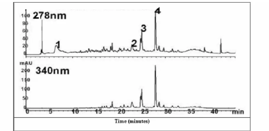
Figure 2. Analytical HPLC chromatogram monitored by UV absorption 278 and 340 for the aqueous alcoholic extract of doum leaves. (1: Gallic acid, 2:Quercetin glucoside,
3:Kaempferol rhamnoglucoside 4: Dimethyoxyquercetin rhamnoglucoside)
Table 1. Phenolic contents (mg/kg) of doum leaves identified by LC/ESI:
Compound mg/kg
Gallic acid 25130
Quercetin glucoside 4721
Kaempferol rhamnoglucoside 10684
Dimethyoxyquercetin rhamnoglucoside 17461
3.3. Identification of compounds 1- 14:
An in-depth phytochemical investigation of the aqueous ethanolic extract of doum leaves using column fractionation on Sephadex LH 20 and paper chromatography resulted in the isolation of 14 compounds: 8-C- -D-glucopyranosyl-5, 7, 4`-trihydroxyflavone (vitexin) 1
[17-19] , 6-C- -D-glucopyranosyl-5, 7, 4`-trihydroxyflavone (iso-vitexin) 2 [16-18]. quercetin 3-O- -4C1-D-glucopyranoside 3 [20] , gallic acid 4 [21] quercetin 7-O
-4
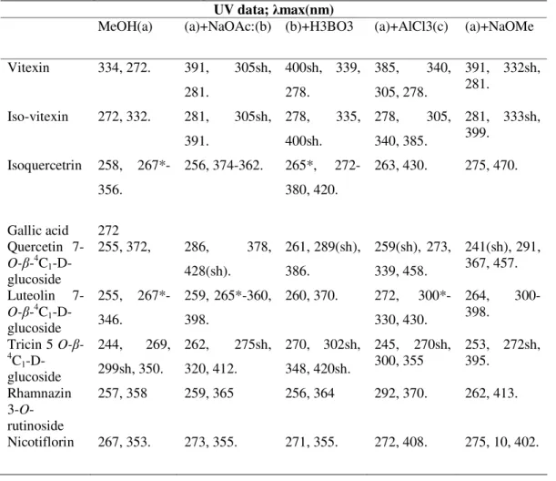
C1-D-glucoside 5 [22] luteolin 7-O- -4C1-D-glucoside 6 [23], tricin 5-O- -4C1-D-glucoside 7 [24], 7, 3` dimethoxy quercetin 3-O-[6''-O- -L-rhamnopyranosyl]- -D-gluco-pyranoside (rhamnazin 3-O-rutinoside) 8 [25], kaempferol-3-O-[6''-O- -L-rhamnopyranosyl]- -D-glucopyranoside (nicotiflorin) 9 [26-27] apigenin 10, luteolin 11, tricin 12 , quercetin 13 and kaempferol 14. The structures of these compounds were unambiguously determined by their chromatographic behaviors as well as spectroscopic analysis via UV (table 2), ESI/MS (table 3) and 1H-NMR (table 4) and 13C–NMR (table 5).
O O OH HO OH O H HO H HO H OH H OH O O OH HO OH O H HO H HO H OH H OH
1 2
O O OH HO O OH O HO HO H OH HO 2 3 4 5 6 7 8 9 10 1' 2' 3' 4' 5' 6' 1'' OH HO OH OH OH O 1 2 3 4 5 6 7 OH O OH O OH OH O O H HO H HO H OH H H OH
3 4 5
H O OH O OH OH O O H HO H HO H OH H H OH HO O O OH O O HO O O OH H OH H H HO H H HO
6 7
O O OH O O OH O HO HO H OH 2 3 4 5 6 7 8 9 10 1' 2' 3' 4' 5' 6' 1'' O OH OH H3C HO O 1''' 6''' O O O OH HO O OH O HO HO H OH 2 3 4 5 6 7 8 9 10 1' 2' 3' 4' 5' 6' 1'' O OH OH H3C
HO
O 1''' 6'''
Table 2. UV-Spectral data for the phenolics of doum leaves.
UV data; max(nm)
MeOH(a) (a)+NaOAc:(b) (b)+H3BO3 (a)+AlCl3(c) (a)+NaOMe
Vitexin 334, 272. 391, 305sh,
281.
400sh, 339,
278.
385, 340,
305, 278.
391, 332sh, 281.
Iso-vitexin 272, 332. 281, 305sh,
391.
278, 335,
400sh.
278, 305,
340, 385.
281, 333sh, 399.
Isoquercetrin 258,
267*-356.
256, 374-362. 265*,
272-380, 420.
263, 430. 275, 470.
Gallic acid 272
Quercetin
7-O- -4C1 -D-glucoside
255, 372, 286, 378,
428(sh).
261, 289(sh),
386.
259(sh), 273,
339, 458.
241(sh), 291, 367, 457.
Luteolin
7-O- -4C1 -D-glucoside
255,
267*-346.
259, 265*-360,
398.
260, 370. 272,
300*-330, 430.
264,
300-398.
Tricin 5 O
-4 C1 -D-glucoside
244, 269,
299sh, 350.
262, 275sh,
320, 412.
270, 302sh,
348, 420sh.
245, 270sh, 300, 355
253, 272sh, 395.
Rhamnazin 3-O -rutinoside
257, 358 259, 365 256, 364 292, 370. 262, 413.
Table 3. ESI / MS data for the phenolics of doum leaves
Compound Vitexin Iso-vitexin Isoquercetrin Quercetin
7-O- -4C1- D-glucoside
Luteolin 7-O- -4C1- D-glucoside
Tricin 5-O- -4C1- D-glucoside
Gallic acid Nicotiflorin Rhamnazin
3-O -rutinoside
m/z [M-1] 431.37 431.37 461.37 463.37 447.37 507.42 169.11 593.51 637.57
Table 4.1HNMR data for the phenolics of doum leaves
Nicotiflorin Rhamnazin
3-O-rutinoside Tricin
5-O- -4C1- D-glucoside Luteolin
7-O- -4C1- D-glucoside Quercetin
7-O- -4C1- D-glucoside Isoquercetrin
Iso-vitexin Vitexin
Pos.
6.17, s 6.37, s 7.53, d, J=7.5 6.83, d, J=7.5 6.83, d, J=7.5 7.53, d, J=7.5
5.31, d, J=7.2 4.39, d, J=8.0 1.16, d, J=6.6 H-6'''[-CH3] 6.57,d, J=1.8
6.64,d, J=1.8
8.42,d, J=1.8 7.43,d, J=8.5 7.96,dd, J=8.5 ,1.8,7-Ome 3.73, s, 3`-OMe 6.31, d, J=7.3 5.35, br s, H-1```(rhamnose) 1.48d, J=6.1, H-6'''[-CH3] 6.83 s
6.13 d, J=2.1 6.39 d, J=2.1 6.89 s
6.89 s
3.73 s, H-3`, 5` of OMe
5.36 d, J= 7.3 6.2, d, J=2.5
6.45, d, J=2.5 7.57, m
6.84, d, J=8.0 7.55, m
5.4, d, J=8.0 6.44, d, J=2.0
6.74, d, J=2.0 7.74, d, J=2.0
6.88, d, J=7.6
7.65, dd,
J=7.6, 2.0
5.05, d, J=7.2 6.2, d, J=2.5
6.45, d, J=2.5 7.57, m
6.84, d, J=8 7.55, m
5.4, d, J=8.o 6.47, s
4.7, d, J=8 7.93, d, J=8 6.92, d, J=8 6.92, d, J=8 7.93, d, J=8
4.7, d, J=8.0
3.00-3.90, m, Other sugar protons 6.77, s
6.21, s
7.93, d, *J=8 6.92, d, J=8 6.92, d, J=8 7.93, d, J=8
4.63, d, J=8
3.1-3.9, m, Other sugar protons 3
6 8 2` 3` 5` 6`
1`` 1```
Table 5. C- NMR data for the phenolics of doum leaves
Vitexin Iso-vitexin
Isoquercetrin Gallic acid Quercetin 7-O- -4C1
-D-glucoside
Luteolin 7-O
-4
C1-D-glucoside
Tricin 5 O-
-4
C1
-D-glucoside
Rhamnazin 3-O-rutinoside Nicotiflorin 1 2 3 4 5 6 7 8 9 10 1` 2` 3` 4` 5` 6` 1`` 2`` 3`` 4`` 5`` 6`` 1``` 2``` 3``` 4``` 5``` 6```` 163.9 102.4 182.0 161.0 98.1 162.5 104.6 155.9 104.0 121.5 128.8 115.7 160.3 115.7 128.8 73.3 70.8 78.8 70.5 81.7 61.3 163.5 102.8 181.9 161.2 108.8 163.2 93.7 156.2 103.4 121.1 128.4 116.0 160.6 116.0 128.4 73.1 70.6 78.9 70.3 81.4 61.4 157.24 133.0 177.40 161.30 99.71 163.08 94.94 156.30 102.03 120.03 115.30 142.0 149.0 77.50 122.20 100.23 73.42 76.77 69.92 116.32 60.95 120.6 108.8 145.5 138.1 145.5 108.8 167.7 147.9 135.9 175.9 160.3 98.9 162.7 94.5 155.7 104.6 121.9 115.5 145.0 147.9 115.4 120.1 100.3 73.2 76.5 69.9 77.2 60.9 164.5 103.20 181.6 161.10 99.70 162.90 94.90 156.90 105.5 121.60 113.70 145.9 149.6 116.1 119.0 100.4 73.30 76.60 70.80 77.30 61.0 162.4 106.3 177.0 158.3 104.3 161.0 98.5 158.5 108.1 120.4 104.4 148.1 139.4 148.1 104.4 104.0 73.6 75.6 69.6 77.5 60.8
The authors are grated to Prof. Dr. R. W. Owen, Division of Toxicology and Cancer Risk Factor, German Cancer Research Center (DKFZ), Heidelberg, Germany, for hosting the antioxidant activity and LC-ESI measurement.
References
[1] D.B.Fanshawe (1966). Hyphaene thebaica (Del.). Mart. East Afr.Agric.For. J 32, 108. [2] R.B. Ledin (1961). Cultivated Palms. Amer. Hort. Mag40 (1), 189.
[3] L.Boulos (1983). Medicinal plants of North Africa, Reference Publication, Algonac, Michigan.
[4] A. S. Sorour, N. Gomaa and M.Youssef (1972). Comparative studies on hypocholesterolemic effect of different fractions of Hyphaene thebaica (Doum) in experimental animals. Qual. Plant. Mater. Veg. XXII, 1, 83.
[5] O.N.Irobi and O. Adedayo (1999). Antifungal activity of aqueous extract of dormant fruits of Hyphaene thebaica (Palmae). Pharm-Biol37(2), 114-117.
[6] A.A.El-egami, A.Z. Almagboul, M.E.A.Omar and M.S. El-Tohami (2001). Sudanese plants used in folkloric medicine: screening for antibacterial activity. Fitoterapia Part X", 72(7); 810-817.
[7] J.A.Cook, D.J. VanderJagt, A.Dasgupta, R.S.Glew, W.Blackwell and R.H. Glew (1998). Use of the trolox assay to estimate the antioxidant content of seventeen edible wild plants of niger. Life-Science63(2), 106-110.
[8] Hsu. Betty, Coupar. Ian M, Ng. Ken (2006). Antioxidant activity of hot water extract from the fruit of the Doum palm , Hyphaene thebaica. Food Chemistry98(2); 317-328.
[9] A. Hashim (1994). Phytochemical investigation of the fruit of Hyphaene thebaica (L) Mart. growing in Egypt family Palmae, thesis for master, National Research Centre, Giza, Egypt.
[10] A.Foschini and A.Usai (1968). Detection of the roasted doum palm nut in powdered roasted coffee. Rassegna Chimica 20(4); 147-51.
[11] E.M.Gaydou, J.P.Bianchini, I.Rabarisoa and G. Ravelojaona (1980). Oil plants native to Madagascar. Study of the fatty acid and sterol composition of some palm species. Oleagineux35(8-9), 413-15.
[12] S.S.Amin and A.M.Paleologou (1973). A study of the polysaccharides of the kernel and endocarp of the fruit of the doum palm (Hyphaene thebaica). Phytochemistry 12( 4) 899-901.
[13]B.Maymone, A.Battaglini and M.Tiberio (1950). The digestibility and nutritive value of residues from the commercial use of vegetable ivory of the ivory nut palms (Phytelephas spp.) and of doum palms (Hyphaene spp.). Annali della Sperimentazione Agraria 4, 603-24.
[14] S.D.Bonde, V.V. Agate and D.K.Kulkarni ( 1990). Nutritional composition of the fruits of doum palms (Hyphaene) from the West Coast of India. Principes34(1), 21-23.
[15] R.W.Owen, B.Spiegelhalder and H.Bartsch (2000). Generation of reactive oxygen species by the faecal matrix. Gut 46, 225-232.
[16] R.W.Owen, W.Mier, A.Giacosa, W.E. Hull, B. Spiegelhalder and H.Bartsch ( 2000). Phenolic compounds and squalene in olive oils: the concentration and antioxidant potential of total phenols, simple phenols, secoiridoids, lignansand squalene. Food and Chemical Toxicology 38, 647-659.
[17] J.B.Harborne (1988). The flavonoids. Chapman and Hall.
[18] A.Numata, K.Hokimoto and H.Yamaguchi (1980). C-Glycosylflavones in Lespedeza cuneata. Chem Pharm Bull28, 964.
[19] P.K.Agrawal (1989). Carbon-13 NMR of Flavonoids. Elsevier Science Publishing Co. Inc.: New York, 283-364.
[20] O.A.Eldahshan (2002). Phytochemical study of certain plants belonging to family Umbelliferae of promising biological activities. Thesis for master, Fac., Phramacy, Ain Shams University, Cairo, Egypt. [21] L.Krenn, A.Miron, E.Pemp, U.Petr and B. Kopp (2003). Flavonoids from Achillea nobilis. Z. Naturforsch;
58c, 11-16.
[22] G.B.Jean, M.Russell, N.Paul, W.Gary, P.Antoine, A.K.Paul and J.Nathalie (2002). Functional expression of human liver cytosolic -glucosidase in Pichia pastoris ,Insights into its role in the metabolism of dietary glucosides. Eur J Biochem26, 249-258.
[24] A.Francis, S.Chul, T.Masami, I.Masahiro and H.Michio (2000). Identification and isolation of the probing stimulants in the rice plant for the White-Black Planthopper, Sogatella furcifera. Biosci. Biotechnol. Biochem.64(2), 443-446.
[25] U.S.Harput , I.S. Glui , and Y. Ogihara ( 2004). Methoxyflavonoids from Pinaropappus roseus. Turk J Chem 28, 761 – 766.
[26] K.Kazuma, N. Noda and M.Suzuki (2003). Malonylated flavonol glycosides from the petals of Clitoria ternatea. Phytochemistry 62, 229-237.
[27] P.K.Agrawal (1992). NMR spectroscopy in the structural elucidation of oligosaccharides and glycosides. Phytochemistry31 (10), 3307-3330.