*Correspondence: Samaneh Zolghadri. Nuclear Science and Technol-ogy Research Institute (NSTRI). 14155-1339-Tehran, Iran. E-mail: [email protected]
Article
vol. 51, n. 2, apr./jun., 2015 http://dx.doi.org/10.1590/S1984-82502015000200011
Production, quality control, biodistribution assessment and
preliminary dose evaluation of [
177Lu]-tetra phenyl porphyrin
complex as a possible therapeutic agent
Samaneh Zolghadri
1,*, Hassan Yousefnia
1, Amir Reza Jalilian
1, Yousef Fazaeli
11Nuclear Science and Technology Research Institute (NSTRI), Tehran, Iran
Due to interesting therapeutic properties of 177Lu and tumor avidity of tetraphenyl porphyrins (TPPs), 177Lu-tetraphenyl porphyrin was developed as a possible therapeutic compound. 177Lu of 2.6-3 GBq/mg
speciic activity was obtained by irradiation of natural Lu2O3 sample with thermal neutron lux of
4 × 10¹³ n.cm-2.s-1. Tetraphenyl porphyrin was synthetized and labeled with 177Lu. Radiochemical purity of
the complex was studied using Instant thin layer chromatography (ITLC) method. Stability of the complex was checked in inal formulation and human serum for 48 h. The biodistribution of the labeled compound
in vital organs of wild-type rats was studied up to 7 d. The absorbed dose of each human organ was calculated by medical internal radiation dose (MIRD) method. A detailed comparative pharmacokinetic study was performed for 177Lu cation and [177Lu]-TPP. The complex was prepared with a radiochemical
purity: >97±1% and speciic activity: 970-1000 MBq/mmol. Biodistribution data and dosimetric results showed that all tissues receive approximately an insigniicant absorbed dose due to rapid excretion of the complex through the urinary tract. [177Lu]-TPP can be an interesting tumor targeting agent due to low
liver uptake and very low absorbed dose of approximately 0.036 to the total body of human.
Uniterms: Radiopharmaceuticals/internal dosimetry. Lutetium-177. 177Lu-tetraphenyl porphyrin/
biodistribution. Porphyrins/biodistribution. Medical internal radiation dose. MIRD.
Devido às propriedades interessantes do 177Lu e da avidez tumoral das tetrafenil poririnas (TPP),
desenvolveu-se a 177Lu-tetrafenil poririna como composto terapêutico potencial. 177Lu de atividade
especíica de 2,6-3 GBq/mg foi obtido por irradiação de amostra de Lu2O3 com luxo térmico de nêutrons
de 4 × 10¹³ n.cm-2.s-1 . Sintetizou-se a tetrafenil poririna e marcou-se com 177Lu. A pureza radioquímica
do complexo foi estudada usando método de Cromatograia Instantânea de Camada Delgada ( ITLC). A estabilidade do complexo foi checada na formulação inal e no ser humano por 48 h. A biodistribuição do composto marcado em órgãos vitais de ratos do tipo selvagem foi estudada por mais de 7 dias. A dose absorvida para cada órgão humano foi calculada pelo método da Dose Médica de Radiação Interna (MIRD). Estudo farmacocinético comparativo detalhado foi efetuado para o cátion 177Lu e para o [177
Lu]-TPP. O complexo foi preparado com pureza radioquímica >97±1% e atividade especíica de 970-1000 MBq/mmol. Os dados de biodistribuição e os resultados dosimétricos mostraram que todos os tecidos receberam uma dose absorvida aproximadamente insigniicante devido à rápida excreção do complexo pelo trato urinário. O [177Lu]-TPP pode ser um agente interessante de direcionamento do tumor devido
à baixa captação pelo fígado e pela dose bem baixa absorvida, de, aproximadamente, 0,036 do corpo
humano total.
Unitermos: Radiofármacos/dosimetria interna. Lutécio-177. 177Lu-tetrafenil poririna/biodistribuição.
INTRODUCTION
Nowadays, radiopharmaceuticals are widely used for diagnostic and therapeutic purposes. An ideal radiopharmaceutical should lead to substantially greater accumulation rate in the target organ while the accumulation in other organ should be as low as possible. Therefore, scientists have paid attention to new ligands to differentiate between malignant and normal cells (Sanderson et al., 1972).
Porphyrin is a heterocyclic macrocycle derived from four pyrrole-like subunits that plays an important role in
biological transfer systems. Various porphyrin complexes
have shown interesting tumor-avid activity in vitro and in
vivo (Subbarayan et al., 2001; Das et al., 2008; Bonnett,
1995; Jori, 1996).
Radiolabeled porphyrins have been developed for the therapeutic purposes such as, 109Pd-protoporphyrins
(Fawwaz et al., 1974), 109Pd-porphyrins (Fawwaz,
Hemphill, Winchell, 1971), 109Pd-derivitized porphyrins (Chakraborty et al., 2007), 188Re-porphyrins (Jia, Deng, Pu, 2007; Sarma et al., 2010), 123I-Porphyrins (Jae Hak
et al., 2007). Various radiolabeled porphyrin complexes
such as 57Co-porphyrins (Hambright et al., 1976),
99mTc-porphyrin (Murugesan et al., 2001; Wang, Lin, Lin,
2010), and 111In-porphyrin (Fazaeli et al., 2012) have also
been introduced for imaging.
Whilethe masive accumulation of 109Pd-porphyrins was indicated in ibrosarcoma tumours(Chakraborty et al., 2007), I-123-labeled porphyrin demonstrated high
focal accumulation in the B16-F10 melanoma tumor (Jae Hak et al., 2007). (188)Re-labeled
5,10,15,20-tetrakis[3,4-bis(carboxymethyleneoxy)phenyl]porphyrin has also
shown specific affinity toward the fibrosarcoma and thymic lymphoma tumors in mice (Sarma et al., 2010).
Accumulation of the radiolabeled prophyrins in tumour is dependent on various parameters such as porphyrin structure, choice of radioisotope, pH, the presence of inflammation and many other factors. However, the balance between hydrophilicity and lipophilicity is also recognized as an important factor in tumour accumulation. Whereas, lipophilicity of the agent plays an important role in tumor accumulation,
hydrophilicity is a signiicant key in the clearance of the
agent from the non-target organs. Therefore, a balance between these two properties is necessary for developing a suitable agent and for contributing to the challenge in designing suitable derivatives of porphyrin (Das et al., 2010).
177Lu decays with a half life of 6.73 d by emission
β-particles with maximum energy of 497 keV (78.6%)
and γ-photons of 112 keV (6.4%) and 208 keV (11%)
to stable 177Hf (TOI, 1993). Due to these good physical
characteristics as well as the feasibility of large-scale
production in adequate speciic activity and radionuclidic purity using a moderate flux reactor, 177Lu has been
considered as a promising radionuclide for developing therapeutic radiopharmaceuticals due to its suitable half-life.
177Lu-radiopharmaceuticals have been developed and
used in the therapy of various diseases and malignancies, such as somatostatin receptor radiotherapy (Bodei et
al., 2009), radioimmunotherapy (Michel et al., 2005),
bone palliation therapy (Chakraborty et al., 2008a)
and radiosynovectomy (Chakraborty et al., 2006;
Chakraborty et al., 2008b). 177 Lu-5,10,15,20-tetrakis[4-carboxymethyleneoxyphenyl] porphyrin have recently
been developed and have shown active tumor uptake in
mice bearing ibrosarcoma tumors (Das et al., 2010).
According to the interesting pharmacological properties of porphyrins such as solubility in serum,
rapid wash-out, tumor avidity and feasible complexation
with various bi/tri-valent metals (Falk, 1975), the idea of developing a possible tumor targeting agent by incorporating 177Lu into a suitable porphyrin ligand, i.e.
TPPH2 was investigated (Figure 1).
As for the amount of energy uptake in any organs by ionizing radiation, the absorbed dose, plays an important role in evaluating the risks associated with the administration of radiopharmaceuticals and thus the
maximum amount of activity that should be undertaken
(Stabin et al., 1999). In nuclear medicine, the most commonly used method for calculation of the internal dose estimates is the one developed by the medical internal radiation dose (MIRD) committee (Stabin, 1996)
summarized in MIRD primer (Loevinger, Budinger,
Watson, 1988).
In this work, we endeavour to report, synthesis, radiolabeling, quality control and biodistribution studies of 177Lu-TPP in wild-type rats. The time/decay diagrams
for the labeled compound in vital organs were plotted
compared to lutetium cation. Also the partition coeficient of the complex was calculated and the absorbed dose to
each organ of human was evaluated by biodistribution studies in rats by MIRD method.
MATERIAL AND METHODS
177Lu was produced at Tehran Research Reactor.
Chemicals were purchased from the Aldrich Chemical Co. (Gemany). NMR spectra were obtained on a FT-80 Varian instrument (80 MHz) with tetramethylsilane as an
internal standard. Infrared spectrum was measured on a
Perkin-Elmer 781 spectrometer by means of a KBr disc.
Mass spectrum was recorded by a Finnigan Mat TSQ-70
Spectrometer. Thin layer chromatography (TLC) for cold
compounds was performed on polymer-backed silica gel
(F 1500/LS 254, 20 × 20 cm, TLC Ready Foil, Schleicher
& Schuell, Germany). Normal saline and sodium
acetate used for labeling were of high purity and had
been iltered through 0.22 µm Cativex ilters. Instant thin
layer chromatography (ITLC) was performed by counting
Whatman No. 2 papers using a thin layer chromatography scanner, Bioscan AR2000, Bioscan Europe Ltd. (France). Biodistribution data were obtained by counting normal
saline washed tissues after weighing on a CanberraΤΜ high purity germanium (HPGe) detector (model
GC1020-7500SL). Radionuclidic purity was checked with the same detector. For activity measurement of the samples
a CRC Capintech Radiometer (NJ, USA) was utilized.
All calculations as well as tissue count were based on the 112 keV peak of 177Lu. Animal studies were performed in accordance with the United Kingdom Biological Council’s Guidelines on the Use of Living Animals in Scientific
Investigations, 2nd ed.
Production and quality control of 177LuCl
3 solution
177Lu was produced by irradiation of natural Lu
2O3 target (1 mg) at a thermal neutron lux of approximately
4 × 10¹³ n/cm2.s for 5 days at Tehran Research Reactor
(TRR) according to the reported procedures (Yousefnia et
al., 2011). The irradiated target was dissolved in 200 µL
of 1.0 M HCl, to prepare 177LuCl
3 and diluted to the
appropriate volume with ultra pure water, to produce a
stock solution of inal volume of 5 mL with approximately
2.8 GBq. The mixture was filtered through a 0.22 µm biological ilter and sent for use in the radiolableing stage
in the process. For radionuclidic purity determination, the sample was checked by gamma-ray spectroscopy on an HPGe detector for 5 h based on two major photons of
177Lu (6.4% of 0.112 MeV and 11% of 0.208 MeV). The
radiochemical purity of the 177LuCl
3 was checked using 2 solvent systems for ITLC (A: 10 mM DTPA pH.4 and B:
ammonium acetate 10%:methanol (1:1)).
Preparation of Tetraphenyl Porphyrin (TPPH2)
This compound was prepared according to the reported method using freshly distilled benzaldehyde,
pyrrole and propionic acid followed by oxidation
(Adler et al., 1967). Yield; 20%, m.p.> 248-250 °C. 1H NMR (CDCl3) δ (ppm) –2.8 (2 H, NH), 7.71-7.82 (12 H), 8.14-8.27 (8 H), 8.85 (8 H). 13C-NMR (CDCl3)
δ (ppm) 120.20 (C), 126.74 (CH), 127.76 (CH),
131.16 (CH), 134.62 (CH), 142.22 (C), 145.6 (C).UV (toluene) λmax (ε) = 418 nm (413200), 514 (19060), 549
(8080), 594 (5380), 648 (3870). IR (KBr) 3320, 3055,
3025, 1595.
Preparation of [177Lu]-TPP
0.2 ml of 177LuCl
3 with 111 MBq radioactivity was
transferred to a 5 mL-borosilicate vial and heated to
dryness by using a low of N2 gas at 50-60 °C, followed by the addition of ifty microliters of TPP in absolute ethanol
(1 mg/mL ≈ 81 nmoles) and 450 microliters of acetate
buffer pH 5 (0.1 M). The mixture vortexed at 25 °C for
4 h. The inal solution was then passed through a 0.22 µm
ilter and the radiochemical purity was checked by ITLC.
For this purpose, 5 µL of the inal solution was spotted on
a chromatography Whatman No. 2 paper, and developed
in two mobile phase mixtures, A: water:acetonitrile (3:1)
and B: water:acetonitrile (1:3).
Determination of partition coefficient
Partition coefficient (log P) of [177Lu]-TPP was
calculated. A mixture of 1 mLof 1-octanol and 1 mL
of isotonic acetate-buffered saline (pH 7) containing
approximately 3.7 MBq of the radiolabeled complex at 37 °C was vortexed 1 min and left 5 min. Following
centrifugation at >1200 g for 5 min, the octanol and aqueous phases were sampled and counted in an automatic
well-type counter. A 500 μL sample of the octanol phase from this experiment was shaken again thrice with fresh
of the second and third extractions from three to four
independent measurements.
Stability tests
The stability of the complex was checked according to the conventional ITLC method. A sample of [177Lu]-TPP
(37 MBq) was kept at room temperature for 2 days while
being checked by ITLC at time intervals in order to check
stability in final product using above chromatography system. For serum stability studies, to 36.1 MBq of [177
Lu]-TPP was added 500 µL of freshly collected human serum
and the resulting mixture was incubated at 37 °C for 5 h;
Aliquots (5 µL) were analyzed by ITLC.
Biodistribution in wild-type rats
The distribution of 177Lucl
3 and the radiolabeled complex among tissues were determined for wild-type
rats. 50–100 µL of 177Lu-TPP or 177LuCl
3 solutions with 1.85 MBq radioactivity were injected intravenously
via their tail veins. The total amount of radioactivity injected into each rat was measured by counting the 1 mL syringe before and after injection in a dose calibrator
with ixed geometry. The animals were sacriiced by CO2 asphyxiation at selected times after injection (2, 4, 24, 48, 120 and 168 h). The tissues (blood, heart, lung, brain,
intestine, feces, skin, stomach, kidneys, liver, muscle and bone) were weighed and rinsed with normal saline and
their speciic activities were determined with an HPGe
detector equipped with a sample holder device as percent of injected dose per gram of tissues.
Dosimetric studies
The absorbed dose of each human organ was calculated by MIRD method based on biodistribution data in wild-type rats. The accumulated activity in animals
was extrapolated to the accumulated activity in humans
by the proposed method of Sparks et al. (eq. 1) (Sparks, Aydogan, 1996).
Ahuman organ= Aanimal organ human human
animal animal
OrganMass OrganMass
/ BodyMass
/ BodyMass (1)
where à is the accumulated activity in the source organs and can be calculated by the equation 2.
1
t
à =
∫
∞A (t) dt (2)It should be noticed that A (t) is the activity of each organ at time t.
The accumulated source activity for each organ of animals was calculated by plotting the percentage-injected dose versus time for each organ and computing the area under the curves. For this purpose, the data points which represent the percentage-injected dose were created.
The curves were extrapolated to infinity by fitting the tail of each curve to a monoexponential curve with the exponential coeficient equal to physical decay constant
of 177Lu. Then the area under the curve was calculated. In order to extrapolate this accumulated activity to human,
the mean weights of each organ for standard human were used (Table I).
The radiation absorbed dose was calculated by
MIRD formulation (Henrichs, Kaul, Roedler, 1982):
k h k h
h
D(r )=
∑
A ×S(r ←r ) (3)where D(rk) is the absorbed dose of the target
organ, and S(rk ←––– rh) called S factor which is deined as the mean absorbed dose to the target region rk per unit
accumulated activity in the source region rh. S factor
represents the physical decay characteristics of the radionuclide, the range of the emitted radiations, and the
organ size and coniguration (Bevelacqua, 2005) expressed
in mGy/MBq.s. The S factors have been taken from the OLINDA software (OLINDA, 2007).
RESULTS AND DISCUSSION
Radionuclide production
The radionuclide was prepared in a research reactor
according to the regular methods with a range of speciic
TABLE I - The mean weights of organs for human with standard weight (ICRP 89, 2001)
Organ Weight (g)
Bone 5500
Heart 330
Stomach 150
Kidneys 310
Small intestine 650
Spleen 150
Muscle 29000
Liver 1800
Lung 500
activity 2.6-3 GBq/mg for radiolabeling use. The obtained
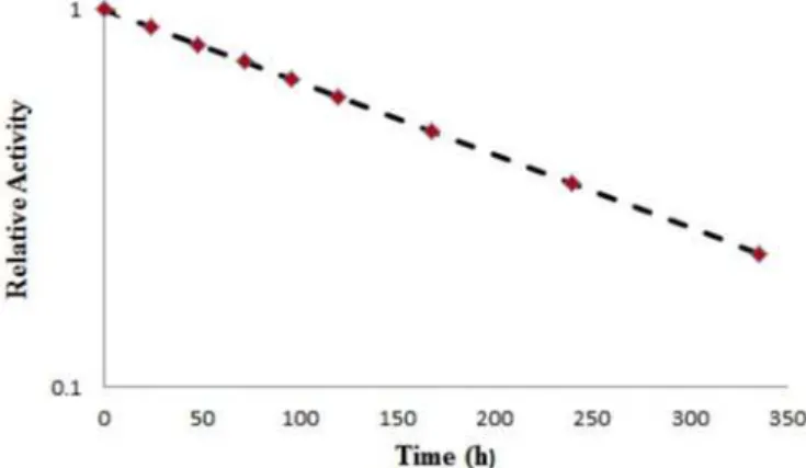
radionuclidic purity was 99.98% (Figure 2). Furthermore,
half-life of the 177Lu was also studied by counting the
sample at different time intervals. The decay scheme for the radionuclide is shown in Figure 3.
The radioisotope was dissolved in acidic media as a starting sample and was further diluted and evaporated for obtaining the desired pH and volume followed by sterile
iltering. The radiochemical purity of the 177Lu solution
was checked in two solvent systems: in 10 mM DTPA, free Lu3+ cation as a complex in more lipophilic LuDTPA form
migrates to higher Rf, while small radioactive fraction remains in its origin which could be related to other Lu
ionic species, not forming LuDTPA complex, such as LuCl4
-, etc. and/or colloids.
FIGURE 2 - Gamma-ray spectrum for 177LuCl
3 solution used
in this study. FIGURE 3- Decay scheme for 177Lu used in this study.
FIGURE 4 - ITLC chromatograms of 177LuCl
3 solution in DTPA solution (pH. 4) (left) and 10% ammonium acetate:methanol (1:1) solution (right) using Whatman No. 2.
On the other hand, 10% ammonium acetate:methanol
mixture was also used for the determination of
radiochemical purity. In this solvent system, the fast eluting species were possibly Lu-177 cations, other than
Lu3+ and the remaining fraction at R
f.0 was a possible mixture of Lu3+ and/or colloids. The difference in values
of impurity in two solvent systems is possibly due to the presence of colloidal impurity in the sample (Figure 4).
Preparation of [177Lu]- TPP
The synthetic scheme for radiolabeling of TPP with 177LuCl
3 is demonstrated in Figure 5. Because of
the engagement of NH polar functional groups in its structure, labeling of TPPH2 with lutetium cation affects
its chromatographic properties and the inal complex is
more lipophilic. Two different chromatographic systems
were used. Using water/acetonitrile (1:3) mixture, free
while the radiolabeled compound migrates to higher Rf.
Using a more polar mobile phase, acetonitrile:water (1:3),
free lutetium cation migrated to a higher Rf, while the radiolabeled compound remained at the origin (Figure 6).
Partition coefficient
As expected, the lipophilicity of the [177Lu]-TPP
compound is rather high. The measured octanol/water
partition coeficient, P, for the complex was found to depend
on the pH of the solution. At the pH 7, the log P was 1.63.
Stability
The stability of [177Lu]-TPP prepared complex
at room temperature was checked up to 48 hours. The radiochemical purity of the complex remained at 98% for 2 days. Also the stability of the complex was determined at 37 °C for 48 h and the data were almost consistent with the inal solution stability.
Biodistribution studies of 177Lucl
3 and 177Lu-TPP in wild-type rats
The animals were sacriiced by CO2 asphyxiation at selected times after injection (2, 4, 24, 48 and 168 h).
The biodistribution data show that the liver uptake of
FIGURE 5 - Synthetic scheme for radiolabeling of TPP with 177LuCl
3.
FIGURE 6 - ITLC chromatograms of 177LuCl 3 and
FIGURE 7 - Percentage of injected dose per gram (ID/g%) of 177LuCl
3 in wild-type rat tissues at 2, 4, 24, 48 and 168 h post injection (ID/g%: percentage of the injected dose per gram of tissue calculated based on the area under curve of 112 keV peak in gamma spectrum) (n=5).
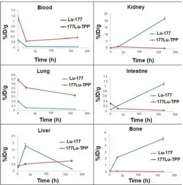
FIGURE 8 - Biodistribution of [177Lu]-TPP (1.85 MBq) in wild type rats 2, 24, 120 and 168 h after iv injection via tail vein (ID/g%: percentage of injected dose per gram of tissue calculated based on the area under curve of 112 keV peak in gamma spectrum) (n=5) the cation is comparable to many other radio-metals
mimicking ferric cation accumulation; about 3% of the
activity accumulates in the liver after 48 h. Binding of 177Lu by transferrin and transport to the liver appears to
be the route of accumulation (Figure 7).
As it can be seen from Figure 5, the blood content is low at all time intervals, which shows the rapid removal of activity in the circulation. The lung, muscle and also
skin do not demonstrate signiicant uptake while it is in
accordance with other cations accumulation. A 5% bone
uptake is observed for the cation at 168 h. The spleen also
has uptake (1%) possibly related to reticuloendothelial uptake. The kidney plays an important role in 177Lu cation
excretion especially after 24 h (1%). Biodistribution of 177Lu-TPP in different organs of wild-type rats is shown in Figure 8.
Comparison of vital organs uptake for 177Lu-TPP and 177LuCl
3 demonstrates kinetic pattern difference for both
species. 177Lu cation is accumulated in the liver within the irst 24 h post injection slightly, while 177Lu-TPP second major excretion route is through the liver and slow uptake of less than 1% is observed in 168 h for the radiolabeled
compound (Figure 9).
Intestinal activity increases in 177Lu3+ after 7 days as a consequence of the liver excretion through GI tract,
The accumulation of the tracer in other tumor models is under investigation due to the low liver uptake and rapid
excertion through the urinary tract. Biodistribution data
and dosimetric results showed that all tissues receive
virtually insigniicant absorbed dose due to rapid excretion of the complex through the urinary tract. [177Lu]-TPP can
be an interesting tumor targeting agent due to the low liver
uptake and very low absorbed dose of approximately 0.036
mSv/MBq to the total body of human.
TABLE ∏ - Absorbed dose in each organ of human after injection of 177Lu-TPP
Organ
Absorbed Dose (mSv/MBq)
Organ
Absorbed Dose (mSv/MBq)
Adrenals 0.005 Ovaries 0.003
Brain 0.001 Pancreas 0.005
Breasts 0.001 Red Mar. 0.069
GB Cont. 0.006 Cort Bone Sur. 0.081
LLI Cont. 0.101 Trab. Bone
Sur.
0.105
SI Cont. 0.003 Cort Bone Vol. 0.039
Stom. Cont. 0.036 Trab. Bone
Vol.
0.099
ULI Cont. 0.003 Spleen 0.149
Heart Cont. 0.007 Testes 0.001
Heart Wall 0.015 Thymus 0.002
Kidneys 0.313 Thyroid 0.002
Liver 0.233 UB Cont 0.002
Lungs 0.089 Uterus 0.002
Muscle 0.033 Tot. Body 0.036
FIGURE 10 - The clearance curves from each organ of the rats.
FIGURE 9 - Comparative organ uptake of 177LuCl 3 and
177 Lu-TPP in wild-type rats.
decreases due to the low liver uptake compared with
urinary excretion. As shown earlier, 177Lu cation is slightly
absorbed in the skeletal system (5%) while the labeled compound almost shows no uptake in the bone.
Since the urinary tract is a major route of excretion
of the porphyrins, the amount of the kidney activity is
maximum for the labeled compound especially after 24 h, however in 7days major urinary excretion is observed
in free cation. The circulation wash-out for the labeled compound is observed while a lesser amount of activity in blood is observed for free Lu cation.
Dosimetric studies
Dosimetric evaluation in human organs was made by MIRD method based on biodistribution in rat organs. The clearance curves from each organ of the rats are shown in Figure 10. The absorbed dose in each organ of human after injection of 177Lu-TPP is given in Table Π.
CONCLUSION
Total labeling and formulation of [177Lu]-TPP took about 4 h (radiochemical purity: >97 ± 1% ITLC, speciic activity, 970-1000 MBq/mmol). The complex was stable in inal formulation and human serum at least for 24 h. The
REFERENCES
A D L E R , A . D . ; L O N G O , F. R . ; F I N A R E L L I , J . D . ; GOLDMACHER, J.; ASSOUR, J.; KORSAKOFF, L. A simpliied synthesis for meso-tetraphenylporphine. J. Org. Chem., v.32, p.476, 1967.
BEVELACQUA, J.J. Internal dosimetry primer. Radiat. Prot.
Manage., v.22, p.7-17, 2005.
BODEI, L.; FERONE, D.; GRANA, C.M.; CREMONESI, M.;
SIGNORE, A.; DIERCKX, R.A.; PAGANELLI, G. Peptide
receptor therapies in neuroendocrine tumors. J. Endocrinol.
Invest., v.32, p.360-369, 2009.
BONNETT, R. Photosensitisers of porphyrin and phthalo-cyanine series for photodynamic therapy. Chem. Rev., v.24, p.19-33, 1995.
CHAKRABORTY, S.; DAS, T.; BANERJEE, S.; BALOGH, L.; CHAUDHARI, P.R.; SARMA, H.D.; POLYÁK, A.; MÁTHÉ, D.; VENKATESH, M.; JANOKI, G.; PILLAI, M.R. 177Lu-EDTMP: a viable bone pain palliative in skeletal metastasis. Cancer Biother. Radiopharm., v.23, p.202-213, 2008a.
CHAKRABORTY, S.; DAS, T.; BANERJEE, S.; SARMA, H.D.; VENKATESH, M. Preparation and preliminary biological evaluation of a novel 109Pd labeled porphyrin derivative for possible use in targeted tumor therapy. Q. J.
Nucl. Med. Mol. Imaging., v.15, p.16-23, 2007.
CHAKRABORTY, S.; DAS, T.; BANERJEE, S.; SARMA, H.D.; VENKATESH, M. Preparation and preliminary biological evaluation of 177Lu-labeled hydroxyapatite as a promising agent for radiation synovectomy of small joints.
Nucl. Med. Commun., v.27, p.661-668, 2006.
CHAKRABORTY, S.; DAS, T.; SARMA, H.D.; VENKATESH, M.; BANERJEE, S. Preparation and preliminary studies on 177Lu-labeled hydroxyapatite particles for possible use in
the therapy of liver cancer. Nucl. Med. Biol., v.35, p.589-597, 2008b.
DAS, T.; CHAKRABORTY, S.; SARMA, H.D.; BANERJEE, S. A novel ]109Pd] palladium labeled porphyrin for possible use in targeted radiotherapy. Radiochim. Acta., v.96, p.427-433, 2008.
DAS, T.; CHAKRABORTY, S.; SARMA, H.D.; BANERJEE,
S.; VENAKATESH, M. A novel 177Lu-labeled porphyrin
for possible use in targeted tumor therapy. Nucl. Med. Bio., v.37, p.655-663, 2010.
FALK, J.E. Porohyrins and metalloporphyrins. New York:
Elsevier Science Publishing, 1975.
FAWWAZ, R.A.; FRYE, F.; LOUGHMAN, W.D.; HEMPHILL, W. Survival of skin homografts in dogs injected with 109 Pd-protoporphyrin. J. Nucl. Med., v.15, p.997-1002, 1974.
FAWWAZ, R.A.; HEMPHILL, W.; WINCHELL, H.S. Potential use of 109Pd-porphyrin complexes for selective lymphatic ablation. J. Nucl. Med., v.12, p.231-236, 1971.
FA Z A E L I , Y. ; J A L I L I A N , A . R . ; A M I N I , M . M . ; ABOUDZADEH-ROVAIS, M.R.; SHAFAEE, K.; MIRZAI, M.; RAHIMINEJAD, A. Radiosynthesis and biological evaluation of [111In]-5,10,15,20-tetrakis(pentaluorophenyl)
porphyrin complex as a possible imaging agent. IJNESE,
v.2, p.28-32, 2012.
HAMBRIGHT, P.; SMART, J.C.; McRAE J.; NOHR, M.L.; YANO, Y.; CHU, P.; BEARDEN, A.J. Tumor imaging with 57cobalt(III)-sandwich complexes and 57 cobalt(III)-porphyrins. Inorg. Nucl. Chem. Letters., v.12, p.217-222, 1976.
HENRICHS, K.; KAUL, A.; ROEDLER, H.D. Estimation of age-dependent internal dose from radiopharmaceuticals.
Phys. Med. Biol., v.27, p.775-784, 1982.
ICRP Publication 89, Basic anatomical and physiological data for use in radiological protection: reference values, 2001.
JAE HAK, L.; BYUNG SEOK, M.; TAE SUP, L.; DAE YOON, C.; KWON SOO, C.; GI JEONG, C. Synthesis and biologic evaluation of I-123-labeled porphyrin derivative as a potential tumor-imaging agent. Cancer. Biother.
Radiopharm., v.22, p.853-862, 2007.
JIA, Z.; DENG, H.; PU, M. Synthesis and preliminary biological studies of the novel conjugate 188 Re-labeledmeso-tetrakis(4-sulfophenyl)porphyrin in mice. Nucl. Med. Bio., v.34, p.643-649, 2007.
JORI, G. Tumour photosensitizers: approaches to enhance selectivity and efficiency of photodynamic therapy. J.
LOEVINGER, R.; BUDINGER, T.; WATSON, E. MIRD primer for absorbed dose calculations. New York: Society of
Nuclear Medicine, 1988. 128 p.
MICHEL, R.B.; ANDREWS, P.M.; ROSARIO, A.V.;
GOLDENBERG, D.M.; MATTES, M.J. 177Lu-antibody
conjugates for single-cell kill of B-lymphoma cells in vitro and for therapy of micrometastases in vivo. Nucl. Med.
Biol., v.32, p.269-278, 2005.
MURUGESAN, S.; SHETTYC, S.J.; SRIVASTAVA, T.S.;
NORONHA, O.P.D.; SAMUEL, A.M. A
technetium-99m-labeled cyclam acid porphyrin (CAP) for tumour imaging.
Applied. Radiat. Isotopes, v.55, p.641-646, 2001.
OLINDA - Organ Level Internal Dose Assessment Code (Version 1.1), copyright Vanderbilt University, (2007).
SANDERSON, D.R.; FONTANA, R.S.; LIPSON, R.L.; BALDES, E.J. Hematoporphyrin as a diagnostic tool.
Cancer, v.30, p.1368-1372, 1972.
SARMA, H.D.; DAS, T.; BANERJEE, S.; VENKATESH, M.;
VIDYASAGAR, P.B.; MISHRA, K.P. Biologic Evaluation
of a Novel 188Re-Labeled Porphyrin in Mice Tumor Model.
Cancer Biother. Radiopharm., v.25, p.47-54, 2010.
SPARKS, R.B.; AYDOGAN, B. Comparison of the effectiveness of some common animal data scaling techniques in estimating human radiation dose. Sixth International Radiopharmaceutical Dosimetry Symposium, Oak Ridge, TN: Oak Ridge Associated Universities, 1996. p.705-716.
STABIN, M.G. MIRDOSE: personal computer software for internal dose assessment in nuclear medicine. J. Nucl. Med., v.37, p.538-546, 1996.
S TA B I N , M . G . ; TA G E S S O N , M . ; T H O M A S , S . R . ; LJUNGBERG, M.; STRAND, S.E. Radiation dosimetry in nuclear medicine. Appl. Radiat. Isot., v.50, p.73-87, 1999.
SUBBARAYAN, M.; SHETTY, S.J.; SRIVASTAVA, T.S.; NORONHA, O.P.D.; SAMUEL, A.M.; MUKHTAR, H. Water-soluble 99mTc-labeled dendritic novel porphyrins tumor imaging and diagnosis. Biochem. Biophys. Res.
Commun., v.281, p.32-36, 2001.
Table of radioactive isotopes, Available at: <http://ie.lbl.gov/ toi/nuclide.asp?iZA=710177>. Accessed on: 03rd Feb 2014.
WANG, A.Y.; LIN, J.L.; LIN, W.C. Studies on the porphine labeled with 99mTc-pertechnetate. J. Radioanal. Nucl. Chem., v.284, p.121-128, 2010.
YOUSEFNIA, H.; RADFAR, E.; JALILIAN, A.R.; BAHRAMI-SAMANI, A.; SHIRVANI-ARANI, S.; ARBABI, A.;
GHANNADI-MARAGHEH, M. Development of 177
Lu-DOTA-anti-CD20 for radioimmunotherapy. J. Radioanal.
Nucl. Chem., v.287, p.199-209, 2011.
![FIGURE 6 - ITLC chromatograms of 177 LuCl 3 and 177 Lu-TTP on Whatman No. 2 paper using water:acetonitrile (1:3) [up (a & b)]](https://thumb-eu.123doks.com/thumbv2/123dok_br/15413672.587238/6.892.65.434.131.401/figure-itlc-chromatograms-lucl-whatman-paper-using-acetonitrile.webp)
![FIGURE 8 - Biodistribution of [ 177 Lu]-TPP (1.85 MBq) in wild type rats 2, 24, 120 and 168 h after iv injection via tail vein (ID/g%:](https://thumb-eu.123doks.com/thumbv2/123dok_br/15413672.587238/7.892.193.702.792.1073/figure-biodistribution-tpp-mbq-wild-type-rats-injection.webp)