The Inflammatory Phenotype in Failed
Metal-On-Metal Hip Arthroplasty Correlates
with Blood Metal Concentrations
Erja-Leena Paukkeri1, Riku Korhonen1, Mari Hämäläinen1, Marko Pesu2,3, Antti Eskelinen4, Teemu Moilanen1,4, Eeva Moilanen1*
1The Immunopharmacology Research Group, University of Tampere School of Medicine and Tampere University Hospital, Tampere, Finland,2Immunoregulation, Institute of Biomedical Technology, University of Tampere, Tampere, Finland,3Department of Dermatology, Tampere University Hospital, Tampere, Finland,
4Coxa Hospital for Joint Replacement, Tampere, Finland
Abstract
Introduction
Hip arthroplasty is the standard treatment of a painful hip destruction. The use of modern metal-on-metal (MOM) bearing surfaces gained popularity in total hip arthroplasties during the last decade. Recently, worrisome failures due to adverse reaction to metal debris (ARMD), including pseudotumor response, have been widely reported. However, the patho-genesis of this reaction remains poorly understood. The aim of the present study was to investigate the ARMD response by flow cytometry approach.
Methods
Sixteen patients with a failed Articular Surface Replacement (ASR) hip prosthesis were included in the study. Samples of pseudotumor tissues collected during revision surgery were degraded by enzyme digestion and cells were typed by flow cytometry. Whole blood chromium and cobalt concentrations were analyzed with mass spectrometry before revision surgery.
Results
Flow cytometry analysis showed that the peri-implant pseudotumor tissue expressed two principal phenotypes, namely macrophage-dominated and T-lymphocyte-dominated response; the average portions being 54% (macrophages) and 25% (T-lymphocytes) in macrophage-dominated inflammation and 20% (macrophages) and 54% (T-lymphocytes) in T-lymphocyte-dominated response. The percentages of B-lymphocytes and granulocytes were lower in both phenotypes. Interestingly, the levels of blood chromium and cobalt were significantly higher in patients with macrophage-dominated response.
Conclusions
The results suggest that the adverse tissue reactions induced by MOM wear particles con-tain heterogeneous pathogeneses and that the metal levels are an important factor in the
a11111
OPEN ACCESS
Citation:Paukkeri E-L, Korhonen R, Hämäläinen M, Pesu M, Eskelinen A, Moilanen T, et al. (2016) The Inflammatory Phenotype in Failed Metal-On-Metal Hip Arthroplasty Correlates with Blood Metal Concentrations. PLoS ONE 11(5): e0155121. doi:10.1371/journal.pone.0155121
Editor:Amir A. Zadpoor, Delft University of Technology (TUDelft), NETHERLANDS
Received:September 3, 2014
Accepted:April 25, 2016
Published:May 26, 2016
Copyright:© 2016 Paukkeri et al. This is an open access article distributed under the terms of the
Creative Commons Attribution License, which permits unrestricted use, distribution, and reproduction in any medium, provided the original author and source are credited.
Data Availability Statement:All relevant data are included in the paper.
Funding:This study was supported by grants from the Academy of Finland and the competitive research funds of Pirkanmaa Hospital District, Tampere, Finland. EP is a student in National FinPharma Doctoral Program. The funders had no role in study design, data collection and analysis, decision to publish, or preparation of the manuscript.
determination of the inflammatory phenotype. The present results support the hypothesis that higher metal levels cause cytotoxicity and tissue injury and macrophages are recruited to clear the necrotic debris. On the other hand, the adverse response developed in associa-tion with lower metal levels is T-lymphocyte-dominated and is likely to reflect hypersensitiv-ity reaction.
Introduction
Total hip arthroplasty (THA) is an established and well documented operative treatment of a painful hip destruction associated with osteoarthritis (OA) or rheumatoid arthritis (RA). Although the conventional hip arthroplasty has a good reputation, the factor limiting its dura-bility especially in younger THA patients has been the wear of polyethylene (PE) socket [1,2]. Metal-on-metal articulation (MOM) has been used for decades as a more wear-resistant alter-native to conventional metal-on-polyethylene bearing in THA [3]. The recent MOM hip boom was initiated by the encouraging early results obtained using hip resurfacing and THA
implants [4,5]. Most major companies introduced their versions of the resurfacing implant, fol-lowed by adaptation of MOM bearing surfaces into conventional stemmed THA devices. Early clinical reports of these second generation MOM hips were promising, and the method was soon widely used: for example, in the US the proportion of MOM bearing surface was 35% of
total hip arthroplasties in 2005–2006 [6].
Only after several years of routine clinical use of MOM hip implants, reports of failures started to emerge. It became apparent that in some patients the metal wear debris induces a
local inflammatory reaction, now known as“adverse reaction to metal debris”(ARMD) [7–
10]. Clinical features associated with ARMD and increased failures of MOM hips include sub-optimal acetabular component positioning [11,12] leading to edge loading and excessive pro-duction of metal wear particles [13] but the reaction may occur also in perfectly well aligned implants. Also, the head size of the MOM implant seems to relate to the risk and the incidence of ARMD has been reported to be higher in females [12,14]. Further, there are clear differences between the MOM implant designs, the Articular Surface Replacement (ASR) being the one associated with most frequent ARMD reactions [12,15,16]. The poor performance of ASR prostheses in the British National Joint Registry prompted a medical device alert by the author-ities, followed by a voluntary recall of the product by the manufacturer DePuy [17,18].
According to the studies analyzing the cases with a clinical failure of a MOM, ARMD is widely present regardless of the type of the implant. At the same time, the inflammatory pro-cess related to the ARMD reaction remains poorly understood in many aspects. Already in 1970s Jones and co-workers reported of fluid-filled capsules and necrotic tissue around metallic McKee devices [19] mimicking the pseudotumour like reaction seen also in association with the failure of many modern MOM devices. Histologically, the tissue displays an inflammatory
appearance with cystic and solid masses with tissue debris and inflammatory cells [7,9,20–23].
The histology of ARMD has been beautifully described [7,20–23,27]. In the present study, we aimed to have another approach to investigate and understand the cell types and mecha-nisms involved in ARMD by utilizing flow cytometry to further characterize the tissue reaction in failed MOM implants using tissue obtained from ASR revisions as an example.
Patients and Methods
Patients
The study was approved by the Ethics Committee of Tampere University Hospital, Tampere, Finland, and complies with the declaration of Helsinki. All patients provided their written informed consent.
Pseudotumor tissue from sixteen consecutive revisions of Articular Surface Replacement (ASR, DePuy, Warsaw, IN, USA) hip arthroplasties carried out at Coxa Hospital for Joint Replacement, Tampere, Finland were collected and analyzed. The clinical characteristics of the
patients are described inTable 1. All primary operations had been performed for the treatment
of end-stage osteoarthritis.
Reasons for Revision Surgery
Revision surgery of a MOM hip was considered if 1) a thick-walled pseudotumour with atypi-cal contents or a solid pseudotumour was seen in cross-sectional imaging regardless of symp-toms and whole blood metal ion levels; or 2) the patient had both elevated metal ion levels and hip symptoms despite a normal finding on cross-sectional imaging; or 3) increasingly and sig-nificantly symptomatic hip regardless of imaging findings or metal ion levels [28]. Symptoms included hip pain, discomfort, sense of instability, and/or impaired function of the hip as well as sounds from the hip. Infection was ruled out by at least five bacterial cultures obtained dur-ing revision surgery.
Cell Isolation
Peri-implant tissue was obtained directly from surgery; the necrotic mass was removed and the tissue was minced and digested fresh with a cocktail of 1 mg/ml of Liberase TM Research
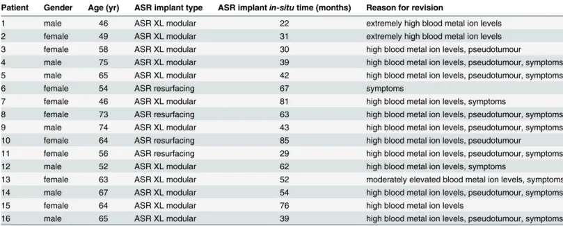
Table 1. Patient Characteristics.
Patient Gender Age (yr) ASR implant type ASR implantin-situtime (months) Reason for revision
1 male 46 ASR XL modular 22 extremely high blood metal ion levels
2 female 49 ASR XL modular 31 extremely high blood metal ion levels
3 female 58 ASR XL modular 30 high blood metal ion levels, pseudotumour
4 male 75 ASR XL modular 39 high blood metal ion levels, pseudotumour, symptoms
5 male 65 ASR XL modular 42 high blood metal ion levels, pseudotumour, symptoms
6 female 54 ASR resurfacing 67 symptoms
7 female 46 ASR XL modular 81 high blood metal ion levels, symptoms
8 female 73 ASR resurfacing 63 high blood metal ion levels, pseudotumour, symptoms
9 male 74 ASR XL modular 43 high blood metal ion levels, pseudotumour, symptoms
10 female 64 ASR resurfacing 85 high blood metal ion levels, pseudotumour
11 female 56 ASR resurfacing 29 high blood metal ion levels, pseudotumour, symptoms
12 male 52 ASR XL modular 62 high blood metal ion levels, symptoms
13 female 63 ASR XL modular 52 moderately elevated blood metal ion levels, symptoms
14 male 67 ASR XL modular 54 high blood metal ion levels, pseudotumour, symptoms
15 female 64 ASR XL modular 76 high blood metal ion levels
16 male 65 ASR XL modular 39 high blood metal ion levels, pseudotumour, symptoms
Grade (Roche, Mannheim, Germany) and 0.04/0.32 U/ml of Collagenase/Dispase (Roche, Mannheim, Germany) in RPMI 1640 (Lonza Group Ltd, Basel, Switzerland) supplemented
with penicillin (100 U/ml), streptomycin (100μg/ml) and amphotericin B (250 ng/ml) (all
from Invitrogen/Life Technologies, Carlsbad, CA, USA) at 37°C for 2 hours. After the diges-tion, the cell suspension was passaged through a cell strainer, washed three times with cold phosphate buffered saline (PBS) containing 5% of heat-inactivated foetal bovine serum (FBS) (Lonza Group Ltd, Basel, Switzerland) and resuspended in PBS supplemented with 5% of FBS for flow cytometry analysis, or in RPMI 1640 supplemented with 10% of FBS and antibiotics (see above) for cell culturing.
Cell Characterization by Flow Cytometry
1 x 10⁶freshly isolated pseudotumor tissue -derived cells were first incubated with purified
human Fc Receptor Binding Inhibitor (eBiosciences, San Diego, CA, USA) for 5 minutes and then stained with a combination of antibodies against the pan-leukocyte marker CD45 (labelled with fluorescein isothiocyanate, FITC; Becton, Dickinson and Company, Franklin Lakes, NJ, USA), macrophage marker CD14 (labelled with phycoerythrin Cy7, PE-Cy7; eBios-ciences), T-lymphocyte marker CD3 (labelled with allophycocyanin Cy7, APC-Cy7; Becton, Dickinson and Company), B-lymphocyte marker CD19 (labelled with allophycocyanin, APC; eBiosciences) and pan-granulocyte marker CD15 (labelled with phycoerythrin, PE; Becton, Dickinson and Company) for 20 minutes. After the incubation, the cells were washed twice with PBS supplemented with 1% of FBS, fixed with 1% of paraformaldehyde for 10 minutes and again washed twice with PBS/1% FBS. The samples were examined by FACSCanto II flow cytometry (Becton, Dickinson and Company) and the data was analyzed using FlowJo software (Tree Star, Inc, Ashland, OR, USA). In the preliminary experiments both anti-CD14 (see above) and anti-CD68 (labelled with fluorescein isothiocyanate, FITC; eBiosciences) were used as macrophage markers; as the results were similar anti-CD14 was chosen for the further experiments.
Cell Culture
Pseudotumor tissue -derived cells were cultured (in two duplicates per patient) in the density
of 5 x 10⁵cells/ml in 6-well plates in RPMI 1640 supplemented with 10% of FBS and antibiotics
(see above) at 37°C in humidified 5% CO2atmosphere. After 42 hours of incubation, the
cul-ture media were collected, the samples were centrifuged at 400 g for 10 minutes and the
super-natants were stored at -20°C until assayed for IL-6 and TNFα.
Enzyme-Linked Immunosorbent Assay (ELISA)
The concentrations of IL-6 and TNFαin the culture media were determined by ELISA using
reagents from R&D Systems Europe, Abingdon, UK and the assays were run in duplicate.
Metal Ion Concentrations
Whole blood samples were collected using twenty-one-gauge needle connected to a Vacutainer TM system (Becton, Dickinson and Company) and trace element blood tubes containing sodium ethylenediaminetetraacetic acid. In the Finnish Institute for Occupational Health (FIOH), standard operating procedures were established for Co and Cr measurement using dynamic reaction cell inductively coupled plasma (quadripole) mass spectrometry (Agilent 7500 cx, Agilent Technologies, Santa Clara, CA, USA). The FIOH laboratory is accredited and
Statistics
Results of normally distributed data are expressed as mean ± standard error of mean (SEM) and non-normally distributed data as median ± quartile range and range of values as indicated. Statistical significance of the differences between groups was calculated either by unpaired t-test (with Welch correction when needed) or by Mann-Whitney t-test where appropriate.
Differ-ences were considered significant at=p<0.05,=p<0.01, and= p<0.001.
Results
The Distribution of the Inflammatory Cells were Polarized either to
Macrophages or T-Lymphocytes
Inflammatory cells present in the pseudotumour tissue were characterized by flow cytometry. CD45 antigen was used as a general hematopoietic cell marker. CD14 (confirmed with similar results with CD68) was used as a marker of macrophages and CD3 and CD19 antigens were used to identify T-lymphocytes and B-lymphocytes, respectively. CD15 was used as a pan-granulocyte marker.
Macrophages and T-lymphocytes were the most dominant subsets of CD45+cells (Table 2).
lymphocytes were less frequent than T-lymphocytes, but in four cases the proportion of B-lymphocytes was more than 10% of all leukocytes, 19.2% being the highest proportion (Table 2). The proportion of granulocytes was generally low (median 1.78%) and only in one case it exceeded 5% (Table 2).
Interestingly, most of the samples were clearly dividable into two groups according to their cell distributions (Fig 1). Seven of the 16 samples displayed macrophage-dominated cell distri-bution and six of the 16 were T-lymphocyte-dominated. In the macrophage-dominated group,
Table 2. Inflammatory Cell Distributions and Whole Blood Metal Levels. Proportions of haematopoietic cells (%)
Patient Macro-phages T cells B cells granulo-cytes B-Cr (μg/l) B-Co (μg/l)
1 45.7 27.0 no data 3.03 28.8 64.9
2 51.2 20.7 no data 2.24 36.2 114.3
3 42.7 42.4 0.09 4.01 13.3 14.2
4 30.4 50.2 1.20 2.06 3.5 13.8
5 53.7 29.9 7.25 1.15 20.6 18.7
6 15.0 48.5 8.92 4.90 2.9 2.5
7 30.4 31.8 19.2 1.44 12.2 20.0
8 56.8 15.8 0.05 13.0 45.2 113.3
9 68.4 18.5 0.34 3.54 9.3 22.1
10 43.9 34.9 12.3 0.73 53.5 96.9
11 55.2 27.0 3.07 1.13 11.8 14.2
12 15.7 54.8 6.44 1.00 8.2 13.2
13 16.3 59.3 0.73 1.17 1.3 5.9
14 25.8 54.2 11.5 1.50 2.7 10.7
15 29.4 31.8 0.72 2.15 21.6 36.3
16 18.1 55.1 12.3 0.98 3.3 10.5
B-Cr = whole blood chromium; B-Co = whole blood cobalt CD45+cells were de
fined as hematopoietic cells, CD45+CD14+as macrophages, CD45+CD3+as T-lymphocytes (T cells), CD45+CD19+as B-lymphocytes (B cells) and CD45+CD15+as granulocytes.
the average (mean ± SEM) proportion of macrophages was 53.5 ± 3.0% and that of T-lympho-cytes 24.8 ± 2.5%. While the corresponding values were 20.2 ± 2.5% (macrophages) and 53.6 ± 1.5% (T-lymphocytes) in the T-lymphocyte-dominated group. There were no overlap-ping cases in the proportions of either macrophages or T-lymphocytes between the two groups. The remaining three samples were classified as mixed reaction as they had about equal percent-age of CD14 and CD3 positive cells. The three groups had comparable percentpercent-ages of B-lym-phocytes and granulocytes (Fig 1).
IL-6 and TNF
α
Production was Elevated in Macrophage-Dominated
Response
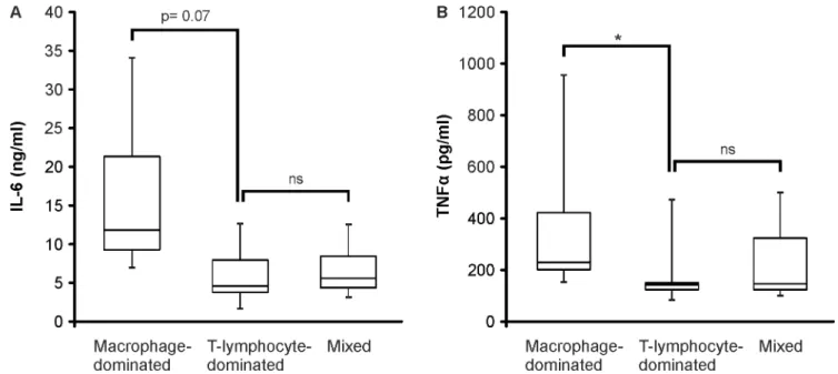
Next, we analyzed the IL-6 and TNFαproduction of the cells derived from pseudotumor tissue.
The cells were cultured for 42 hours and the levels of IL-6 and TNFαwere measured in the
cul-ture medium by ELISA. TNFαproduction was higher in the macrophage-dominated group
than in the T-lymphocyte-dominated group (p<0.05) and IL-6 production showed a similar
trend (p = 0.07) as seen inFig 2.
Blood Metal Ion Concentrations were Related to the Inflammatory Cell
Distribution in the Pseudotumor Tissue
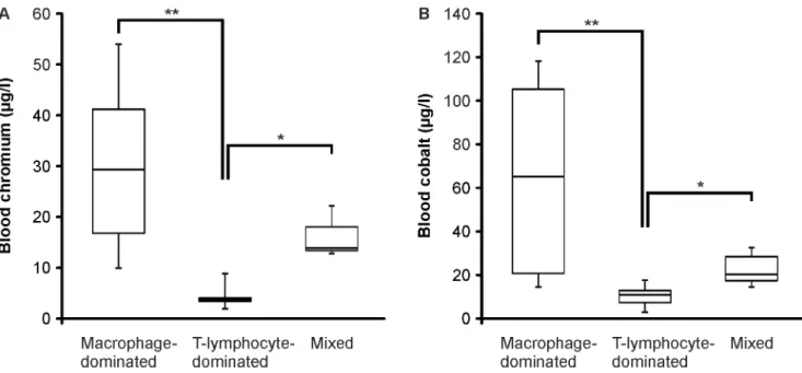
All of the patients had increased blood metal ion levels with a significant variation between the patients. The metal ion levels of either blood chromium or cobalt exceeded the action level of
Fig 1. Flow Cytometry Analysis of CD45+Cells Derived from Pseudotumor Tissue.CD45+cells were gated and proportions of CD14+, CD3+, CD15+and CD19+cells were analyzed. The cases were divided into three groups according to CD14+and CD3+cell proportions, namely macrophage-dominated (B), T-lymphocyte-dominated (C) and mixed (D) groups. Average values (mean + SEM) of the proportions of different cell types in the three different phenotypes of inflammation (A) with representative flow cytometry blots (B-D) are shown. n = 7 in macrophage-dominated group, n = 6 in T-lymphocyte-dominated group and n = 3 in mixed group.***p<0.001
doi:10.1371/journal.pone.0155121.g001
Fig 2. The Production of IL-6 and TNFαby Cells Derived from Pseudotumor Tissue.Samples of pseudotumor tissue were degraded by enzyme digestion and cell suspensions were cultured for 42 hours. Thereafter IL-6 (A) and TNFα(B) accumulated into the culture medium were
measured by ELISA. Boxplots represent medians and interquartile ranges and whiskers indicate range of values. n = 7 in macrophage-dominated group, n = 5 in T-lymphocyte-macrophage-dominated group and n = 3 in mixed group.*p<0.05.
7 ppb proposed in the Medicines and Healthcare Products Regulatory Agency (MHRA, UK) guidelines [31] in 14 out of 16 patients. The median (range) of blood chromium concentration
was 12.0 (1.3–53.5)μg/l and the median of blood cobalt level was 16.5 (2.5–114.3)μg/l
(Table 2).
When the levels of blood chromium and cobalt in patients with macrophage or T-lympho-cyte -dominated inflammatory phenotype were compared, a remarkable difference between the groups was found. Both the chromium and cobalt levels were significantly higher in patients with macrophage-dominated inflammation than in the patients with
T-lymphocyte-dominated response with the median levels being 28.8μg/l (Cr) and 64.9μg/l (Co) in the
mac-rophage-dominated group and 3.1μg/l (Cr) and 10.6μg/l (Co) in the
T-lymphocyte-domi-nated group (Fig 3). The small group of patients with similar macrophage and T-lymphocyte
abundance had median blood chromium level of 13.3μg/l and cobalt level of 20.0μg/l.
Discussion
The adverse tissue reactions related to MOM implants are presently a major topic in hip arthroplasty debates, but the understanding on the detailed cellular mechanisms eventually resulting in the development of the reaction remains limited and various hypotheses have been
presented [7,20,23–27]. The present study shows that the inflammatory activation related to
failed ASR implants is heterogeneous and can be characterized either as s macrophage or as a T-lymphocyte dominated reaction. Interestingly, the two types of responses differed also according to the blood metal ion concentrations in a manner that the macrophage-dominated phenotype was associated with higher blood levels of chromium and cobalt.
The histology of ARMD has been described in a number of publications, e.g. [7,20–23,27].
The present study extends the previous data by utilizing flow cytometry analysis in the charac-terization of cell types in the pseudotumor tissue of revised MOM hips. The pseudotumour tis-sue was degraded by enzyme digestion and the resulting cell suspension was analyzed by flow
Fig 3. The Blood Chromium and Cobalt Levels.Whole blood samples were taken preoperatively from the patients and chromium (A) and cobalt (B) concentrations were analyzed by inductively coupled plasma mass spectrometry. Boxplots represent medians and interquartile ranges and whiskers indicate range of values. n = 7 in macrophage-dominated group, n = 5 in T-lymphocyte-dominated group and n = 3 in mixed group.*p<0.05,**p<0.01
cytometry. Therefore, results on the cell typing are more representative for the entire pseudotu-mor tissue reaction than the usual histology which is limited to the sections investigated. Fur-ther, the flow cytometric data can be analyzed in a more quantitative manner.
Our results show that the leukocyte populations in the ARMD are dominated either by mac-rophages or by T-lymphocytes but the percentage of the polymorphonuclear leukocytes (i.e. CD15 positive cells including both neutrophils and eosinophils) remained rather low, the median being less than 2% of all leukocytes. Eosinophils have sometimes been noted in the histological studies [7,9] but in the present study we did not specify the granulocyte subtypes. However, the low percentage of granulocytes suggests that neither direct eosinophil mediated allergic response nor neutrophil mediated innate immune response plays any major role in the pathogenesis of the ARMD in most patients. Further, infection was ruled out with multiple bacterial cultures of sam-ples collected during the revision surgery. Intriguingly, also the percentage of B-lymphocytes was lower than that of T-lymphocytes in all patients supporting a major role of the latter in the lym-phocyte mediated responses. However, in four patients the proportion of B-lymlym-phocytes exceeded 10% of all leukocytes. Based on a recent study [27], also B-lymphocytes may have a sig-nificant role in the pathogenesis of ARMD, at least in some patients as discussed below.
The present results show that macrophage-dominated ARMD was typical for those patients who had higher blood metal ion concentrations. This suggests that the metal debris induces cytotoxicity or other phenomena that attract macrophages to the pseudotumor tissue. Cobalt-chromium alloys are especially hard, but some amounts of Cobalt-chromium and cobalt particles or soluble metal are released from hip devices by the wear or corrosion of the implant [32]. This is
reflected as increased chromium and cobalt concentrations in the circulation [28–30,33,34] as
also detected in the present study. In contrast to the micrometer-sized wear debris particles produced by polyethylene bearings, the wear particles of MOM articulations have been found to be primarily less than 50 nm in size [35,36]. The physiochemical and biological corrosion also results in the release of soluble metal ions, from the larger particles and from the devices, and the blood metal concentrations depict the concentrations of soluble metal ions surround-ing the MOM device [37].
Activated macrophages phagocytize wear particles, which in the case of ARMD is evidenced by metal particles within the cells [23,26,32]. Cobalt and chromium ions can also be trans-ported into different cell types via non-specific anion carriers. Inside the cells, both cobalt and chromium can induce the formation of reactive oxygen species, DNA and chromosome
dam-age, and cytotoxicity. [32,38]In vitrostudies show that cobalt is more toxic than chromium:
cobalt nanoparticles have been reported to induce direct cell death at concentrations of 1 x 10¹²
particles/ml and cobalt ions at concentrations of 1000μM [39]. Based on electron microscopic
examinations, cobalt cytotoxicity is associated with damage in mitochondria and cellular mem-branes as well as disruption of cellular metabolism [39,40].
Furthermore, cobalt is known to induce cellular changes mimicking hypoxia, mainly
through enhancing the levels of transcription factor hypoxia inducible factor 1 alpha (HIF-1α)
[41,42]. Therefore it is likely that cobalt ions released from MOM devices induce inflammatory reactions and cytotoxicity typical for hypoxia. In support, it was recently reported based on
immunohistochemical staining that HIF-1αlevels were increased in the peri-implant tissue of
failed MOM implants as compared to that of failed MOP implants [43]. Further, enhanced
HIF-1αexpression was associated with increased production of reactive oxygen species and the
inflammatory cytokine TNFα. Cobalt ions may also have a direct stimulatory effect on
The data described above give a potential explanation to our present finding that high blood cobalt and chromium concentrations are associated with macrophage-dominated inflamma-tory response in the pseudotumor tissue around failed MOM hips. Taken together, we hypoth-esize that high blood metal levels reflect high peri-implant tissue levels of nanometer-sized metal particles and metal ions, especially cobalt, which cause tissue injury and cell necrosis around MOM hips. The damaged tissue recruits macrophages, which are the professional cells to phagocytize necrotic cells and clean the tissue debris. This assumption is supported by the histological finding that macrophages in the pseudotumor tissue contain small droplike inclu-sions which resemble phagocytized organic material but not implant-derived wear debris [7]. In addition, the macrophages infiltrated into the peri-implant tissue are further activated by the metal nanoparticles and soluble metal ions released locally from the MOM devices. That further magnifies the inflammatory reaction in the pseudotumor tissue, e.g. through increased
production of proinflammatory cytokines IL-6 and TNFαas shown in the present study. This
sequence of events and the cytotoxic mechanisms of the metal debris and released metal ions described above are proposed to form the basis of the pathogenesis of the macrophage-domi-nated type of ARMD found in the present study.
A perivascular lymphocyte reaction is another characteristic feature in ARMD response [7,20,23]. Our results suggest that the response is T-cell-dominated; and in about half of the cases the T-lymphocyte accumulation is a predominant feature over the macrophage infiltra-tion. According to the hypothesis of delayed hypersensitivity reaction, the metal ions released from the implant can form complexes with tissue proteins and the aberrant metal-protein com-plexes may be recognized by the immune system as foreign antigens and result in the activation of the adaptive immune system. Lymphocyte infiltration into pseudotumor tissue supports this hypothesis, but the results of hypersensitivity skin-tests performed to the patients with failure
of MOM implant have been contradictory [19,24,45–47]. However, it is challenging to establish
the connection between peri-implant tissue reaction and hypersensitivity measured by skin tests since the immunological reactions evoked by transient cutaneous exposure to a pre-deter-mined allergen may differ from that induced by the constant exposure to an orthopaedic implant which may give rise to development of uncharacterized metal-modified endogenous molecules [24,48]. Other methods to measure hypersensitivity, like lymphocyte transformation test or migration test have still been only in a minor use [24]. In our study, when an inflamma-tory pseudotumor reaction developed in patients who had only slightly increased whole blood metal concentrations, the response showed T-lymphocyte-dominated phenotype. That points primarily to metal-induced hypersensitivity reaction as the pathogenesis of the inflammatory reaction around the hip arthroplasties rather than to metal-induced cytotoxicity.
In histological studies of ARMD, lymphocyte aggregates containing B-lymphocytes in addi-tion to T-cells have been detected in a subpopulaaddi-tion of patients [7,27]; and this is supported by the present finding that a quarter of our patients had moderate levels of B-lymphocytes. Fur-thermore, the T and B-lymphocyte containing lymphoid aggregates were reported to display features typical for tertiary lymphoid organs [27]. This finding suggests that also autoimmunity to metal-carrier complexes may be involved in the pathogenesis of lymphocyte-dominated ARMD [23,27].
susceptible individuals can develop delayed hypersensitivity reaction to altered metal-conju-gated host proteins formed in the presence of MOM-released ions or nanoparticles. Also, auto-immune mechanisms may contribute to the lymphocyte-mediated response. However, the proposed hypothesis on the dominant role of the metal concentrations in the inflammatory phenotype of ARMD needs to be further investigated since on the basis of our results, it is not possible to conclude if these two inflammatory conditions are distinct from each other or if they are interactive or consecutive processes. Also, the number of patients in our study was rel-atively small.
In conclusion, the present results show that there are distinct inflammatory phenotypes characterizing the adverse reaction to metal debris in patients with a failed ASR metal-on-metal hip which may reflect different disease mechanisms. The macrophage-dominated response was associated with higher blood metal ion concentrations and cytokines typical for innate immune response. This is proposed to depict metal-induced cytotoxicity resulting in massive macrophage infiltration and macrophage-mediated clearance of the necrotic tissue. While ARMD reaction associated with lower metal concentrations was characterized by T-lymphocyte-dominated tissue response applicable to hypersensitivity response.
Acknowledgments
Ms. Meiju Kukkonen, Ms. Sanna Hämäläinen, Mrs. Mirva Järvelä-Stolting, Mrs. Salla Hieta-kangas, Mr. Jan Koski and Ms. Ella Lehto are acknowledged for their excellent technical assis-tance and Mrs. Heli Määttä for her skilful secretarial help.
Author Contributions
Conceived and designed the experiments: EP RK MH MP AE TM EM. Performed the experi-ments: EP MH MP. Analyzed the data: EP MH MP EM. Contributed reagents/materials/analy-sis tools: EP MH MP AE TM EM. Wrote the paper: EP RK AE TM EM.
References
1. Schmalzried TP, Huk OL. Patient factors and wear in total hip arthroplasty. Clin Orthop Relat Res. 2004; 418: 94–97. PMID:15043099
2. Eskelinen A, Remes V, Helenius I, Pulkkinen P, Nevalainen J, Paavolainen P. Total hip arthroplasty for primary osteoarthrosis in younger patients in the Finnish arthroplasty register. 4,661 primary replace-ments followed for 0–22 years. Acta Orthop. 2005; 76: 28–41. PMID:15788305
3. Santavirta S, Bohler M, Harris WH, Konttinen YT, Lappalainen R, Muratoglu O, et al. Alternative materi-als to improve total hip replacement tribology. Acta Orthop Scand. 2003; 74: 380–388. PMID:
14521286
4. Daniel J, Pynsent PB, McMinn DJ. Metal-on-metal resurfacing of the hip in patients under the age of 55 years with osteoarthritis. J Bone Joint Surg Br. 2004; 86: 177–184. PMID:15046429
5. Migaud H, Jobin A, Chantelot C, Giraud F, Laffargue P, Duquennoy A. Cementless metal-on-metal hip arthroplasty in patients less than 50 years of age: comparison with a matched control group using ceramic-on-polyethylene after a minimum 5-year follow-up. J Arthroplasty. 2004; 19(Suppl 3): 23–28. 6. Bozic KJ, Kurtz S, Lau E, Ong K, Chiu V, Vail TP, et al. The epidemiology of bearing surface usage in
total hip arthroplasty in the United States. J Bone Joint Surg Am. 2009; 91: 1614–1620. doi:10.2106/
JBJS.H.01220PMID:19571083
7. Willert HG, Buchhorn GH, Fayyazi A, Flury R, Windler M, Koster G, et al. Metal-on-metal bearings and hypersensitivity in patients with artificial hip joints. A clinical and histomorphological study. J Bone Joint Surg Am. 2005; 87: 28–36.
9. Pandit H, Glyn-Jones S, McLardy-Smith P, Gundle R, Whitwell D, Gibbons CL, et al. Pseudotumours associated with metal-on-metal hip resurfacings. J Bone Joint Surg Br. 2008; 90: 847–851. doi:10.
1302/0301-620X.90B7.20213PMID:18591590
10. Langton DJ, Jameson SS, Joyce TJ, Hallab NJ, Natu S, Nargol AV. Early failure of metal-on-metal bearings in hip resurfacing and large-diameter total hip replacement: A consequence of excess wear. J Bone Joint Surg Br. 2010; 92: 38–46. doi:10.1302/0301-620X.92B1.22770PMID:20044676 11. Williams S, Leslie I, Isaac G, Jin Z, Ingham E, Fisher J. Tribology and wear of metal-on-metal hip
pros-theses: influence of cup angle and head position. J Bone Joint Surg Am. 2008; 90(Suppl 3): 111–117.
doi:10.2106/JBJS.H.00485PMID:18676945
12. Langton DJ, Joyce TJ, Jameson SS, Lord J, Van Orsouw M, Holland JP, et al. Adverse reaction to metal debris following hip resurfacing: the influence of component type, orientation and volumetric wear. J Bone Joint Surg Br. 2011; 93: 164–171. doi:10.1302/0301-620X.93B2.25099PMID:
21282753
13. Fisher J. Bioengineering reasons for the failure of metal-on-metal hip prostheses: an engineer's per-spective. J Bone Joint Surg Br. 2011; 93: 1001–1004. doi:10.1302/0301-620X.93B8.26936PMID:
21768619
14. Langton DJ, Jameson SS, Joyce TJ, Gandhi JN, Sidaginamale R, Mereddy P, et al. Accelerating failure rate of the ASR total hip replacement. J Bone Joint Surg Br. 2011; 93: 1011–1016. doi:
10.1302/0301-620X.93B8.26040PMID:21768621
15. Australian Orthopaedic Association. Annual Reports 2008 and 2009. Available at:https://aoanjrr.dmac. adelaide.edu.au/annual-reports-2014.
16. Seppänen M, Mäkelä K, Virolainen P, Remes V, Pulkkinen P, Eskelinen A. Hip resurfacing arthroplasty: short-term survivorship of 4,401 hips from the Finnish Arthroplasty Register. Acta Orthop. 2012; 83: 207–213. doi:10.3109/17453674.2012.693016PMID:22616745
17. Medicines and healthcare products regulatory agency. Medical device alert: all metal-on-metal (MOM) hip replacements (MDA/2010/033). Available at:http://www.mhra.gov.uk/publications/safetywarnings/ medicaldevicealerts/CON79157.
18. National Joint Registry for England and Wales. 8th Annual Report. Available at:http://www.njrcentre. org.uk.
19. Jones DA, Lucas HK, O'Driscoll M, Price CH, Wibberley B. Cobalt toxicity after McKee hip arthroplasty. J Bone Joint Surg Br. 1975; 57: 289–296. PMID:1158940
20. Campbell P, Ebramzadeh E, Nelson S, Takamura K, De Smet K, Amstutz HC. Histological features of pseudotumor-like tissues from metal-on-metal hips. Clin Orthop Relat Res. 2010; 468: 2321–2327.
doi:10.1007/s11999-010-1372-yPMID:20458645
21. Natu S, Sidaginamale RP, Gandhi J, Langton DJ, Nargol AV. Adverse reactions to metal debris: histo-pathological features of periprosthetic soft tissue reactions seen in association with failed metal on metal hip arthroplasties. J Clin Pathol. 2012; 65: 409–418. doi:10.1136/jclinpath-2011-200398PMID:
22422805
22. Grammatopoulos G, Pandit H, Kamali A, Maggiani F, Glyn-Jones S, Gill HS, et al. The correlation of wear with histological features after failed hip resurfacing arthroplasty. J Bone Joint Surg Am. 2013; 95: e81. doi:10.2106/JBJS.L.00775PMID:23783212
23. Revell PA. Biological causes of prosthetic joint failure. In: Revell PA, editor. Joint replacement technol-ogy. Elsevier; 2014. pp. 298–369.
24. Cousen PJ, Gawkrodger DJ. Metal allergy and second-generation metal-on-metal arthroplasties. Con-tact Dermatitis. 2012; 66: 55–62. doi:10.1111/j.1600-0536.2011.01970.xPMID:21957973
25. Cobelli N, Scharf B, Crisi GM, Hardin J, Santambrogio L. Mediators of the inflammatory response to joint replacement devices. Nat Rev Rheumatol. 2011; 7: 600–608. doi:10.1038/nrrheum.2011.128
PMID:21894210
26. Daniel J, Holland J, Quigley L, Sprague S, Bhandari M. Pseudotumors associated with total hip arthro-plasty. J Bone Joint Surg Am. 2012; 94: 86–93. doi:10.2106/JBJS.J.01612PMID:22218386 27. Mittal S, Revell M, Barone F, Hardie DL, Matharu GS, Davenport AJ, et al. Lymphoid aggregates that
resemble tertiary lymphoid organs define a specific pathological subset in metal-on-metal hip replace-ments. PLoS One. 2013; 8: e63470. doi:10.1371/journal.pone.0063470PMID:23723985
28. Reito A, Puolakka T, Elo P, Pajamäki J, Eskelinen A. High prevalence of adverse reactions to metal debris in small-headed ASR hips. Clin Orthop Relat Res. 2013; 471: 2954–2961. doi:
10.1007/s11999-013-3023-6PMID:23637059
29. Reito A, Moilanen T, Puolakka T, Pajamäki J, Eskelinen A. Repeated metal ion measurements in patients with high risk metal-on-metal hip replacement. Int Orthop. 2014; 38: 1353–1361. doi:10.1007/
30. Hart AJ, Sabah SA, Sampson B, Skinner JA, Powell JJ, Palla L, et al. Surveillance of Patients with Metal-on-Metal Hip Resurfacing and Total Hip Prostheses: A Prospective Cohort Study to Investigate the Relationship Between Blood Metal Ion Levels and Implant Failure. J Bone Joint Surg Am. 2014; 96: 1091–1099. PMID:24990974
31. Medicines and healthcare products regulatory agency. Medical device alert: All metal-on-metal (MoM) hip replacements (MDA/2012/036). Available at:http://www.mhra.gov.uk/publications/safetywarnings/ medicaldevicealerts/CON79157.
32. Keegan GM, Learmonth ID, Case CP. Orthopaedic metals and their potential toxicity in the arthroplasty patient: A review of current knowledge and future strategies. J Bone Joint Surg Br. 2007; 89: 567–573.
PMID:17540737
33. Coleman RF, Herrington J, Scales JT. Concentration of wear products in hair, blood, and urine after total hip replacement. Br Med J. 1973; 1: 527–529. PMID:4692678
34. Savarino L, Granchi D, Ciapetti G, Cenni E, Nardi Pantoli A, Rotini R, et al. Ion release in patients with metal-on-metal hip bearings in total joint replacement: a comparison with metal-on-polyethylene bear-ings. J Biomed Mater Res. 2002; 63: 467–474. PMID:12209889
35. Firkins PJ, Tipper JL, Saadatzadeh MR, Ingham E, Stone MH, Farrar R, et al. Quantitative analysis of wear and wear debris from metal-on-metal hip prostheses tested in a physiological hip joint simulator. Biomed Mater Eng. 2001; 11: 143–157. PMID:11352113
36. Brown C, Williams S, Tipper JL, Fisher J, Ingham E. Characterisation of wear particles produced by metal on metal and ceramic on metal hip prostheses under standard and microseparation simulation. J Mater Sci Mater Med. 2007; 18: 819–827. PMID:17171457
37. Cadosch D, Chan E, Gautschi OP, Filgueira L. Metal is not inert: role of metal ions released by biocorro-sion in aseptic loosening—current concepts. J Biomed Mater Res A. 2009; 91: 1252–1262. doi:10.
1002/jbm.a.32625PMID:19839047
38. Valko M, Rhodes CJ, Moncol J, Izakovic M, Mazur M. Free radicals, metals and antioxidants in oxida-tive stress-induced cancer. Chem Biol Interact. 2006; 160: 1–40. PMID:16430879
39. Gill HS, Grammatopoulos G, Adshead S, Tsialogiannis E, Tsiridis E. Molecular and immune toxicity of CoCr nanoparticles in MoM hip arthroplasty. Trends Mol Med. 2012; 18: 145–155. doi:10.1016/j.
molmed.2011.12.002PMID:22245020
40. Papageorgiou I, Brown C, Schins R, Singh S, Newson R, Davis S, et al. The effect of nano- and micron-sized particles of cobalt-chromium alloy on human fibroblasts in vitro. Biomaterials. 2007; 28: 2946–
2958. PMID:17379299
41. Goldberg MA, Schneider TJ. Similarities between the oxygen-sensing mechanisms regulating the expression of vascular endothelial growth factor and erythropoietin. J Biol Chem. 1994; 269: 4355–
4359. PMID:8308005
42. Wang GL, Semenza GL. Purification and characterization of hypoxia-inducible factor 1. J Biol Chem. 1995; 270: 1230–1237. PMID:7836384
43. Samelko L, Caicedo MS, Lim SJ, Della-Valle C, Jacobs J, Hallab NJ. Cobalt-alloy implant debris induce HIF-1alpha hypoxia associated responses: a mechanism for metal-specific orthopedic implant failure. PLoS One. 2013; 8: e67127. doi:10.1371/journal.pone.0067127PMID:23840602
44. Tyson-Capper AJ, Lawrence H, Holland JP, Deehan DJ, Kirby JA. Metal-on-metal hips: cobalt can induce an endotoxin-like response. Ann Rheum Dis. 2013; 72: 460–461. doi:
10.1136/annrheumdis-2012-202468PMID:23076072
45. Evans EM, Freeman MA, Miller AJ, Vernon-Roberts B. Metal sensitivity as a cause of bone necrosis and loosening of the prosthesis in total joint replacement. J Bone Joint Surg Br. 1974; 56-B: 626–642.
PMID:4452710
46. Brown GC, Lockshin MD, Salvati EA, Bullough PG. Sensitivity to metal as a possible cause of sterile loosening after cobalt-chromium total hip-replacement arthroplasty. J Bone Joint Surg Am. 1977; 59: 164–168. PMID:845199
47. Thomas P, Braathen LR, Dorig M, Aubock J, Nestle F, Werfel T, et al. Increased metal allergy in patients with failed metal-on-metal hip arthroplasty and peri-implant T-lymphocytic inflammation. Allergy. 2009; 64: 1157–1165. doi:10.1111/j.1398-9995.2009.01966.xPMID:19220218