Diversity of Cultivated Fungi Associated with
Conventional and Transgenic Sugarcane and
the Interaction between Endophytic
Trichoderma virens
and the Host Plant
Aline Silva Romão-Dumaresq1, Manuella Nóbrega Dourado1,2, Léia Cecilia de Lima Fávaro1,3, Rodrigo Mendes1,4, Anderson Ferreira1,5, Welington Luiz Araújo1,2*
1Department of Genetics, Escola Superior de Agricultura“Luiz de Queiroz”(ESALQ), University of São Paulo, São Paulo, Brazil,2Laboratory of Molecular Biology and Microbial Ecology, Department of Microbiology, Institute of Biomedical Sciences, University of São Paulo, São Paulo, Brazil,3Brazilian Agricultural Research Corporation, Embrapa Agroenergy, Brasília, Distrito Federal, Brazil,4Brazilian Agricultural Research Corporation, Embrapa Environment, Jaguariuna, São Paulo, Brazil,5Brazilian Agricultural Research Corporation, Embrapa Agrosilvopastoral, Sinop, Mato Grosso, Brazil
Abstract
Plant-associated fungi are considered a vast source for biotechnological processes whose potential has been poorly explored. The interactions and diversity of sugarcane, one of the most important crops in Brazil, have been rarely studied, mainly concerning fungal commu-nities and their interactions with transgenic plants. Taking this into consideration, the pur-pose of this study was, based on culture dependent strategy, to determine the structure and diversity of the fungal community (root endophytes and rhizosphere) associated with two varieties of sugarcane, a non-genetically modified (SP80-1842) variety and its genetically modified counterpart (IMI-1, expressing imazapyr herbicide resistance). For this, the sugar-cane varieties were evaluated in three sampling times (3, 10 and 17 months after planting) under two crop management (weeding and herbicide treatments). In addition, a strain of Tri-choderma virens, an endophyte isolated from sugarcane with great potential as a biological control, growth promotion and enzyme production agent, was selected for the fungal-plant interaction assays. The results of the isolation, characterization and evaluation of fungal community changes showed that the sugarcane fungal community is composed of at least 35 different genera, mostly in the phylum Ascomycota. Many genera are observed at very low frequencies among a few most abundant genera, some of which were isolated from specific plant sites (e.g., the roots or the rhizosphere). An assessment of the possible effects upon the fungal community showed that the plant growth stage was the only factor that significantly affected the community’s structure. Moreover, if transgenic effects are present, they may be minor compared to other natural sources of variation. The results of interaction studies using the Green fluorescent protein (GFP)-expressingT.virensstrainT. v.223 revealed that this fungus did not promote any phenotypic changes in the host plant and was found mostly in the roots where it formed a dense mycelial cover and was able to a11111
OPEN ACCESS
Citation:Romão-Dumaresq AS, Dourado MN, Fávaro LCdL, Mendes R, Ferreira A, Araújo WL (2016) Diversity of Cultivated Fungi Associated with Conventional and Transgenic Sugarcane and the Interaction between EndophyticTrichoderma virens
and the Host Plant. PLoS ONE 11(7): e0158974. doi:10.1371/journal.pone.0158974
Editor:Krishnendu Acharya, University of Calcutta, INDIA
Received:March 29, 2016
Accepted:June 24, 2016
Published:July 14, 2016
Copyright:© 2016 Romão-Dumaresq et al. This is an open access article distributed under the terms of theCreative Commons Attribution License, which permits unrestricted use, distribution, and reproduction in any medium, provided the original author and source are credited.
Data Availability Statement:All relevant data are within the paper and its Supporting Information files.
Funding:This work was supported by FAPESP (http://www.fapesp.br/), grant number: 2002/14143-3 and 2003/13834-5.
penetrate the intercellular spaces of the root epidermis upper layers. The ability ofT.virens to colonize plant roots suggests a potential for protecting plant health, inhibiting pathogens or inducing systemic resistance.
Introduction
Sugarcane (Saccharumsp.) is an important crop in Brazil, mainly for the production of ethanol, a biofuel, as a renewable energy source that is already an alternative to petrol in Brazilian cars. To increase productivity and decrease costs, researchers are investing in new sugarcane varie-ties developed by classical breeding as well as genetically modified plants, searching for charac-teristics such as insect resistance, herbicide resistance, a higher sugar content, etc [1,2]. Therefore, studies on biosecurity and environmental impact should be performed before releasing the genetically modified plants for commercial production
The microbial community of the host plant can have a positive, negative or neutral effect on the plant, caused mainly by those microorganisms that live in intimate contact with the plant tissues, such as endophytes or rhizoplane microbes [3]. Studies reported the effects of the genetically modified plant on the microbial community associated [4–8], but these effects were minor in comparison with the effect caused by cultivar [5,9], soil type [10,11], crop location [12,13] and plant growth stage [5,14,15,16,17].
Microbial diversity studies can help elucidate the role of native microorganisms in the eco-system and the association of biological changes with microbial diversity [18,19]. A few studies have been performed with transgenic sugarcane, mainly involving beneficial fungi and plant interactions [20,21]. Fungi play a key role in several ecological processes essential for ecosystem maintenance [22]. Endophytic fungi colonize plant tissue internally without causing damage to the plant host [23], establishing a balanced and mutualistic interaction [24]. The fungus receives nutrients while the plant benefit from the inhibition of pathogens and stress resistance as well as increased growth [25].
On the other hand, the rhizosphere is influenced by root exudates and is intensely colonized by microbial communities [26], which have direct effects on plant growth and nutrient avail-ability or protecting against pathogens [27,28]. Several rhizosphere fungi degrade toxic com-pounds, such as xenobiotic and aromatic molecules, and are essential for the survival of plants in contaminated soils [29]. Other fungi, such asFusarium oxysporum, a nonpathogenic strain, can suppress pathogenic strains [30]. Many fungi, such as members of theTrichodermagenus, can inhibit phytopathogens and act as a biological control [31]. Among these,Trichoderma virenshas been reported to be aggressive mycoparasite [32,33], able to parasitize not only hyphae but also fungal resistance structures [32,33,34]. In addition to its mycoparasitic activ-ity, it can also produce extracellular chitinase [35] and several antibiotics and can induce the production of plant fitoalexins [36].T.virenshas been considered a versatile and effective bio-logical control agent. Moreover,T.virenshas been reported to be a plant endophyte [37], able to asymptomatically colonize the host plant and occupy the same niche as phytopathogens. In addition,Trichodermaspp. strains have been used to promote plant growth [31,38,39] and show a great potential for agricultural use.
stage) and crop management (weeding and herbicide application) on the structure and diver-sity of the cultivable fungus communities associated with sugarcane plants. Rhizosphere and root endophytic fungi were isolated from two sugarcane varieties, a non-transgenic (SP80-1842) and a genetically modified (IMI-1, expressing imazapyr herbicide resistance) variety, treated and untreated with the herbicide imazapyr, at three different growth stages over a two-year period.Trichoderma, a genus abundant in sugarcane, which has potential agricultural use, was selected for these plant interaction studies.
Material and Methods
The sugarcane orchard was not in a protected area. Sugarcane plants (Saccharum officinarum) were grown in the experimental area of the Centro de Tecnologia Canavieira S.A. (CTC), a pri-vate company that present the CQB (Certificate for Biosecurity Quality) for the cultivation area, in Piracicaba, São Paulo, Brazil (22° 41’S e 47° 33’O), under supervision of Dr. Eugenio Cesar Ulian a Director of the CTC company. Therefore, the only one permission that we need for this experiment permission was gave by the experimental Company Supervisor, Dr. Euge-nio Cesar Ulian."
Sugarcane experiments
Sugarcane plants (Saccharum officinarum) were grown in the experimental area of the Centro de Tecnologia Canavieira S.A. (CTC), Piracicaba, São Paulo, Brazil (22° 41’S e 47° 33’O) under supervision of Dr. Eugenio Cesar Ulian. We used the transgenic sugarcane IMI-1, which expresses resistance to the herbicide imazapyr, and the non-transgenic isoline cultivar SP80-1842. Three treatments were used in a randomized experimental design with four replicates: i) the non-transgenic sugarcane SP80-1842 + weeding (NW), ii) the transgenic sugarcane IMI-1 + weeding (TW) and iii) the transgenic sugarcane IMI-1 with imazapyr herbicide applied two months after planting (TH). In this experimental design the TW x TH comparison allowed evaluate the crop management (weeding x herbicide application) and NW x TW comparison allowed evaluate the plant genotype (non-transgenic X genetically modified variety). Samples were collected at 3, 10 and 17 months after planting (sampling times), and each sample was comprised by four replicates (with 3 plants each).
Isolation of sugarcane endophytic and rhizosphere fungi
Endophytic and rhizosphere fungi were isolated from sugarcane according to Stuart et al. [20]. For endophytic fungi isolation, roots were surface sterilized and for each three treatments (NW, TW and TH), the roots of three plants (from each replicate) were cut in 21 fragments (5 mm x 5mm each) and transferred onto Potato Dextrose Agar (PDA, Merck, Kenilworth, NJ, USA) with tetracycline (50μg mL-1) to inhibit bacterial growth. For rhizosphere fungi isola-tion, 5 g of rhizosphere soil was diluted in 50 mL of PBS buffer (NaCl 140 mM, KCl 3 mM, Na2HPO410 mM and KH2PO42 mM, pH = 7.4) and shaken with glass beads for 1 h at 150
rpm. The suspension was diluted and the 10−3and 10−1dilutions were plated on PDA with
DNA extraction and molecular identification of the endophytic and
rhizosphere fungi
DNA extraction of the isolated fungi was performed according to Raeder & Broda [40] with modifications. The fungi were grown in 50 mL of PDB (Potato Dextrose Broth) for 5 days at 28°C. The fungal suspension was filtered, and the mycelia were ground in liquid nitrogen. The grounded mycelia (200 mg) were transferred to 1 mL of extraction buffer (SDS 1%, EDTA 25 mM, NaCl 250 mM and Tris-HCl 200 mM [pH 8,0]), incubated at 65°C for 20 min and centri-fuged at 16,000 Xgfor 10 min at 4°C. The DNA was purified using phenol/chloroform and precipitated with isopropanol. After the DNA was extracted, the DNA was quantified and its integrity was assessed by electrophoresis in a 0.8% of agarose gel; the DNA was stored at -20°C.
The ITS1-5.8S-ITS2 region of rDNA was amplified using ITS-1 and ITS-4 primers [41]. After electrophoresis, the fragments were purified and sequenced at the Centro de Estudos do Genoma Humano (University of São Paulo, São Paulo, Brazil). To identify the fungal isolates, the sequences were compared to the GenBank database via BLASTn (http://www.ncbi.nlm.nih. gov/BLAST). Partial DNA sequences of the ITS1-5.8S-ITS2 region were deposited in the Gen-Bank database under the accession numbers GQ495269 and GU973612-GU973859.
Sequence analysis and phylogeny
All the chromatograms were first trimmed for high quality bases (80% of bases with
quality>20) by means of Phred software, and the trimmed sequences were used for
compari-son in the GenBank database (nr/nt). The best hits of well-characterized strains were retrieved from the databases and subsequently used for alignment and phylogeny analysis with MEGA 4.0 version software [42]. The evolutionary history was inferred through the Neighbor-Joining method [43] and evolutionary distances were computed using the Jukes-Cantor method.
Determination of endophytic and rhizosphere fungal community
structures
The diversity and richness of the fungal community in the three treatments (NW, TW and TH) for three isolation periods (3, 10 and 17 months) were quantified using the Shannon-Wienner (H’) and Chao1 index, respectively. These indices were calculated using the DOTUR program [44], which also generated rarefaction data used to infer coverage considering the dif-ferent similarity levels of the ITS1-5.8S-ITS2 sequence of fungus rDNA. The similarity matrix was generated using the DOTUR program, aligning the sequence and calculating the distance matrix using the Phylip DNADist program (http://bioweb.pasteur.fr/seqanal/interfaces/ dnadist-simple.html). The taxonomic levels were calculated with the following cutoffs: 100% (strains), 99% (species) and 97%-95% (genera). The similarity coefficient of Jaccard (J) was cal-culated, considering the generic level.
AMOVA was performed to estimate the genetic variation among the different fungal com-munities and to determinate how much of the genetic variation could be attributed to the dif-ferences among the sugarcane genotypes (conventional vs. transgenic), crop management (weeding vs. imazapyr), the sampling period (growth stages) and the rhizosphere and endo-phytic fungal community. All analyses of the DNA sequences from the fungal isolates were per-formed using the Arlequin program version 3.1 [45], considering 16.000 permutations.
Agrobacterium-mediated transformation
The isolateT.virensstrainT.v.223 (GenBank access number GQ495269) obtained from inner root tissues was selected to further studies. For this, transformation was carried out according to a previously described method [47] with modifications [21,48]. Briefly,A.tumefaciens
EHA105 transformed with the plasmid pFAT-gfp[49] was grown at 28°C for 16–20 h. The cul-ture suspension was diluted to aλ
660= 0.15 in an induction medium broth (10 mM K2HPO4;
10 mM KH2PO4; 2.5 mM NaCl; 2 mM MgSO4; 0.7 mM CaCl2; 9 mM FeSO4; 4 mM NH4SO4;
10 mM glucose; 0.5% glycerol; 40 mM 2-morpholinoethanesulfonic acid, pH 5.3) supple-mented with acetosyringone (200 mM) and incubated until aλ660= 0.6 was reached. The cell
suspension was mixed 1:1 with a conidial suspension (106or 107conidia. mL-1) fromT.virens
strainT.v.223. Aliquots were spread on different types of membranes (cellophane, filter paper, nitrocellulose, or nylon) on induction medium agar (5 mM glucose instead of 10 mM) supple-mented with 200 mM or 400 mM of acetosyringone. After co-cultivation at 25°C for 24 h or 48 h, the membranes were transferred onto PDA or M-100 [50] media amended with hygromycin B (200μg mL–1; to select transformants) and cefotaxime (200μg mL–1; to eliminate the bacteria).
Individual transformants were subsequently transferred to PDA supplemented with 200μg mL-1hygromycin. The mitotic stability of the integrated T-DNA was tested by sub culturing five-fold randomly selected transformants on PDA supplemented with 200μg mL-1of hygro-mycin incubated at 28°C for 5 days.
Molecular and morphological characterization of transformants
The genomic DNA of several selected transformants was extracted according to Raeder and Broda [40], and the presence of thegfpgene that express the green fluorescent protein was
veri-fied by PCR. We used the primersglGFP5 (5’-GCCGGAATTCATGAGCAAGGGCGAGGAACT
GTTC-3’) and glGFP3 (5’ -GCCGAGCTCAGATCTCACTTGTACAGCTCGTCCATGCC-3’)that amplified 700 pb ofgfpgene [49].
To check the number of inserts in the genome, four transformants (T2, T7, T10 and T20) and the wild type strain (used as a negative control) were analyzed by Southern blotting. The DNA (15μg) of each strain was cleaved with 10 UμL-1EcoRI (Invitrogen, Life Technologies) incubated at 37°C for 12 h [49]. The digested DNA was electrophoresed in a 1% agarose gel. The probe used in the Southern blot analysis, the 700 pb sequence ofgfpgene, was labeled using a Gene Images™AlkPhos Direct™Labelling and Detection System (GE Healthcare) kit according to the manufacturer’s protocol.
The gel was washed with a 0.25 M HCl solution for 10 min, followed by distilled water, a denaturing solution (NaOH 0.5 M, NaCl 1.5 M) for 30 minutes, distilled water, and two washes with a neutralization solution (Tris-HCl 0.5 M; NaCl 1.5 M; EDTA 0.001 M [pH = 7.2]). The DNA in the gel was transferred to a nylon membrane (Hybond N+, Amersham) for 12 h in transference solution SSC 20X (NaCl 3 M; sodium citrate 0.3 M [pH = 7.0]). The membrane was dried for 2 h at 80°C and stored at 4°C.
Morphological characterization was performed to compare the wild type and mutant strains on the basis of growth rate and morphology (sporulation rate and color). The strains were grown for 5 days in PDA at 28°C, and the colony diameter was measured after 24, 48, 72 and 96 h. All tests were performed in triplicate.
Trichoderma virens
–
host plant interaction
days to obtain conidia.In vitrosugarcane plants (variety SP80-1842) from Centro de Tecnolo-gia Canavieira (CTC, Piracicaba, SP, Brazil), were transferred to glass flasks containing 20 mL of MS liquid broth [51] with 106conidia mL-1of mutant T20. The plants were incubated at 28°C under a 16 h light and 8 h dark cycle. After 48 h, the plants were transferred to pots con-taining a commercial substrate PlantMax (Eucatex) and placed in a greenhouse to acclimatize. Non-inoculated sugarcane plants (variety SP80-1842) were similarly treated as a control. After 20, 40 and 60 days in the greenhouse, the sugarcane plants were sampled and leaves, stem and roots were surface-disinfected according to Stuart et al. [20]. After processing, the leaf and stem tissues were cut in fragments of approximately 100 mm2(10 mm x 10 mm) and roots were cut in 5 mm fragments. A total of 1512 plant fragments (504 fragment per sampling time) were transferred to PDA (Merck) with tetracycline (100μg mL-1) to inhibit bacterial growth. Hygromycin B (200μg mL-1) was also added to the culture medium for the T20 mutant. The isolation plates were incubated at 28°C and periodically evaluated up to 14 days. After growth, the total fungal colonies andTrichodermacolonies were counted, andT.virensstrain T20 were confirmed bygfpfluorescence and hygromycin B resistance.
Microscopic analysis ofTrichoderma virensand sugarcane interaction. Axenic sugar-cane plants (variety SP80-1842) were inoculated withT.virens strainT20conidia (with the
gfpgene) and incubated at 28°C under a 16 h light and 8 h dark cycle. After 24, 48, 72 and 168 h, the roots were processed according to Ferreira et al. [52] and observed under an optical microscope (Zeiss Axiophot-2) using two filters, FITC (459–490 nm) and Rhodamine (546 nm), to differentiate sugarcane fluorescence from GFP fungus fluorescence. Images were cap-tured separately with each filter and then overlapped using the ISIS software (Meta Systems, Germany).
The same roots were collected after 12, 24, 48 and 72 h, and fixed with Karnowsky solution and processed according to Rossetto et al. [53]. In addition to the colonization of the root sur-face, the inner root tissues were visualized via a freeze-fracture technique by fixing the sample with Karnowsky solution and washing with 0.05 M cacodylate buffer. The roots were treated with 30% glycerol for 2 h, wrapped in tissue paper and frozen in liquid nitrogen. The roots were fractured and dehydrated in ethanol, subjected to critical point drying and coated with metal. The samples were observed in a scanning electron microscope (LEO-Zeiss) at the Núcleo de Apoio à Pesquisa em Microscopia Eletrônica (NAP/MEPA), University of São Paulo.
Trichoderma virens–plant interaction assessment. The effect of theT.virenswild type
strain (T.v.223) and a strain containinggfp(T20)on sugarcane (variety SP80-1842) growth were measured. The plants were inoculated with 106conidia mL-1of the wild type strain (T.
v.223) or thegfp-tagged strain (T20)in a commercial PlantMax substrate (Eucatex). Non-inoc-ulated plants were used as a control and were maintained in a greenhouse for two months, after which the plant fresh and dry mass were determined. A total of 23 plants per treatment were used in a completely randomized experimental design. To confirm fungal colonization,T.
virenswas reisolated from the inoculated plants as previously described.
Statistical analysis
Results
Isolation of fungi associated with sugarcane
To analyze the potential effects of genetically modified sugarcane on fungal frequency (Ef) (in root endophyte) and density (Df) (in rhizosphere), we isolated fungi from the rhizosphere and endorhizosphere of genetically modified plants that expressed an imazapyr resistance gene (IMI-1) and the conventional isoline (SP80-1842) for three different treatments (NW, TW and TH) at three growth stages of sugarcane plants (3, 10 and 17 months).
The experiment evaluated the effects of plant genotype, crop management and growth stage on the fungal community associated with sugarcane. Sugarcane roots had a high number of endophytic fungi, and from 756 root fragments (252 fragment per sampling time), 2,236 fungal colonies we isolated, being 781 from 3month-old plants, 659 from 10 month old plants and 796 from 17-month-old plants (Fig 1). Similarly, the plant rhizosphere was also found to be highly colonized by fungi, being isolated 1,224 fungi colonies from this plant site. The number
Fig 1. Filamentous fungal isolation of root entophytes frequency (a) and rhizosphere density (b).The bars indicate the average of each effect in each variation factor: treatments (NW-non-transgenic plants with weeding, TW- genetically modified plants with weeding and TH-genetically modified plants treated with herbicide); sampling time (3, 10 and 17 months after planting). Different letters represent difference at 5% probability in the Tukey test. Comparison among sampling time (3, 10 and 17 months) we used letter a and b and comparison of treatments (NW, TW, TH) we used letters x and y.
of rhizosphere fungi ranged from 4.25 105CFU g-1of soil at 17 month-old plants to 8.20 105 CFU g-1of soil at 10 month-old plants (Fig 1). The ANOVA for the different treatments (NW, TW and TH) and sampling time (3, 10 and 17 months) was performed on the fungus fre-quency (endophytic communities) and density (rhizosphere communities) data (S1 Table).
The ANOVA results showed that regardless the fungal community evaluated (root endo-phyte or rhizosphere), the genetically modified sugarcane did not induce a significant change (P>0.05) in the fungal frequency or density (S1 Table), suggesting that neither the evaluated
plant genotype nor crop management had a significant effect on the sugarcane fungal commu-nity. The frequency of root endophytic fungi was significantly (P = 0.0256) affected by sam-pling time (S1 Table), being the frequency 17 month-old>10-month-old>3-month-old
sugarcane plants (Fig 1). Curiously, such growth effects were not observed in the density of the rhizosphere community (P>0.05) (S1 Table). Additionally, the ANOVA revealed the presence
of an interaction (P = 0.0305) between treatment and sampling time for endophytic fungi, so the effect of the different treatments on the fungal frequency could depended on the plant growth stage.
Composition and distribution of the fungal community associated with
sugarcane
Due to the large number of isolates obtained, the fungi were grouped according to morphologi-cal characteristics (type of mycelium, color, presence of spores), and representative samples were selected for subsequent analysis. The morphological diversity of fungus associated with sugarcane displayed a great variation in mycelial color, including white, beige, yellow, green, pink, lilac, brown, gray and black (S1 Fig). In addition to the variety of colors, the fungus also differed in mycelia growth habits (cottony, submerged aerial hyphae), the presence of repro-ductive structures (spores), the growth rate and the secretion of compounds that changed the color of the growth medium.
Therefore, a total of 249 fungi comprising 16 morphotypes (S1 Fig) from all three treat-ments were selected for molecular identification by sequencing of the ITS1-5.8S-ITS2 region of rDNA. The sequences obtained were used to evaluate the effects of genotype, management and growth stage using a diversity and richness index. The fungal isolates were taxonomically iden-tified at the genus level via BLASTn by comparison with the ITS1-5.8S-ITS2 rDNA sequences available in GenBank and confirmed by morphological characteristics.
Analysis of the ITS1-5.8S-ITS2 sequences showed that the fungal community associated with sugarcane comprised at least 35 different genera (S2 Table). Overall, the phylum Ascomy-cote predominated among the fungi identified (96.0%), whereas the phyla Zygomycota/Mucor-omycotina and Basidiomycota represented only 2.4% and 1.6% of the community, respectively. In the phylum Ascomycote, Eurotiomycetes (43.0%), the classes Sordariomycetes (38.5%) and Dothydeomycetes (7.6%) were predominant, and the most frequent genera werePenicillium
(33.3%),Fusarium(16.9%),Aspergillus(7.2%) andTrichoderma(4.4%) (S2 Table).
Other genera were reported at a low frequency (<2.8%), and 16 (Acremonium,Alternaria, Cladophialophora,Colletotrichum,Curvularia,Exophiala,Mariannaea,Myrmecridium, Myr-othecium,Paecilomyces,Paraphaeosphaeria,Phoma,Phomopsis,Pyricularia,Saccharicolaand
Sagenomella) were represented by only one isolate.
The genusPenicillium(teleomorphsEupenicilliumorTalaromyces), represented 33.3% of the fungi isolated from both the roots and the rhizosphere. A phylogenetic analysis indicated the similarity of 17 different species (P.restrictum,E.katangense,P.janthinellum,T.flavus,E.
marneffei), and fourteen isolates were classified asT.trachyspermus; three isolates were classi-fied asP.pinophilumand two isolates were classified asE.javanicum(S2A Fig).
The genusFusarium(teleomorphGibberella) was the second most frequent genus and included 16.9% of the isolates, and five different species were highly similar (F.oxysporum,F.
acutatum,F.solani,F.dlaminiiandG.moniliformis). In this group, 19 isolates were classified asF.oxysporum, and the remaining 23 isolates were classified only at the genus level (S2B Fig).
The third most frequent genus, with 7.2% of the isolates, wasAspergillus, which had 7 differ-ent species that were highly similar (A.brasiliensis,A.flavus,A.oryzae,A.versicolor,A.niger,
A.terreusandA.fumigatus). The phylogenetic analysis showed that six isolates were classified asA.brasiliensis;three isolates were identified asA.terreusand one isolate was identified asA.
fumigatus(S2C Fig).
The generaTrichodermaandEpicoccumoccurred less frequently than other genera. Tricho-derma(teleomorphHypocrea), the fourth most frequent genus, was represented by 11 isolates corresponding to 4.4% of fungus community. The phylogenetic analysis allowed the classifica-tion of two isolates asH.virens, three isolates asT.asperellumand six isolates asTrichoderma
sp. (S2D Fig). The genusEpicoccumwas the fifth most abundant genus and represented 3.6% of the total isolates.
In addition to the most frequent genera (Penicillium,Fusarium,Aspergillus,Trichoderma
andEpicoccum), the fungal community associated with sugarcane included 28 other genera in the phylum Ascomycota, the genusResinicium(with 3 isolates) in the phylum Basidiomycota and the genusCunninghamella(with 3 isolates) in the phylum Zigomycota/Mucoromycotina.
In the phylum Ascomycota, the class Sordariomycetes comprised a large number of the iso-lates (96), with at least 17 genera identified (Bionectria,Chaetomium,Chaetosphaeria, Colleto-trichum,Diaporthe,Fusarium,Mariannaea,Microdochium,Myrmecridium,Myrothecium,
Nigrospora,Paecilomyces,Phomopsis,Pyricularia,Thielavia,ThozetellaandTrichoderma). Moreover, nine isolates were not similar to any reported genus, so they were classified asnot identified(N.I.) (S2 Table).
In the phylum Ascomycota, besides the class Sordariomycetes, the fungal community asso-ciated with sugarcane was characterized by isolates from the classes Dothideomycetes, Eurotio-mycetes, Leotiomycetos and mitosporic fungi. The class Dothideomycetes comprised 19 isolates, classified in six different genera (Alternaria,Cladosporium,Curvularia,Epicoccum,
ParaphaeosphaeriaandSaccharicola). The Eurotiomycetos class comprised 107 isolates; how-ever, 94.4% were classified in the generaAspergillusandPenicillium, and the other 6 isolates were identified asCladophialophora,Exophiala, andSagenomella. The class Leotiomycetes had only two representatives, both in the genusAcephala.
Sugarcane fungal community structure
Rarefaction curves were constructed to verify whether the sampling of the present work was adequate to assess the population diversity. Overall, the rarefaction curves of different popula-tions using similarity levels of 95% and 97% were sufficient to encompass the diversity of fungal population (S3 Fig). Therefore, richness and diversity comparisons based on 95% and 97% sim-ilarities were performed.
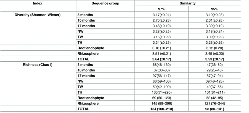
The diversity (Shannon-Wienner) and richness (Chao1) index for the fungal population associated with sugarcane are presented inTable 1. The results show that the fungal commu-nity associated with sugarcane has great richness and diversity; the total diversity and richness indices (H’(95%)= 3.53;H’(97%)= 3.64;Chao1(95%)= 98;Chao1(97%)= 134) are considered high
Moreover, the richness and diversity of the fungal community associated with sugarcane changed according to the sampling time; 17-month-old sugarcane plants had the highest diver-sity and richness indices. Plants 17 and 10 months old had significantly different diverdiver-sity and richness indices (Table 1). In addition, impact of plant genotype (NW x TW) and crop man-agement (TW x TH) on fungal richness and diversity was not observed.
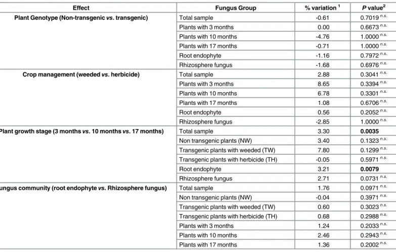
The AMOVA allowed an assessment with a defined statistical probability of how much of the variation in the fungal community associated with sugarcane is due to genetically modified sugarcane planting (genotype effects), weed management (management effects), sampling time or isolation site (root endophyte or rhizosphere). Analysis of the results showed that plant genotype, crop management and the fungal community did not have significant effects on fun-gal community (P>0.05). However, the plant growth stage caused significant variation,
con-tributing to 3.3% of the total variation (P = 0.0035) (Table 2). This variation was mainly related to the root endophytic community (3.21%, P = 0.0079); other groups did not contribute signifi-cant variation (P>0.05) (Table 2). These data suggest that plant age affects the endophytic root
community but not the rhizosphere community.
Correlation between genera and plant niche (root
vs. rhizosphere)
The taxonomic similarities between fungi isolated from the roots (endophytes) and the rhizo-sphere were assessed by the Jaccard coefficient of similarity (J), considering the genera that occurred in each community. The value obtained (J = 0.343) indicated that these communities should have a greater number of genera that occurred infrequently than common genera.
The proportion of rhizosphere and root endophytes in the community profile can be observed inFig 2. The figure shows, for example, that 8 genera (Acephala,Alternaria,Myrothecium,
Nigrospora,Phomopsis,Saccharicola,ThielaviaandThozetella) were observed only in roots and that 15 genera (Acremonium,Cladophialophora,Cladosporium,Colletotrichum, Cunningha-mella,Curvularia,Exophiala,Mariannaea,Myrmecridium,Paecilomyces,Paraphaeosphaeria,
Table 1. Diversity and Richness index of all sequence groups at 97% and 95% similarity.
Index Sequence group Similarity
97% 95%
Diversity (Shannon-Wiener) 3 months 3.17(±0.24) 3.10(±0.23)
10 months 2.75(±0.28) 2.61(±0.28)
17 months 3.48(±0.19) 3.39(±0.19)
NW 3.28(±0.25) 3.18(±0.24)
TW 3.16(±0.22) 3.09(±0.22)
TH 3.34(±0.25) 3.28(±0.26)
Root endophyte 3.16 (±0.21) 3.12 (0.20)
Rhizosphere 3.51 (±0.21) 3.45 (±0.20)
TOTAL 3.64 (±0.17) 3.53 (±0.17)
Richness (Chao1) 3 months 68(46–130) 47(36–80)
10 months 37(30–63) 29(25–46)
17 months 97(56–147) 57(47–94)
NW 88(59–166) 69(48–126)
TW 59(42–109) 49(37–86)
TH 133(74–295) 101(61–211)
Root endophyte 69 (50–123) 52 (42–85)
Rhizosphere 145 (88–296) 121 (76–244)
TOTAL 134 (100–210) 98 (80–141)
Phoma,Pyricularia,SagenomellaandScolecobasidium) were observed only in the rhizosphere. Moreover, some of the most frequent genera (such asAspergillus,Penicillium,Fusariumand Tri-choderma) were predominant in the roots or the rhizosphere. The genusAspergilluswas observed at a frequency two-fold higher in the rhizosphere than in the roots; on the other hand, 80% of the isolates identified asFusariumwere root endophytes (Fig 2), although this genus was also isolated from rhizosphere.
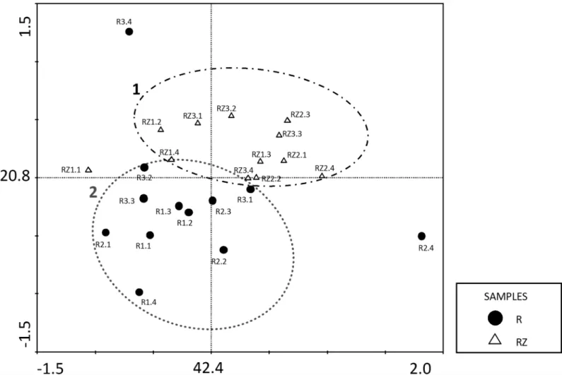
A principal component analysis (PCA) confirmed that the fungal communities in the roots and the rhizosphere were different (Fig 3). Although some fungi genera were shared by both inner roots and rhizosphere, the two principal components, which explain 63.2% of the vari-ance, clustered the samples in two groups, being the first composed by rhizosphere community (RZ); and the second by endophytic community (R). These results indicated that the isolation site determined the variation of the frequency and the occurrence of such genera in the isolated fungal community.
Genetic transformation of
T.
virens
mediated by
A.
tumefaciens
The number of transformants that grew on selective medium was counted every 5 days for 30 days. The tests indicated that the optimal conditions for the transformation ofT.virensfungus
Table 2. Analysis of molecular variance (AMOVA) for different fungus populations according to the evaluated effect.
Effect Fungus Group % variation1 Pvalue2
Plant Genotype (Non-transgenicvs. transgenic) Total sample -0.61 0.7019n.s.
Plants with 3 months 0.00 0.6673n.s.
Plants with 10 months -4.76 1.0000n.s.
Plants with 17 months -0.71 1.0000n.s.
Root endophyte -1.16 0.7972n.s.
Rhizosphere fungus -1.68 0.6976n.s.
Crop management (weededvs. herbicide) Total sample 2.88 0.3041n.s.
Plants with 3 months 8.65 0.3394n.s.
Plants with 10 months 6.78 0.3301n.s.
Plants with 17 months 1.08 0.6706n.s.
Root endophyte 0.56 0.2052n.s.
Rhizosphere fungus -2.85 1.0000n.s.
Plant growth stage (3 monthsvs. 10 monthsvs. 17 months) Total sample 3.30 0.0035
Non transgenic plants (NW) 3.40 0.1323n.s. Transgenic plants with weeded (TW) 7.80 0.1299n.s. Transgenic plants with herbicide (TH) -0.05 0.5971n.s.
Root endophyte 3.21 0.0079
Rhizosphere fungus 2.71 0.0731n.s.
Fungus community (root endophytevs. Rhizosphere fungus) Total sample 1.76 0.0971n.s. Non transgenic plants (NW) -0.04 0.3971n.s. Transgenic plants with weeded (TW) 0.60 0.3023n.s. Transgenic plants with herbicide (TH) 0.68 0.2988n.s.
Plants with 3 months 1.24 0.2033n.s.
Plants with 10 months 2.46 0.2943n.s.
Plants with 17 months 1.36 0.2002n.s.
1Percentage of variation between analyses groups; 2P
–probability of a variation component higher than the observed for thefixation index;
n.s.
–non significantly values at the level of 5% probability.
withAgrobacteriumwere the use of filter paper, inoculated with 107conidia mL-1, in PDA medium and co-cultivated for 48 h. Other conditions, such as two selection conditions were tested (normal–no addition of culture media to the membrane after the transfer to selective medium andoverlay–the addition of culture media to the membrane) as well as two concentra-tions (200 and 400μM) of the bacterial virulence inductor acetosyringone (AS).
The results showed that transformants were obtained under all tested conditions with simi-lar transformation efficiencies. Overall, the average number of transformants obtained was 12 transformants per 107conidia. mL-1, and we obtainedT.virenstransformants with the chosen transformation conditions.
Analysis of transformants
Mitotic stability. The mitotic stability of 20 random transformants was evaluated after 5 successive generations in non-selective medium. Of the transformants tested, 70% maintained hygromycin B resistance and green fluorescence emission (observed under FITC filter–459 to 490 nm). Moreover, we did not observe sector emission in the evaluated transformants.
Molecular analysis. Eight transformants (T2, T4, T7, T9, T10, T15, T20, T22) resistant to hygromycin B were selected to confirm the presence ofgfpgene by PCR using the glGFP5 and glGFP3 primers. All transformants tested and the plasmid pFAT-gfp(positive control) led to the amplification of a 700 pb fragment. The wild type strainT.v.223 was used as a negative con-trol and did not allow amplification of this fragment.
Four transformants (T2, T7, T10 and T20) were randomly picked for Southern blot analysis from the eight transformants obtained to confirm the integration of the T-DNA genome and determine the number of copies inserted using a probe containing thegfpsequence (700 pb) (S4 Fig). The results confirmed that T-DNA was integrated into the genomes of allT.virens
transformants and indicated the random occurrence of this event because different band sizes indicated different integration events. Transformants T20 and T2 had only one copy inserted,
Fig 2. Percentage of the occurrence of each genus observed in the sugarcane fungal community and their relative proportions according to isolation location (rhizosphere or roots).
whereas the remaining transformants had more than one copy of T-DNA integrated into the genome. The wild type strainT.v.223 used as a negative control and the pFAT-gfpvector used as a positive control showed no bands and a 15 kb band, respectively.
Comparison of the transformants and the wild typeT.virens. The mitotically stable T20 transformant that had a single copy inserted was selected for further analysis. The morphologi-cal and physiologimorphologi-cal similarities with the wild type strainT.v.223were evaluated. After 5 days of growth in PDA at 28°C, the morphological characteristics of both isolates were similar. Both showed weak and diffuse growth, with cottony mycelia and green conidia. The growth rates were similar; both grew approximately 2.7 cm per day in a Petri dish with PDA medium. More-over, theT.v. T20 transformant and the wild typeT.v.223 had similar conidial production (1.31 x 10⁷conidia cm-2and 1.71 x 107cm-2, respectively).
Interaction of
T.
virens
with the host plant
Reisolation ofT.virensstrain T20 from sugarcane plants. The results show that strain T20 endophytically colonized plant roots and stems but scarcely colonized plant aerial tissues. Root and stem colonization by T20 occurred at a low frequency (Fig 4) and did not lead to
Fig 3. Principal Component Analysis (PCA) of the occurrence of the different fungus genera in the root endophytic community (●R) and the fungal rhizosphere community (ΔRZ).The values on both axes indicate the percentage of variance explained by sample distribution on each respective axis.
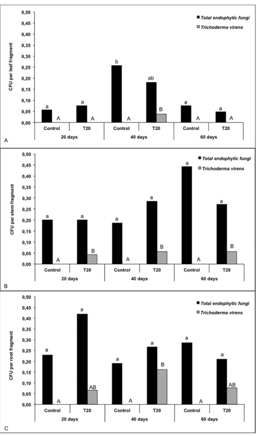
Fig 4. Isolation frequency of total endophytic fungi andT.virensstrain T20 from leaves (a), stems (b) roots (c) of sugarcane plants at 20, 40 and 60 days.The same letter indicates that treatments are not significantly different according to Tukey’s test at 5% significance.
phenotypic changes in the plants. Moreover, the presence of T20 did not significantly affect the total frequency of endophytic fungi on the various plant tissues (Fig 4). Although this fungus is naturally found in sugarcane, in the present experimentT.virenscould not be isolated from control (uninoculated) plants under any of the conditions tested (Fig 4); it was found only in inoculated plants.
The statistical analysis of the fungal reisolation frequency from sugarcane leaves showed that“total endophytic fungi”did not differ significantly between the inoculated and non-inocu-lated plants (S3 Table). However, a significant difference (P = 0.0418) was observed for the iso-lation of endophytic fungi in sugarcane plants at 20, 40 and 60 days; after forty days, plants showed a higher frequency of endophytic fungi.
A statistically significant difference for“Total Trichoderma virens”isolation was observed between the inoculated and uninoculated plants (P = 0.0415) and among the different plant ages (P = 0.0198), and their interaction was also significant (P = 0.0198). These differences are shown inFig 4A, which shows that T20 was presented in inoculated plants only after 40 days. Similar results were observed in the stems and the roots. For both roots and shoots the“Total endophytic fungus”were not significantly different for either inoculated or non-inoculated plants and different isolation data. The variable“Total Trichoderma virens”showed significant differences between inoculated and non-inoculated plants, with a probability of P = 0.0222 for stems and P = 0.0005 for roots, indicating that that the frequency ofT.virensreisolated from the inoculated plants was significantly higher than for the control plants becauseT.virens
could not be isolated from the control plants (Fig 4B and 4C). Moreover, the reisolation fre-quency was not significantly different for different plant ages.
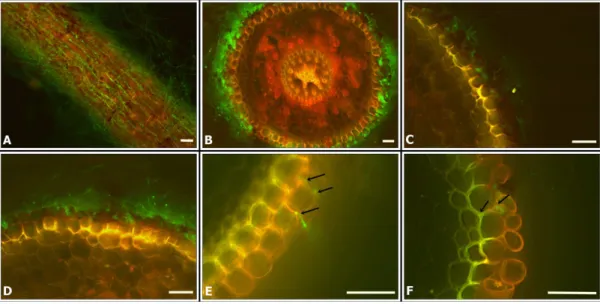
Monitoring of fungal colonization by fluorescence microscopy (FM) and scanning elec-tronic microscopy (SEM). The superficial and internal colonization of sugarcane root tissue byT.virensfungus were observed by optical fluorescence microscopy (FM) and scanning elec-tronic microscopy (SEM). The transformant T20 was selected for the microscopic analysis because it had a similar morphology, growth rate and sporulation as the wild type. Root tissue had an intense auto fluorescence, so two different filters were used: FITC (459–490 nm) and Rhodamine (546 nm). The fungus fluoresces only under the FITC filter, so root tissue could be distinguished from the fungus in an overlaid image. The conidia and hyphae of T20 transfor-mant emit an intense green fluorescence (Fig 5), whereas the mycelium of the wild type did not. We were able to visualize dense mycelia in sugarcane roots inoculated withT.v. T20 (Fig 5A, 5B, 5C, 5D and 5F).
Conidial production was also observed in samples collected after 4 days. In addition to root surface colonization, in some cross sections, we were able to observe intercellular colonization of the outer root layers (Fig 5).
Sugarcane root tissue infected withT.virenswere also analyzed by SEM, which revealed interesting aspects of fungal behavior during plant colonization (Fig 6). First, we were able to observe the conidial germination on the root tissue surface and the elongation of hyphae adhered to the cuticle (Fig 6C). Moreover, the hyphae showed preferential growth toward the cellular junctions (Fig 6C and 6E). We could not see any appressorium or alteration of the plant surface in the presence of the fungus. Seven days after conidial inoculation, we were able to visualize dense colonization on the root surface, hyphae ramification, conidiospore forma-tion and conidial producforma-tion (Fig 6A, 6D and 6E). Observation of roots fractured with liquid nitrogen revealed the presence of fungal hyphae in the root tissue, in the intercellular space (Fig 6F).
no significant differences were observed in shoots, roots or the entire plant in any treatment (Table 3). These results show that neither wild type nor transformantT.virensdid not affect the growth of sugarcane under the conditions tested. Moreover, genetic alterations present in strain T20 apparently did not interfere with its interaction with sugarcane compared to the wild type strain.
Fig 5. Optical fluorescence microscopy (OFM) with a FITC filter (459–490 nm), Rhodamine filter (546 nm) and an overlapped image with both filters, showing sugarcane root colonization (variety SP80-1842) by T20. (A)
Sugarcane root with T20;(B)Cross section of sugarcane root with T20(C, D)A longitudinal section of sugarcane colonized by T20(E, F)Cross section of sugarcane root with T20; the arrows indicate possible fungal penetration in the outer layers of the root epidermis. Scale bars = 50μm.
doi:10.1371/journal.pone.0158974.g005
Fig 6. Scanning electronic microscopy images (SEM) showing sugarcane root colonization (variety SP80-1842) byT.virensstrain T20.(A, B) Root surface colonization by T20; (C)T.virensconidia germination adhered to the plant host cuticle; (D) Asexual reproduction structure (conidiophore and conidia) ofT.virensfungi; (E) Mycelia growth toward the cellular junctions and (F) Inner root segment showing the presence of fungal hyphae in the intercellular space. Scale bars are indicated in the figure.
To confirm that the fungal inoculation of the soil was sufficient to promote sugarcane endo-phytic colonization and that the effects observed occurred in the presence ofT.virens, some plants were sampled and endophytic fungi were isolated from sugarcane. Reisolation showed a low frequency of endophyticTrichodermain the leaves and stems, similar to the previous reiso-lation results.T.virenswas isolated only from the roots of inoculated plants, indicating that inoculation of the soil leads to the colonization of plant roots byT.virensbut does not translo-cate to the shoot nor affect the plant biomass.
Discussion
The indirect effect of genetically modified plants on ecosystem communities and functions are still being discussed [4,54]. There is great concern about potential effects on non-target organ-isms such as mycorrhizae, rhizobium and other microorganorgan-isms that protect plants, as well as on phosphate solubilizing, decomposition and growth promotion.
The transgenic sugarcane plants in this study that are resistant to imazapyr (IMI-1) carry a plasmid containing a mutated wheatAHASgene, which confers imazapyr resistance, that is under the control of a constitutive sugarcane promoter. The herbicide imazapyr inhibits the enzymeAHAS, which is responsible for the synthesis of some essential amino acids. This enzyme occurs in bacteria, fungi and plants, and has a similar amino acid sequence in these organisms, which suggests a common ancestor [55] and the potential that imazapyr could affect organisms other than weeds. Some studies have reported that nitrogen fertilizers affect the microbial community [56], as do imidazolinone-derived herbicides [20,57], and transgen-esis [20,58,59]; however, the results of this study indicated that neither the plant genotype (transgenic or non-transgenic) nor crop management (weeding or herbicide application) sig-nificantly changed the fungal community of cultivated sugarcane.
Interestingly, Stuart et al. [20] used the same field experiments as in the present study but evaluated only the first two growth stages (3 and 10 months) and observed an effect on the endophytic fungal community that colonized the leaves of the transgenic variety IMI-1 man-aged with imazapyr. They reported that the herbicide led to a rapid and transient alteration of the fungal community, whereas the transgenic variety displayed a slow and persistent change in the leaf fungal community [20]. Therefore, the effects of management and plant genotype seem to depend on the fungal community and probably on the plant growth stage.
However, similar to the present study, the lack of significant herbicide and/or genetically modified plants effects on the fungal community has been previously reported [5,16,60–62]. Indeed, the effect of genetic modification is low compared to that of other factors, such as the differences between plant varieties, which can be greater than those between genetically modi-fied plants and their non-transgenic counterparts [63–65]. Therefore, it is likely that these stud-ies did not detect any effect of plant genotype because other factors, such as the plant growth
Table 3. Effect of the inoculation of strainsT.v.223 andT.vT20 on the fresh and dry weight accumulation on root and shoot of sugarcane plants (variety SP80-1842) cultivated for 60 days in greenhouse.
Treatment Fresh mass (g)1 Dry mass (g)1
Root Shoot Total Root Shoot Total
SP80-1842 (control) 5.12a 2.90a 8.02a 0.51a 0.83a 1.34a
SP80-1842 +T.v.strain223 4.75a 2.53a 7.27a 0.43a 0.68a 1.12a
SP80-1842 +T.v. strain T20 4.66a 2.66a 7.33a 0.47a 0.69a 1.16a
1Values are the average of 23 repetitions;
Treatments with the same letters do not statistically differ in Tukey test, at 1% of significance.
stage or crop management also significantly affected the microbial community, not mentioning other environmental factors, masking the lesser changes resulting from genetically modified plants.
Differently from genotype and crop management effects, the plant sampling time (3, 10 and 17 months) significantly affected the sugarcane endophytic fungal community, resulting in changes in the fungal frequency and the diversity and richness indices. This effect was also observed on endophyte in leaves by Stuart et al. [20]. Actually, the plant growth stage was pre-viously reported to determine the microbial community structure, overcoming even the effects of genetically modified plants [11,15,66,67]. Curiously though, the fungal community of the rhizosphere did not exhibit obvious changes in the parameters evaluated (fungal density, diver-sity and richness indices and molecular variance). Although culture dependent method could mask the presence of some important fungi, if the plant genotype or crop management had sig-nificant effect on these parameters, shifts in the fungi community would be observed.
The identification of the fungal community that colonizes the sugarcane roots and rhizo-sphere allowed us to calculate the diversity and richness indices. The results revealed that root endophytic fungi are less diverse than rhizosphere fungi, which we expected in view of the enormous fungal density in soil and the rhizosphere effect. The rhizosphere effect was demon-strated by Gomes et al. [68], who compared the DGGE profiles of the rhizosphere and soil fun-gal communities of maize and showed a significant increase in the relative abundance of fungi in the rhizosphere. Although the endophytic fungal diversity was lower than in the rhizosphere, the diversity index obtained (H’= 3,12) is similar to those reported in studies that evaluated endophytes from other host plants, such asGuarea guidonia[69].
The fungal community are structured by the isolation site [70–72], and it is likely that the community differs due to the different micro-environments in each plant tissue, soil type and rhizosphere. In overall, it was observed that the cultivable fungal community associated with sugarcane comprised at least 35 different genera, distributed in the phyla Ascomycota (96.0%), Zygomycota/Mucoromycotina (2.4%) and Basidiomycota (1.6%). The predominance of culti-vable Ascomycota fungi associated with plants has been previously reported [73–77], whereas the lack of basidiomycetes is probably because methods that use plant fragments plated in growth broth do not favor their isolation [78]. In fact, several fungi cannot be retrieved with cultivable isolation methods, in this way the characterization of fungal communities by either dependent or independent cultivation methods can generate different taxonomic community profiles [58,79], but both method can support conclusion about the impact assessment.
In Ascomycota population, two classes (Eurotiomycetes, Sordariomycetes) represented approximately 80% of the fungal community, and among these, 65.4% of the isolates belongs toPenicillium,Fusarium,Aspergillus,TrichodermaandEpicoccumgenera, corroborating previ-ous studies which showed that the fungal community associated to the host plant is composed by a great number of species, but are dominated by few species [80–82]. The genusEpicoccum, the fifth most frequent associated with sugarcane, was previously reported as an endophyte in banana [83] and apple trees [84], in the wheat rhizosphere [85] and in sugarcane leaves [86]. The generaPenicillium,Fusarium,AspergillusandTrichodermahave been isolated both as endophytes [87–89] and from the rhizosphere of different host plants [90,91] and are usually the most frequent genera.
The data collected in this study together with the literature on the sugarcane fungal commu-nity revealed the presence of some ubiquitous fungal genera associated with the culture of this plant. For example, common genera in the rhizosphere fungal community wereAcremonium,
Aspergillus,Cladosporium,Chaetomium,Cunninghamella,Curvularia,Fusarium,Penicillium,
sugarcane fungal community could not be assigned using ITS sequencing to any known fungal genera opens the possibility of the discovery of novel fungal species because the great diversity of higher plants has long been considered the greatest reservoir of new fungi [93].
The generaPenicillium(teleomorphTalaromycesorEupenicillium) andAspergillusare ascomycota of Trichocomaceae family. Several species of this family have medical, industrial and biotechnological importance [94]. These genera can contain plant pathogens, generally not of economic importance but also harbour endophytes in several host plants [72,87,88,89]. In this study, at least three differentAspergillus(A.brasiliensis,A.fumigatusandA.terreus) and
Penicilliumspecies (P.pinophilum,E.javanicumandT.trachyspermus) were observed. Fusar-iumgenera (teleomorfeGibberella) have also been reported to be plant pathogens and to cause great economic losses. In sugarcane,F.verticillioides,F.moniformeandF.subglutinansspecies have been reported as the casual agents ofPokkah-boengandFusariumrot, which remain latent until stress conditions trigger the plant disease [95,96]. This genus has also been reported to contain endophytes, for example,F.mangiferaeandF.sterilihyphosumin mango [97],F.solaniin palm trees [98],F. cf.avenaceum,F.decemcellulareandF.solaniinManilkara bidentata[99] andF.graminearumin wheat [89]. Moreover, some endophytic strains ofF.
oxysporumhave great potential as biological control agents, for example, in cucumber against the pathogenPythium ultimum[100]. In this study, 42 isolates were classified asFusarium, andF.oxysporumwas the predominant species.
The genusTrichoderma(teleomorphHypocrea) is known for rapid growth, its ability to use different substrates and resistance to toxic compounds [101] showing a great plasticity which features could increase the fitness during plant interaction. These fungi colonize soil, roots and leaves, and have a ubiquitous distribution, predominating in soils in various climates [102,
103]. They have also been reported as endophytes in cacao [104,105],Azadirachta indica[88], banana [106] and fig trees [107]. Some species are of great economic importance due to their ability to produce enzymes and antibiotics, their potential as biological control agents and the capacity to induce plant growth [108,109]. Specifically, in sugarcane, cellulase production was tested inT.virideto deconstruct the lignocellulose biomass fromSaccharum spontaneumby fermentation [110] and the production of ethanol directly from sugarcane bagasse by a recom-binantT.reesei[111]. This species has been reported to be an aggressive mycoparasite, able to synthesize antibiotics, induce phytoalexin production and plant host resistance [36], as well as to produce endochitinases used for biocontrol [112] and, in some cases, to promote plant growth [113–114]. Because of these features,T.virens, one of the most abundant species that is not described as a toxigenic fungus, was chosen to be used in the studies to understand the interaction of this sugarcane plants and the fungi communities, allowing not only to study the impact of plant genotype, sampling time and crop management, but also how these fungi may interact with the host plant.
The first step to study theTrichoderma-sugarcane interaction involved the generation of hygromycin-resistant fungal mutants expressing GFP by AMT transformation which has been used for transformation of several fungi [21,47,48]. A high mitotic stability was observed in the mutants we generated, similar to reports of transformations usinggfpgene with the Agro-bacteriumsystem in other fungal species [21,49,115].
(this study). Likewise, preferential root colonization has been reported for otherTrichoderma
species [116,117]. In fact, different plant tissue preferences by an endophytic fungus have been shown to be related to specific conditions present in each plant organ [21,24,71]. Overall, sug-arcane colonization by theT.virensT20 mutant occurred at a low frequency and did not lead to plant phenotypic alterations or disease signs and did not affect the total endophytic fungal frequency. Given that endophytic colonization can be affected by the inoculum type and by the plant growth substrate [104], perhaps the low frequency of the T20 mutant may have resulted from the conditions used for the interaction study. Interestingly, in this pot interaction study, the fungal frequency varied according to plant age as previously observed in the field study, corroborating the fact that plant growth stage may significantly affect the fungal community. In addition, it was observed that the inserted genes (hphandgfp) were maintained in the T20 mutant even after colonizing plant tissues in the pot interaction experiment, confirming the high genetic stability of this strain.
Specific aspects of the colonization ofT.virensin sugarcane roots were explored by micros-copy, in which OFM and SEM were used to monitor fungal colonization of superficial and inner tissues during plant growth. The results showed that this fungus colonized the root sur-face, forming a dense mycelial mass. In addition, the observation of root transverse sections revealed thatT.virensalso entered the host plant tissues and colonized the intercellular space of the outer layers of the root epidermis. SomeTrichodermastrains have been reported to be able to colonize only specific parts of the roots [118], although rhizosphere strains are known to be able to colonize the entire root surfaces for several weeks [119] or months [120].
Generally, the penetration ofTrichodermaspp. into plant root tissue is limited to the outer cell layers [118,121] and does not lead to disease signs [116]. The first step of an endophytic colonization may occur when the fungus enters and forms specialized structures such as an apressorium, as observed inDiscula umbrinellaon the leaf surface [122] and inPiriformospora indicaon the root surface [123]. Curiously, in the present work, we did not observe apressor-ium formation or the presence of any other specialized structures, but growth proceeded toward the cell junction, suggesting thatT.virensmay penetrate plant root tissues by other mechanisms. Endophytic fungi, for instance, can enter the host plant through natural openings such as stomata or between epidermal cells through the cuticle via the action of lytic enzymes, as has been reported for the fungusBeauveria bassiana[124] andE.nigrum[21], and which could also occur withT.virens.
In addition to being largely known as biological control agents,Trichodermaspecies are also known for other indirect effects in plants [116], including plant growth promotion and raising productivity [120,125]. In this regard, the effect of theT.virensfungus in the host plant has been broadly studied in cotton [36], but to date, no such results have been reported for sugar-cane. This study also evaluated the effects ofT.virenson sugarcane growth and the dry mass of the shoots and the roots. The results showed that the presence of this fungus on the roots did not affect the phenotypical characteristics, plant growth or dry mass accumulation. In addition,
T.virensT20 imposed similar effects on sugarcane plants compared to the wild type strain. Transgenic strains do not always maintain similar characteristics in their interactions with the plant host, and changes of the fungal life style (from mutualistic to parasitic) during the inter-action with the plant have been reported for the endophytic fungusEpichloë festucaedue to a mutation in one gene (noxA) [126].
interaction between genetically modified sugarcane and the root endophytic and rhizospheric fungi communities. In addition, a large number of fungal species was isolated, generating a rich strains collection, which could be used in future studies that seeking to explore fungus-plant interactions and for the evaluation of potential fungal isolates that have biotechnological and agronomic potential.
Supporting Information
S1 Fig. Morphological diversity observed in fungal community associated to sugarcane. Fungal colonies grown in PDA broth at 28°C for 5–10 days. (a)Epicoccumsp., (b)Penicillium
sp., (c)Chaetomiumsp., (d)Fusariumsp., (e)Trichoderma virens, (f) Not identified root endo-phyte fungi, (g)Eupenicillium javanicum, (h)Acremoniumsp., (i)Talaromyces trachyspermus, (j)Aspergillus niger, (k)Penicilliumsp., (l)Epicoccum nigrum, (m)Mariannaeasp., (n) Fusar-iumsp., (o)Bionectriasp., (p)Myrmecridium schulzeri.
(DOCX)
S2 Fig. Phylogenetic tree built byNeighbor-joiningmethod using Jukes and Cantor model for ITS1-5,8S-ITS2 sequence isolated from sugarcane root and rhizosphere.Reference sequences of GenBank were used and the sequences obtained in the present work were signaled with the following symbol♦.Bootstrapvalues (n = 1000) lower than 50 are represented in nods. a) 70 fungi ofPenicilliumgenera, using the ascomycotaBuergenerula spartinaeas out-group. b) 39 fungi ofFusariumgenera, using the ascomycotaBionectria ochroleucaas out-group. c) 17 fungi ofAspergillusgenera, using the ascomycotaPenicillium pinophilumas outgroup. d) 11 fungi ofTrichodermagenera, using the ascomycotaHypomyces aurantiusas outgroup.
(DOCX)
S3 Fig. Rarefaction analysis showing the number of expected phylotypes associated to sug-arcane fungal community, for the different levels of similarity (100%, 99%, 97% and 95%) of ITS1-5,8S-ITS2 sequence.(a) Comparison between the expected number of phylotype for endophytic and root fungal population; (b) Number of phylotype expected for all fungal com-munity associated to sugarcane (root endophyte + rhizosphere fungi).
(DOCX)
S4 Fig.Southern blothybridization of four randomly pickedT.virens gfp-tagged strains. Genomic DNA of the isolates were digested with restriction enzymeEcoRI, which cuts T-DNA twice and do not cutgfpgene sequence, a electrophoresis were performed in 0.8% agarose gel, transferring to nylon membrane and hybridized with the 700 pb fragment (gfpgene) labeled with digoxigenine. Column 1 positive control (pFAT-gfp plasmid); column 2 is the negative control (Wild strainT.v.223); columns 3–6 are the transformed strains (T20, T10, T7 and T2). Visualized bands are indicated with arrow.
(DOCX)
S1 Table. ANOVA for the treatments factors and growth period. (DOCX)
S2 Table. Taxonomic classification of the 249 evaluated fungus at genera level, distributed according to the isolation place (root or rhizosphere, plant growth (3, 10 or 17 months) and treatment (NW, TW, TH).
S3 Table. ANOVA for factors Strain (L) and isolation period (I) for both variable, total endophytic fungi and totalTrichoderma.Experimental design totally randomized. Analysis were performed separately for each part of the plant (leave, stem and root).
(DOCX)
Acknowledgments
The Centro de Tecnologia Canavieira (CTC), for hosting the crop experiment performed by Dr. Eugênio César Ulian. Dra. Sabrina M. Chabregas (Centro de Tecnologia Canavieira—CTC, Pir-acicaba, SP, Brazil) for the courtesy of thein vitrosugarcane plants (variety SP80-1842). We thank Dr. K. M. Plummer (La Trobe University, Australia) for providing the pFAT-gfpplasmid to Dra. Léia C. L. Fávaro. Núcleo de Apoio à Pesquisa em Microscopia Eletrônica (NAP/MEPA) and Laboratório de Citogenética Molecular de Plantas, coordinated by Prof. Margarida L.R.A. Perecin, both at ESALQ-USP (Piracicicaba, SP, Brazil) for the SEM and fluorescence optical microscopy images, respectively.
Author Contributions
Conceived and designed the experiments: ASR RM LCLF WLA. Performed the experiments: ASR LCLF AF. Analyzed the data: ASR MND. Contributed reagents/materials/analysis tools: ASR MND WLA. Wrote the paper: ASR MND WLA.
References
1. Dal-Bianco M, Carneiro MS, Hotta CT, Chapola RG, Hoffmann HP, Garcia AA, et al. Sugarcane improvement: how far can we go? Curr Opin Biotechnol. 2012; 23(2):265–70. doi:10.1016/j.copbio. 2011.09.002PMID:21983270
2. Wu H, Altpeter F. Sugarcane (Saccharumspp. hybrids). Methods Mol Biol. 2015; 1224: 307–316. doi:
10.1007/978-1-4939-1658-0_24PMID:25416267
3. Hardoim PR, van Overbeek LS, Berg G, Pirttilä AM, Compant S, Campisano A, et al. The Hidden World within Plants: Ecological and Evolutionary Considerations for Defining Functioning of Microbial Endophytes. Microbiol Mol Biol Rev. 2015; 79.
4. Singh AK, Singh M, Dubey SK. Changes in Actinomycetes community structure under the influence of Bt transgenic brinjal crop in a tropical agroecosystem. BMC Microbiol. 2013: 29; 13:122. doi:10. 1186/1471-2180-13-122PMID:23718216
5. Singh AK, Singh M, Dubey SK. Rhizospheric fungal community structure of a Bt brinjal and a near iso-genic variety. J Appl Microbiol. 2014: 117(3),750–765. doi:10.1111/jam.12549PMID:24848712 6. Danielsen L, Thürmer A, Meinicke P, Buée M, Morin E, Martin F, et al. Fungal soil communities in a
young transgenic poplar plantation form a rich reservoir for fungal root communities. Ecol Evol. 2012; 2(8):1935–1948. doi:10.1002/ece3.305PMID:22957194
7. Andow DA, Zwahlen C. Assessing environmental risks of transgenic plants. Ecol Lett. 2006; 9: 196–
214. PMID:16958885
8. Widmer F. Assessing effects of transgenic crops on soil microbial communities. Adv Biochem Eng Biot. 2007; 107: 207–234.
9. Dunfield KE, Germida JJ. Diversity of bacterial communities in the rhizosphere and root interior of field-grown genetically modifiedBrassica napus. FEMS Microbiol Ecol. 2001; 38: 1–9.
10. Rasche F, Hodl V, Poll C, Kandeler E, Gerzabek MH, Van Elsas JD, et al. Rhizosphere bacteria affected by transgenic potatoes with antibacterial activities compared with the effects of soil, wild-type potatoes, vegetation stage and pathogen exposure. FEMS Microbiol Ecol. 2006; 56: 219–235. PMID:
16629752
11. Rasche F, Velvis H, Zachow C, Berg G, Van Elsas JD, Sessitsch A. Impact of transgenic potatoes expressing anti-bacterial agents on bacterial endophytes is comparable with the effects of plant geno-type, soil type and pathogen infection. J Appl Ecol. 2006; 43: 555–566.
13. Lamarche J, Hamelin RC. No evidence of an impact on the rhizosphere diazotroph community by the expression ofBacillus thuringiensisCry1Ab toxin byBtwhite spruce. Appl Environ Microbiol. 2007; 73: 6577–6583. PMID:17660307
14. Liu W, Lu HH, Wu W, Wei QK, Chen YX, Thies JE. TransgenicBtrice does not affect enzyme activi-ties and microbial composition in the rhizosphere during crop development. Soil Biol Biochem. 2008; 40: 475–486.
15. Van Overbeek L, Van Elsas JD. Effects of plant genotype and growth stage on the structure of bacte-rial communities associated with potato (Solanum tuberosumL.). FEMS Microbiol Ecol, 2008; 64: 283–296. doi:10.1111/j.1574-6941.2008.00469.xPMID:18355298
16. Hannula SE, de Boer W, van Veen J. A 3-year study reveals that plant growth stage, season and field site affect soil fungal communities while cultivar and GM-trait have minor effects. PLoS One. 2012; 7(4):e33819. doi:10.1371/journal.pone.0033819PMID:22529898
17. Lee SH, Kim CG, Kang H. Temporal dynamics of bacterial and fungal communities in a genetically modified (GM) rice ecosystem. Microb Ecol. 2011; 61(3): 646–659. doi:10.1007/s00248-010-9776-5
PMID:21128072
18. Atlas RM, Horowitz A, Krichevsky M, Bej AK. Response of microbial populations to environmental dis-turbance. Microbial Ecol. 1991; 22: 249–256.
19. Turco RP, Kennedy AC, Jawson MD. Microbial indicators of soil quality. In: DORAN J. W.; COLEMAN D. C.; BEZDICEK D. F.; STEWART B. A. (Ed.). Defining soil quality for a sustainable environment. Madison: American Society of Agronomy Special Publication, 1994. p. 73–90.
20. Stuart RM, Romão AS, Pizzirani-Kleiner AA, Azevedo JL, Araújo WL. Culturable endophytic filamen-tous fungi from leaves of transgenic imidazolinone-tolerant sugarcane and its non-transgenic isolines. Arch Microbiol. 2010; 192(4):307–313. doi:10.1007/s00203-010-0557-9PMID:20191263
21. Fávaro LCL, Sebastianes FLS, Araújo WL.Epicoccum nigrumP16, a sugarcane endophyte, pro-duces antifungal compounds and inpro-duces root growth. PLoS One. 2012; 7(6):e36826. doi:10.1371/ journal.pone.0036826PMID:22675473
22. Anderson IC, Campbell CD, Prosser JI. Potential bias of fungal 18S rDNA and internal transcribed spacer polymerase chain reaction primers for estimating fungal biodiversity in soil. Environ Microbiol. 2003; 5: 36–47. PMID:12542711
23. Azevedo JL, Araújo WL. Diversity and applications of endophytic fungi isolated from tropical plants. In: GANGULI B. N.; DESMHMUKH S. K. (Ed.). Fungi: multifaceted microbes. Boca Raton: CRC Press. chap. 12, 2007; p. 191–209.
24. Schulz B, Boyle C. The endophytic continuum. Mycol Res. 2005; 109: 661–686. PMID:16080390 25. Saikkonen K, Faeth SH, Helander M, Sullivan TJ. Fungal Eendophytes: a continuum of interactions
with host plants. Annu Rev Ecol Syst. 1998; 29: 319–343.
26. Sorensen J. The rhizosphere as a habitat for soil microorganisms. In: VAN ELSAS J. D.; TREVORS J. T.; WELLINGTON E. M. H. (Ed.). Modern soil microbiology. New York: Marcel Dekker, 1997;p. 21–
45.
27. Atkinson D, Watson CA (2000) The beneficial rhizosphere: a dynamic entity. Appl Soil Ecol. 15: 99–
104.
28. Sylvia DM, Chellemi DO. Interactions among root inhabiting fungi and their implications for biological control of root pathogens. Adv Agron. 2001; 73: 1–33.
29. Harvey PJ, Campanella BF, Castro PM, Harm H, Lichtfouse E, Schaffner AR, et al. Phytoremediation of polyaromatic hydrocarbons, anilines and phenols. Environ Sci Pollut Res Int. 2002; 9: 29–47. PMID:11885416
30. Fravel D, Olivain C, Alabouvette C.Fusarium oxysporumand its biocontrol. New Phytologist. 2003; 157: 493–502.
31. Whipps JM, Lumsden RD. Commercial use of fungi as plant disease biological control agents: status and prospects. In: Butt T, Jackson C, Magan N. (Ed.). Fungal Biocontrol Agents: progress, problems and potential. Wallingford: CABI Publishing, 2001;p. 9–22.
32. Howell CR. Effect ofGliocladium virensonPythium ultimum,Rhizoctonia solani, and damping-off of cotton seedlings. Phytopathology. 1982; 72: 496–498.
33. Tu JC.Gliocladium virens, a destructive mycoparasite ofSclerotinia sclerotiorum. Phytopathology. 1980; 70: 670–674.
34. Aluko MO, Herring TF. The mechanisms associated with the antagonistic relationship between Corti-cium solaniandGliocladium virens. T Brit Mycol Soc. 1970; 55: 173–179.