663
Rev Bras Cir Cardiovasc | Braz J Cardiovasc Surg
Rev Bras Cir Cardiovasc 2014;29(4):663-6 Schaitza GA, et al. - Surgical treatment of a giant left ventricular aneurysm-
A case report
RBCCV 44205-1604 DOI 10.5935/1678-9741.20140107
Surgical treatment of a giant left ventricular
aneurysm- A case report
Tratamento cirúrgico do aneurisma gigante de ventrículo esquerdo - Relato de caso
Gustavo Alves Schaitza
1, MD; José Rocha Faria Neto
2, MD, PhD; Julio Cesar Francisco
2, PhD;
Cristiana Pellegrino Baena
2, MD, PhD; Helcio Giffhorn
3, MD, MsC; Bruna Olandoski
4; Leanderson
Franco de Meira
2, Me; Luiz César Guarita-Souza
5, MD, MsC, PhD
1Universidade Federal do Paraná (UFPR), Curitiba, PR, Brazil and Hospital de Clínicas da Universidade Federal do Paraná (HC/UFPR), Curitiba, PR, Brazil. 2Pontifícia Universidade Católica do Paraná (PUCPR), Curitiba, PR, Brazil. 3Hospital Nossa Senhora do Pilar (HP), Curitiba, PR, Brazil.
4Faculdade Evangélica do Paraná (FEPAR), Curitiba, PR, Brazil.
5InCor-Hospital das Clínicas da Faculdade de Medicina da Universidade de São Paulo(HCFMUSP), São Paulo, SP, Brazil, Pontifícia Universidade Católica do Paraná (PUCPR), Curitiba, PR, Brazil and Universidade Federal do Paraná (UFPR), Curitiba, PR, Brazil.
This study was carried out at Hospital Nossa Senhora do Pilar (HP), Curitiba, PR, Brazil, Pontifícia Universidade Católica do Paraná (PUCPR), Curitiba, PR, Brazil and Universidade Federal do Paraná (UFPR), Curitiba, PR, Brazil.
No inancial support.
Correspondence address: Luiz César Guarita-Souza
Pontifícia Universidade Católica do Paraná – PUCPR
Rua Imaculada Conceição, 1155 – Prado Velho, Curitiba, PR, Brazil Zip code: 80215-901
E-mail: [email protected]
Article received on May 7th, 2014 Article accepted on September 2nd, 2014 HOW TO DO IT
Abstract
An aneurysm of the left ventricle is a complication of acute myocardial infarction. We report a case of a giant aneurysm of the left ventricle after myocardial infarction in a 59 year-old male patient. The surgery to correct the aneurysm was performed with the use of cardiopulmonary bypass under normothermia. A bovine pericardial patch was used for the geometric reconstruc-tion of the ventricular wall affected by the aneurysm. After the procedure, echocardiography and magnetic resonance imaging revealed improvement in left ventricular ejection fraction and volume reduction.
Descriptors: Aneurysm. Heart Aneurysm. Myocardial Infarction.
Resumo
O aneurisma de ventrículo esquerdo é uma complicação do infarto agudo do miocárdio. Relatamos um caso de um aneurisma gigante de ventrículo esquerdo pós-infarto de miocárdio em um paciente de 59 anos do sexo masculino. A cirurgia para correção do aneurisma foi realizada com uso de circulação extracorpórea sob normotermia. Utilizou-se uma placa de pericárdio bovino para a reconstrução geométrica da parede ventricular acometida
pelo aneurisma. Após o procedimento, ecocardiograia e resso -nância magnética revelaram melhora da fração de ejeção com redução do volume ventricular esquerdo.
Descritores: Aneurisma. Aneurisma cardíaco. Infarto do Miocárdio.
INTRODUCTION
Although a left ventricular aneurysm is a common com-plication following a myocardial infarction, its incidence has declined, primarily due to the treatment of a myocardial
infarc-tion with coronary angioplasty performed in the acute phase of
the event. The condition can be classiied as a true aneurysm
when the aneurysm forms at the damaged wall of the myocar-dium and as a pseudoaneurysm when the cardiac rupture is contained by adherent pericardium or scar tissue[1,2].
Watch the videos acessing the link below:
664
Rev Bras Cir Cardiovasc | Braz J Cardiovasc Surg
Rev Bras Cir Cardiovasc 2014;29(4):663-6 Schaitza GA, et al. - Surgical treatment of a giant left ventricular aneurysm-
A case report
Abbreviations, acronyms & symbols
CPB Cardiopulmonary bypass
MRI Magnetic resonance imaging
NYHA New York Heart Association TEE Transesophageal echocardiography
The main complications of a left ventricular aneurysm are heart failure, ventricular arrhythmias, systemic embolization, cerebrovascular accident, and ventricular rupture. The main surgical indications occurring in patients with a true aneu-rysm, intractable ventricular arrhythmias and heart failure unresponsive to drug treatment. Other possible indications include refractory angina and systemic embolization in pa-tients who cannot take oral anticoagulants. In cases of pseu-doaneurysm, surgical treatment is the best option, given its high probability of symptom dissolution[2,3].
Surgical techniques currently in use for correction of a left ventricular aneurysm are based on reconstruction of the left ventricle or a reduction of its volume with the goal of restoring normal cardiac geometry[4,5].
The present article reports a case of a giant ventricular aneurysm post-myocardial infarction in a 59 year-old male patient and shows an example of a positive outcome of surgi-cal correction with the ventricular remodeling technique. The case report contains full imaging documentation with cardiac magnetic resonance imaging and transesophageal echocardi-ography images.
CASE REPORT
A 59 year-old male patient suffered from hypertension and dyslipidemia. He was a smoker and had a positive fam-ily history for coronary artery disease. Following an acute myocardial infarction in February 2013, he underwent a
cir-cumlex coronary stent implantation. Twenty-ive days after
stent implantation, the patient presented with acute coronary symptoms, which were found to be due to stent occlusion; however, another angioplasty proved to be impossible due to
technical dificulties.
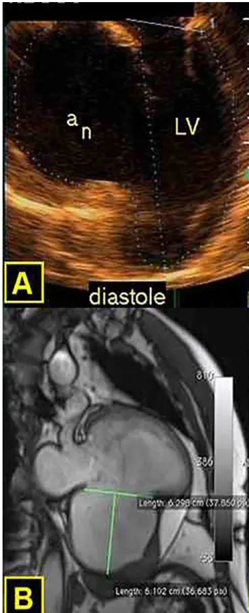
In August 2013, the patient suffered heart failure, functional class III (NYHA). A giant aneurysm of the left ventricle was present. Transesophageal echocardiography (TEE) and cardiac magnetic resonance imaging (MRI) were performed (ejection fraction: 19% [Simpson]; left- end diastolic volume: 402.7 cm3; left-end systolic volume:
324 cm3; ejection fraction: 19%; left-end diastolic volume:
490 ml; left-end systolic volume: 398 ml). Left ventricle weight was 144 gm2. The aneurysm was 7.3 x 6.4 x 7.5 cm
with tapered walls towards the base of the left ventricle; a thrombus was present (Figures 1A and 1B).
665
Rev Bras Cir Cardiovasc | Braz J Cardiovasc Surg
Rev Bras Cir Cardiovasc 2014;29(4):663-6 Schaitza GA, et al. - Surgical treatment of a giant left ventricular aneurysm-
A case report
The patient underwent repair surgery of the left ventri-cle with geometric correction through a median sternotomy (video 1). Cardiopulmonary bypass (CPB) from the aorta to the right atrium was established under normothermia (Figure 2A). Myocardial protection was held with anterograde and retrograde cardioplegia under continuous normothermic es-molol, potassium, and magnesium.
After incising the aneurysm (video 2) and extracting a large thrombus (video 3) measuring 8 x 3 cm (Figure 2B), a 7 x 5 cm
bovine pericardial patch was placed and anchored with Telon
wires (videos 4 and 5). A transition zone was established
be-tween the healthy myocardium and an area of ibrosis (video 6)
using 2.0 ethibond thereby excluding the infarcted region and a geometric correction was performed (Figure 2C). The mitral valve was competent. Cardiopulmonary bypass time was 56 minutes and the aorta was clamped for 48 minutes. The patient was weaned from the CPB with a low dose of intravenous dobu-tamine, which was maintained until closure of the incision.
A new transesophageal echocardiography was performed and revealed a 30% (Simpson) ejection fraction; left-end di-astolic volume of 138.6 cm3, and left-end systolic volume of
96.87 cm3 (Figure 3A).
The patient was extubated in the operating room and trans-ferred to the intensive care unit where he remained for 36 hours. Intraoperative blood loss was 450 ml. He was discharged 72 hours later with prescriptions for carvedilol 12.5 mg daily and acetylsalicylic acid 100 mg daily. At the one month follow-up examination, the patient was at functional class I (NYHA). He
underwent an MRI that identiied: ejection fraction of 41%,
left-end diastolic volume of 198 ml, left ventricular systolic volume of 115 ml, and left ventricular weight of 144 gm2 (Figure 3B).
DISCUSSION
Although left ventricular aneurysm is a common compli-cation following myocardial infarction, its incidence has
de-Fig. 2 – A) Giant aneurysm of the left ventricle after the establishment of cardiopulmonary bypass.
Fig. 2 – B) Aneurismectomy performed with removal of thrombus and
identiication of the transition zone between the healthy myocardium and ibrotic area.
Fig. 2 – C) Pericardial patch implanted in the transition zone between healthy myocardium and ibrotic area.
666
Rev Bras Cir Cardiovasc | Braz J Cardiovasc Surg
Rev Bras Cir Cardiovasc 2014;29(4):663-6 Schaitza GA, et al. - Surgical treatment of a giant left ventricular aneurysm-
A case report
clined, primarily due to the treatment of myocardial infarction with coronary angioplasty performed in the acute phase of the
event. The condition can be classiied as a true aneurysm when
the aneurysm forms at the damaged wall of the myocardium and as a pseudoaneurysm when the cardiac rupture is con-tained by adherent pericardium or scar tissue[1,2].
The main complications of a left ventricular aneurysm are heart failure, ventricular arrhythmias, systemic embolization, cerebrovascular accident, and ventricular rupture. The main surgical indications occur in patients with a true aneurysm; include intractable ventricular arrhythmias and heart failure not responsive to drug treatment. Other possible indications are refractory angina and systemic embolization in patients who cannot take oral anticoagulants. In cases of pseudoan-eurysm, surgical treatment is the best option, given its high probability of symptom dissolution[2,3].
Surgical techniques currently in use for correction of a left ventricular aneurysm are based on reconstruction of the left ventricle or a reduction of its volume with the goal of restoring the normal cardiac geometry[4-6]. This case exempliies a posi
-tive outcome of surgical correction with the ventricular remod-eling technique. When appropriate indications are present, the procedure can result in improved ejection fraction of the left ventricle and ventricular volume reduction.
Authors’ roles & responsibilities
GAS Conception and study design, performing the procedures and/ or experiments, writing of the manuscript or review of its content
JRFN Conception and study design, performing the procedures and/ or experiments
JCF Drafting of the manuscript or review of its content CPB Drafting of the manuscript or review of its content HG Performing the procedures and/or experiments BO Performing the procedures and/or experiments
LFM Final approval of the manuscript, performing the procedures and/or experiments
LCGS Final approval of the manuscript, performing the procedures and/or experiments
Video 1 - Giant left ventricular aneurysm before the establishment of the cardiopulmonary bypass. The aneurysm
is clearly delimited by the surgeon ingers.
Video 2 - The aneurysm wall is opened revealing its extension.
Video 3 - A large thrombus measuring 8 x 3 cm is removed from the aneurysm wall.
Video 4 - After the thrombus removal, the bovine pericardial
patch was placed and anchored with Telon wires in order to
reconstruct the geometry of the ventricular wall impaired by the aneurysm formation.
Video 5 - Bovine pericardial patch fully anchored to the wall.
Video 6 - A transition zone was established with 2.0 Ethibond
between the healthy myocardium and an area of ibrosis,
excluding the infarcted region.
REFERENCES
1. Vijayvergiya R, Pattam J, Rana SS, Singh JD, Puri GD, Singhal M. Giant left ventricular pseudoaneurysm presenting with hemoptysis. World J Cardiol. 2012;4(6):218-20.
2. Inan MB, Yazicioglu L, Acikgoz B, Tasoz R, Ozyurda, U. Giant posterolateral left ventricular aneurysm diagnosed 6 weeks after incomplete surgical revascularization. Ann Thoracic Surg. 2012;93(3):980-2.
3. Antman EM, Anbe DT, Armstrong PW, Bates ER, Green LA, Hand M, et al.; American College of Cardiology; American Heart Association Task Force on Practice Guidelines; Canadian Cardiovascular Society. ACC/AHA guidelines for the management of patients with ST-elevation myocardial infarction: a report of the American College of Cardiology/ American Heart Association Task Force on Practice Guidelines (Committee to Revise the 1999 Guidelines for the Management of Patients with Acute Myocardial Infarction). Circulation. 2004;110(9):e82-292.
4. Jatene AD. Left ventricular aneurysmectomy: resection or reconstruction. J Thorac Cardiovasc Surg. 1985;89(3):321-31.
5. Dor V, Saab M, Coste P, Kornaszewska M, Montiglio F. Left ventricular aneurysm: a new surgical approach. Thorac Cardiovasc Surg. 1989;37(1):11-9.