Expression and Activity of Matrix
Metalloproteinase-2 (MMP-2) in the
Development of Rat First Molar Tooth Germ
Paola COTRIM1
Cleverton Roberto de ANDRADE1 Sergio LINE2
Oslei Paes de ALMEIDA1 Ricardo D. COLETTA1
1Discipline of Oral Patology and 2Discipline of Histology, Faculty of Dentistry of Piracicaba, UNICAMP,
Piracicaba, SP, Brazil
Tooth germ development is associated with morphological and biochemical changes of the dental papilla and enamel organ. Enzymes with gelatinolytic activities were studied by semiquantitative reverse transcriptase-polymerase chain reaction (RT-PCR) and enzymography in tooth germ of newborn to 15-day-old rats. Three major bands with gelatinolytic activity were detected at all periods and characterized as the latent and active forms of MMP-2 using their molecular weight and activity dependent on Zn++ and Ca++ ions as criteria.
Expression and activity of MMP-2 increased progressively from 0 to 15 days after birth. Mechanical separation of the tooth germ from 10-day-old rats showed that the gelatinolytic activity was localized mainly in the dental papilla and not the dental organ. These data indicate that the expression and activity of MMP-2 varies during the development and maturation of rat first molar tooth germ.
Key Words: matrix metalloproteinase-2, tooth germ, rat.
Correspondence: Prof. Ricardo D. Coletta, Disciplina de Patologia Oral, Faculdade de Odontologia de Piracicaba, UNICAMP, Av. Limeira 901, 13414-900 Piracicaba, SP, Brasil. Tel/Fax: +55-19-3430-5318. E-mail: [email protected]
INTRODUCTION
The development and differentiation of the tooth germ is accompanied by rapid changes in its extracellu-lar matrix (1). Protein synthesis, degradation and re-sorption occur intensely during amelogenesis and den-tinogenesis, with participation of ameloblasts, odonto-blasts and possibly other epithelial and mesenchymal cells (2,3). There are many reports indicating the par-ticipation of proteases during tooth formation, but their origin and characteristics must be better determined. Serine proteases appear to be the most important en-zymes to degrade amelogenin, the main enamel matrix protein (4). A novel enzyme named enamelysin (MMP-20) was recently cloned from tooth tissues and was later characterized as a member of matrix metalloproteinase (MMPs) group (5). MMPs are endopeptidases capable of degrading various macromolecules of extracellular ma-trix. They are secreted into the extracellular matrix in
MATERIAL AND METHODS
Specimen Collection and MMPs Extraction
First molars were dissected from the maxillas and mandibles of newborn, 1, 3, 5, 7, 10 and 15-day-old Wistar rats. MMPs were extracted according to a modi-fied protocol of Robinson et al. (11). Briefly, first molars from 3 animals per period were ground in a glass homogenizer in buffer containing 50 mM Tris-HCl, pH 7.4, 10 mM CaCl2, 2 mM phenylmethylsufonyl fluoride (PMSF, Sigma Chemical Co., St. Louis, MO, USA), 2 mM N-ethylmaleimide (NEM, Sigma) and 0.2% Triton X-100. The homogenate was centrifuged at 6000 g for 20 min at 4ºC, and the pellet was resuspended in the same solution, except with 100 mM CaCl2. After incu-bation at 40ºC for 30 min, and centrifugation at 6000 g
for 20 min, the supernatant was collected and frozen in liquid nitrogen.
Enzymography
Gelatinolytic activity was examined on 10% polyacrylamide gel containing 1.6 mg/ml of gelatin. The protein concentration of each sample was deter-mined as described by Bradford (12) using bovine serum albumin (Sigma) as standard. Equivalent amounts (0.4 µg) of proteins from tooth germ extracts were mixed with an equal volume of sample buffer (2% SDS, 125 mM Tris-HCl, pH 6.8, 10% glycerol and 0.001% bromophenol blue) and then electrophoresed. After eletrophoresis, the gel was incubated twice in 2% Tri-ton X-100 for 20 min at room temperature and then incubated at 37ºC for 16 h in 10 mM Tris-HCl buffer (pH 8.0) containing 5 mM CaCl2 (Tris-CaCl2). Gels were stained with 0.05% Coomassie blue R250 (BioRad, Richmond, CA, USA). Gelatinolytic activity was de-tected as unstained bands. Densitometric analysis was performed on a BioRad GS-700 imaging densitometer (BioRad). The relative molecular weights of proteases were determined by the relation of log Mr to the relative mobility of Sigma SDS-PAGE LMW marker proteins.
Effect of pH Variation on Zymographic Activity
After substrate gel electrophoresis, gels were washed with 2% Triton X-100 as described above and incubated at 37ºC for 16 h in buffer at pH ranging from
5.0 to 10.0. The buffers used were 50 mM sodium acetate plus 5 mM CaCl2 adjusted to pH 5.0 or 6.0, and 10 mM Tris-HCl, pH 7.0, 8.0, 9.0 or 10.0, with 5 mM CaCl2.
Effect of Proteinase Inhibitors
After electrophoresis, the gels were incubated in Tris-CaCl2 buffer at 37ºC for 16 h in the presence of the following inhibitors: 0.5 mM 1,10-phenanthroline (Sigma), a specific inhibitor of MMPs, 0.5 mM EDTA (Reagen, Brazil), a divalent ions quelant, 0.5 mM NEM (Sigma), a thiol-proteinase inhibitor, and 0.5 mM PMSF (Sigma), a serine-proteinase inhibitor.
Localization of MMPs
Dental papilla was mechanically removed from the dental organ. Microscopic examination showed that the odontoblastic layer and portions of dentine matrix were part of the dental organ, as well as enamel epithe-lium and matrix. MMPs were extracted and examined as previously described.
Expression of MMP-2
polymerase (Gibco BRL). ß-actin was used as a stan-dard housekeeping gene. Primers for MMP-2 were sense 5’ CCA CGT GAC AAG CCC ATG GGG CCC C 3’ and antisense 5’ GCA GCC TAG CCA GTC GGA TTT GAT G 3’ and for ß-actin were sense 5’ TCA GAA GGA CTC CTA TGT GG 3’ and antisense 5’ TCT CTT TGA TGT CAC GCA CG 3’. After denatur-ation for 3 min at 93ºC, 40 cycles of amplificdenatur-ation were performed using a model 9700 thermocycler (Perkin Elmer, Foster City, CA, USA), followed by final exten-sion of 5 min at 72ºC. The cycling parameters were: denaturation for 45 s at 93ºC, annealing for 45 s at 58ºC, extension for 1.5 min at 72ºC. After amplification, 3 µl of PCR product was electrophoresed on a 5% non-denaturing polyacrylamide gel and the PCR products stained with silver as described by Sanguinetti et al. (15). The PCR amplification yield of target sequences was expressed in arbitrary units as the ratio of optical density of MMP-2/ß-actin electrophoretic bands.
RESULTS
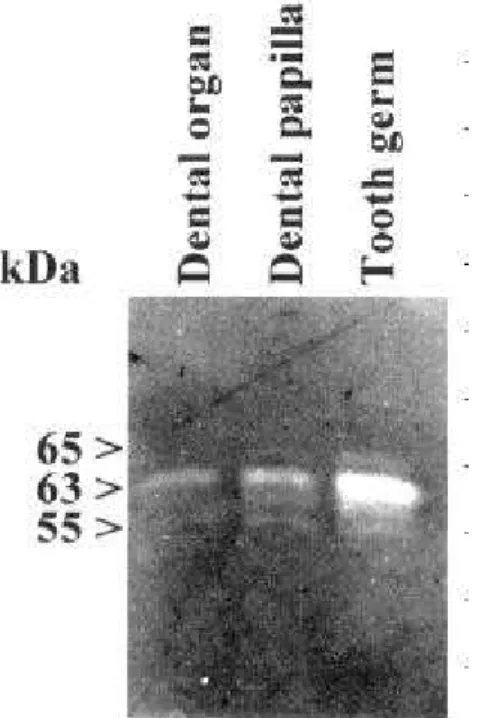
All tooth germ extracts electrophoresed on gela-tin showed three unstained bands, with Mr = ~65, ~63
and ~55 kDa (Figure 1A). These enzymes were charac-terized MMPs because their activities were inhibited by 1,10-phenanthroline and EDTA but not by PMSF and NEM, and were active in the pH range of 6.0-9.0, with an optimum activity at pH 8.0 (data not shown). These bands corresponded to the latent (65 kDa) and active (63 and 55 kDa) forms of MMP-2 (16). The amount of these gelatinolytic enzymes increased steadily during tooth germ development as shown by densitometric analysis (Figure 1B). Mechanically separated 10-day-old tooth germ expressed higher quantities of MMP-2 in the dental papilla than in the dental organ (Figure 2) To confirm these findings, RT-PCR for MMP-2 was employed. The integrity of the extracted RNA and the subsequent fidelity of first-strand cDNA synthesis for all cDNA preparations were assessed by PCR using the primers for ß-actin, a standard housekeeping gene. Only those samples which yielded the predicted 506 base pair DNA fragment were amplified with the MMP-2 specific primers (Figure 3A). Scanning densitometry of PCR products, after correction by the value from ß-actin, demonstrated that the levels of expression of MMP-2 increased in a constant manner from day 0 to 15 days (Figure 3B).
DISCUSSION
Extracts of molar tooth germ of 0 to 15-day-old rats demonstrated three gelatinolytic enzymes by enzymography assay that were characterized as MMP-2, because they were inhibited by 1,10-phenanthroline and EDTA, exhibited activities at a neutral pH and were extracted from the extracellular matrix containing a fraction of tooth germ (Triton X-100 insoluble).
The expression and activity of MMP-2 increased progressively with the development of the tooth germ, and were found mainly in the dental papilla. This gradual increase of MMP-2 correlates with a period of rapid morphological alterations in the dental papilla, which changes from a soft myxoid-type connective tissue found in neonates to a more fibrous stroma present in 15-day-old rats (17). Because the enzymes were detected in extracts, they were not topographi-cally determined, but they were probably associated with fibroblasts and odontoblasts. MMP-2 can play a fundamental role during tooth germ development in dentine deposition by odontoblasts, basement mem-brane between dental papilla and dental organ degradation and in increased collagen formation in the Figure 2. Localization of the MMP-2. Dental papilla and dental
organ extracts of 10-day-old tooth germs were electrophoresed on 10% polyacrylamide gel containing gelatin as substrate. Activity of MMP-2 was localized mainly in the dental papilla and only small amounts of MMP-2 were observed in the dental organ.
dental papilla. Reponen et al. (8) demonstrated that MMP-2 is expressed in the mesenchyme of developing organs including early developmental stages of the tooth, and it is strongly upregulated in differentiated odontoblasts at the time of basement membrane degra-dation. After the complete removal of the basement membrane between the newly secreted dentine and enamel matrices, MMP-2 expression is downregulated. Similar results were described by Heikinheimo and Salo (18) using in situ hybridization assays. They dem-onstrated that MMP-2 but not MMP-9 may participate in remodeling and degradation of basement membrane and dental papilla during early stages of human tooth morphogenesis.
MMP-2 and MMP-9 are thought to be essential to complete the degradation of type I collagen frag-ments generated by MMP-1 cleavage. Synergistic action of MMP-2, MMP-9 and MMP-1 is likely to play an important role in the degradation of type I collagen (19). Although several studies have reported the ex-pression of MMP-9 in dental papilla during tooth germ development (3,18), this enzyme was not detected in our assays. This could be explained by methodology differences, because the other studies used more sensi-tive methods.
Two major groups of dental organ proteinases have been identified in gelatin enzymography. The first group is formed by two serine proteinases with appar-ent molecular weight of 32-35 kDa. It has been demonstrated that these enzymes are able to degrade amelogenin (20). The second group is formed by three calcium-dependent enzymes migrating with Mr = ~43, ~60 and ~68 kDa. The enzyme with ~43 kDa was characterized from porcine enamel organ and named enamelysin (MMP-20) (5). The other two enzymes were shown to cleave the carboxy-terminal region of amelogenin (20). These enzymes possibly correspond to the latent and active forms of MMP-2 detected in small amounts in the dental organ after mechanical separation of the tooth germ. Interestingly, when tooth germ extract from all experimental periods was eletrophoresed on the gelatin containing gels and incu-bated in 10 mM Tris buffer at 37ºC without calcium ions, no gelatinolytic bands were detected (data not shown).
In summary, our results show that the expression and activity of MMP-2 varies during the morphological periods of development of rat first molar tooth germ.
Further research is required to establish the exact role of this enzyme in the morphogenesis of the rat tooth.
RESUMO
Cotrim P, de Andrade CR, Line S, de Almeida OP, Coletta RD. Expressão e atividade da metaloproteinase de matriz-2 (MMP-2) durante o desenvolvimento do germe dental do primeiro molar de ratos. Braz Dent J 2002;13(2):97-102.
O desenvolvimento do germe dental está associado a alterações morfológicas e bioquímicas da papila dental e do órgão do esmalte. A expressão e atividade de enzimas com atividade gelatinolítica de germes dentais dos primeiros molares de ratos recém nascidos e com 1, 3, 5, 7 10 e 15 dias de vida foram analisadas pelo método semiquantitativo da transcriptase reversa-reação da polimerase em cadeia (RT-PCR) e por enzimografia. Três enzimas com atividade gelatinolítica foram detectadas em todos os períodos e caracterizadas como sendo as formas latente e ativa da metaloproteinase de matriz-2 (MMP-2), utilizando-se como critérios à massa molecular e a atividade dependente dos íons Zn++ e Ca++. A expressão e atividade de MMP-2 aumentaram
progressivamente do nascimento até o 15o dia de vida. A
separação mecânica do germe dental demonstrou que a expressão de MMP-2 é maior na região da papila dental quando comparada ao órgão dental. Estes resultados indicam que a expressão e atividade de MMP-2 se apresentam variáveis durante o desenvolvimento e maturação do germe dental do primeiro molar de ratos.
Unitermos: metaloproteinase de matriz-2, germe dental, rato.
REFERENCES
1. Andujar MB, Magloire H, Hartmann DJ, Ville G, Grimaud J-A. Early mouse molar root development: cellular changes and distri-bution of fibronectin, laminin and type IV collagen. Differentia-tion 1985;30:111-122.
2. Fukae M, Kaneko I, Tanabe T, Shimizu M. Metalloproteinases in the mineralized compartments of porcine dentine as detected by substrate-gel electrophoresis. Archs Oral Biol 1991;36:567-573.
3. Sahlberg C, Reponen P, Tryggvason K, Thesleff I. Association between the expression of murine 72 kDa type IV collagenase by odontoblasts and basement membrane degradation during mouse tooth development. Archs Oral Biol 1992;37:1021-1030. 4. Bartlett JD, Simmer JP. Proteinases in developing dental enamel.
Crit Rev Oral Biol Med 1999;10:425-441.
5. Bartlett JD, Simmer JP, Xue J, Margolis HC, Moreno EC. Mo-lecular cloning and mRNA tissue distribution of a novel matrix metalloproteinase isolated from porcine enamel organ. Gene 1996;183:123-128.
6. Quinn CO, Scott DK, Brinckerhoff CE, Matrisian LM, Jeffrey JJ, Partridge NC. Rat collagenase. J Biol Chem 1990;265:22342-22347.
7. Birkedal-Hansen H. Role of matrix metalloproteinases in human periodontal diseases. J Periodontol 1993;64:474-484.
collagenase and its expression during mouse development. J Biol Chem 1992;267:7856-7862.
9. Chin JR, Murphy G, Werb Z. Stromelysin, a connective tissue degrading metalloendopeptidase secreted by stimulated rabbit synovial fibroblasts in parallel with collagenase. J Biol Chem 1985;260:12367-12376.
10. Caron C, Xue J, Bartlett JD. Expression and localization of membrane type 1 matrix metalloproteinase in tooth tissues. Ma-trix Biol 1998;17:501-511.
11. Robinson PJ, Siew C, Gruninger SE, Chang S-B, Turner DW, Harper DS. Transamidase and collagenase activity in healthy and diseased human gingival tissues. J Oral Pathol 1992;21:471-476. 12. Bradford MM. A rapid and sensitive method for the quantifica-tion of microgram quantities of protein utilizing the principle of protein-dye binding. Anal Biochem 1976;72:248-254.
13. Chomczynski P, Sacchi N. Single-step method of RNA isolation by acid guanidinium thiocyanate-phenol-chloroform extraction. Anal Biochem 1987;162:156-9.
14. Onisto M, Garbisa S, Caenazzo C, Freda MP, Di Francesco C, Nitti D, Liotta LA, Stetler-Stevenson WG. Reverse transcription-polymerase chain reaction phenotyping of metalloproteinases and inhibitors involved in tumor matrix invasion. Diagn Mol Pathol
1993;2:74-80.
15. Sanguinetti CJ, Dias Neto E, Simpson AJ. Rapid silver staining and recovery of PCR products separated on polyacrylamide gels. Biotechniques1994;17:914-921.
16. Fukuda Y, Masamichi I, Kudoh S, Masanori K, Yamada N. Localization of matrix metalloproteinases-1, -2, and -9 and tissue inhibitor of metalloproteinase-2 in interstitial lung diseases. Lab Invest 1998;78:687-698.
17. Coletta RD, Veiga SS, Line SR. Immunohistochemical and bio-chemical analysis of laminin in neonatal rat first molars. J Nihon Univ Sch Dent 1997;39:176-181.
18. Heikinheimo K, Salo T. Expression of basement membrane type IV collagen and type IV collagenase (MMP-2 and MMP-9) in human fetal teeth. J Dent Res 1995;74:1226-1234.
19. Okada Y, Naka K, Kawamura K, Matsumoto T, Nakanishi I, Fujimoyo N, Sato H, Seiki M. Localization of matrix metalloproteinase 9 (92 kilodalton gelatinase/type IV collagenase gelatinase B) in osteoclast: implications for bone resorption. Lab Invest 1995;72:311-322.
20. Moradian-Oldak J, Sarte PE, Fincham AG. Description of two classes of proteinases from enamel extracellular matrix cleaving a recombinant amelogenin. Connect Tissue Res 1996;35:231-238.