Braz Dent J 13(2) 2002 Effects of LLLT on laryngeal carcinoma cells 109 Braz Dent J (2002) 13(2): 109-112
Does LLLT Stimulate Laryngeal Carcinoma Cells?
An
In Vitro
Study
Antonio Luiz Barbosa PINHEIRO1 Silene Carneiro do NASCIMENTO2 Alessandro Leonardo de Barros VIEIRA3
Aluízio Barros ROLIM3 Pedro Soriano da SILVA4
Aldo BRUGNERA Jr.5
1Faculty of Dentistry, Federal University of Bahia, Salvador, BA, Brazil and
IP&D, University of Vale do Paraíba, São José dos Campos, SP, Brazil
2Department of Antibiotics, Federal University of Pernambuco, Recife, PE, Brazil 3Private Dental Practice, Recife, PE, Brazil
4Course of Medicine, Federal University of Pernambuco, Recife, PE, Brazil 5Laser Center, UNICASTELO, São Paulo, SP, Brazil
Low level laser therapy (LLLT) has been used successfully in biomedicine and some of the results are thought to be related to cell proliferation. The effects of LLLT on cell proliferation is debatable because studies have found both an increase and a decrease in proliferation of cell cultures. Cell culture is an excellent method to assess both effects and dose of treatment. The aim of this study was to assess the effect of 635nm and 670nm laser irradiation of H.Ep.2 cells in vitro using MTT (3-[4,5-dimethylthiazol-2-yl]-2,5-diphenyltetrazolium bromide). The cells were obtained from squamous cell carcinoma (SCC) of the larynx and were routinely processed from defrost to the experimental condition. Twenty-four hours after transplantation the cells were irradiated with doses ranging from 0.04 to 0.48J/cm2 for seven consecutive days (5 mW diode lasers: 635nm or 670nm, beam cross-section ~1mm) at local
light doses between 0.04 and 0.48J/cm2. The results showed that 635nm laser light did not significantly stimulate the proliferation of
H.Ep.2 cells at doses of 0.04J/cm2 to 0.48J/cm2, However, 670nm laser irradiation led to an increased cell proliferation when compared
to both control and 635nm irradiated cells. The best cell proliferation was found with 670nm laser irradiated cultures exposed to doses of doses of 0.04 to 0.48J/cm2. We conclude that both dose and wavelength are factors that may affect cell proliferation of H.Ep.2 cells.
Key Words:cell proliferation, hazards, stimulatory effect.
Correspondence: Dr. Antonio Luiz B. Pinheiro, Av Araújo Pinho, 62, Canela, 40110-150 Salvador, BA, Brasil. e-mail: [email protected]
ISSN 0103-6440
INTRODUCTION
Low level laser therapy (LLLT) has been used successfully in many medical and dental specialities. The effects of LLLT on wound healing is often attrib-uted to increased cell proliferation (1-3). There is still controversy concerning the effect of LLLT on cell proliferation because there are reports of both stimula-tory and inhibistimula-tory action of visible laser light on cell cultures (4-6).
In the last 25 years, in vitro research has become
one of the most used methods for laboratory experi-ments worldwide, mainly for studies involving drug interaction, anti-tumoral procedures and recently the
laser. This method will continue to be the method of choice for research due to difficulties of both human and animal experimentation, because this method al-lows the study of the effect of several agents on cells of different origins and also LLLT action on different cell lines (1,4,5,7).
Braz Dent J 13(2) 2002
110 A.L.B. Pinheiro et al.
malignant cells because of the possibility of exposition to laser light during clinical treatment.
The use of malignant cells in cell cultures has been reported previously (8,9). Increased cell prolifera-tion of cultures from melanoma, cervical and breast cancer, after irradiation with the argon laser (4.2 and
150KJ/m2, 35 to 500W/m2) has been reported (10). A
study using 805nm laser light (4J/cm2 and 20J/cm2) on
gingival SCC cultured cells (ZMK), urothelial carci-noma (J82) and normal urothelial cells (HCV 29) observed increased and decreased number of mitosis of HCV29 and J82 with the above mentioned doses, re-spectively. On the other hand, ZNK cells presented decreased mitotic activity with both doses (11). Stimu-lation of synthesis of both RNA and DNA was also
analyzed using He-Ne laser radiation (102J/m2) on HeLa
cells (12). This study concluded that there is expressive cell proliferation on the exponential phase and also increased cell adhesivity to the substrate of human
fibroblasts irradiated with He-Ne laser (15 min, 1W/m2)
(4).
There is controversy about the results observed previously because visible laser light may present in-hibitory or stimulatory effects on the dependence of photoreceptors which are part of the respiratory chain and depend upon external factors such as the amount of energy, wavelength, potency and irradiation time, mean-ing that the effect is dose-dependent (4). The reduction of the amount of energy has a positive influence on cell metabolism, because increased amounts of energy and products resulting from photosensitization lethally
dam-age cells(4).
The aim of this study was to assess the effect of 635nm and 670nm laser light on H.Ep.2 cultured cells.
MATERIAL AND METHODS
H.Ep.2 cultured cells (Departamento de
Antibióticos/UFPE) were kept frozen at -80oC (10)in a
medium containing Dubecco’s modified Eagle’s me-dium, 10% fetal bovine serum (FBS), 1% L-glutamine and a 1% antibiotic solution (250 µg/ml streptomycin and 80 mg/ml gentamycin sulfate. Twenty percent FBS was used after defrosting and the pH was kept at 7.8 using a 10% sodium bicarbonate solution. The cells were cultured on plastic culture flasks as described previously (13,14). Cell cultures were re-planted twice a week. Before any experimentation, all cultures were
observed under light microscopy and the viability of the culture was confirmed using the Tryptan exclusion test, because this substance easily penetrates on non-viable cells and normal cells remain colorless (15). In order to calculate cell viability the following equation was used: viability = number of living cells x 100/number of dead cells. Twenty-four hours before irradiation a cell
sus-pension (5 x 10⁴ cells/ml) was prepared. Prior to
irradiation, 100 µl of the solution was placed in 24 wells of a 96-well culture plate in order to avoid light disper-sion to others. The suspendisper-sion was then irradiated with
doses ranging from 0.03 to 0.48J/cm2 for seven
con-secutive days (5 mW diode lasers: 635nm or 670nm, beam cross-section ~1mm) at local light doses between
0.04 and 0.48J/cm2. The assessment of cell
prolifera-tion was carried out using the MTT method. Spectroscopy (540nm) was used to determine the opti-cal density (OD) of both irradiated and control samples as described by Nascimento (14). Analysis of variance was used for statistical evaluation.
RESULTS
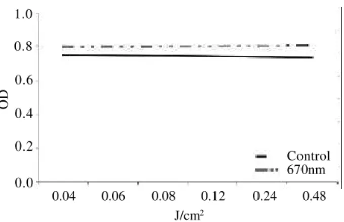
The proliferation of non-irradiated H.Ep.2 cul-tures remained as reported in the literature (14) during the experimental period. However, all irradiated cell cultures behaved differently. Figure 1 shows a similar tendency for cell proliferation for both 635nm irradi-ated and control groups. The average OD of irradiirradi-ated cultures was 0.72 and controls 0.71. Analysis of vari-ance did not show significant differences between the two groups (p = 0.455). On the other hand, there was a tendency to increased cell proliferation of samples irradiated with 670nm laser light when compared to controls (Figure 2). The average OD of these cultures was 0.80 compared to 0.74 for the controls. Analysis of variance showed a significant difference between groups (p = 0.014). When the two irradiated groups were compared, there was a difference for the tendency of cell proliferation (Figure 3). The average OD of 635nm irradiated samples was 0.72, and 0.80 for 670nm irradi-ated samples. Analysis of variance also showed a significant difference between the two irradiated groups
(p = 0.004).
DISCUSSION
Braz Dent J 13(2) 2002 Effects of LLLT on laryngeal carcinoma cells 111
use of LLLT on H.Ep.2 cells. Although there is consen-sus amongst authors regarding the contra-indication of the use of LLLT on proliferative lesions and glandular tissue, we must also consider the inclusion of premalig-nant lesions and lesions which may not be diagnosed properly at an early stage. This aspect increases the possibility of such lesions being irradiated during LLLT. It is understood that the behavior of cancerous cells significantly differs from that of normal ones. It is also well accepted that the observation of LLLT benefi-cial effects requires some type of deficiency because normal cells do not show any changes in their function after irradiation. In this study we used optimal condi-tions for experimentation using normal cell cultures in order to avoid additional variables that would prevent a more precise analysis of the data as abnormal cells have an atypical behavior.
Although our data show that the proliferation of non-irradiated H.Ep.2 cells followed the same pattern observed in previous studies (14), others have reported a different pattern (8,16). Our results also show that H.Ep.2 irradiated cell cultures behave differently from non-irradiated ones. We agree with Karu (4) that LLLT effects on cells is still a controversial topic. Our data also demonstrate that cell proliferation is influenced by the amount of energy given to the medium as suggested by Karu (12). We were also able to detect differences regarding the wavelength used for irradiation of the cells (4).
Wavelength influence indicates that 635nm laser light does not significantly stimulate the proliferation of
H.Ep.2 when using doses ranging from 0.04J/cm2 to
0.48J/cm (16). However, other doses may present differ-ent results. On the other hand, the data on the proliferation
of 670nm irradiated samples (0.05 to 0.6J/cm2) did show
differences when compared to 635nm irradiated and non-irradiated cells. When comparing the non-irradiated groups, there was a tendency to increased proliferation of cells
irradiated with 670nm from doses of 0.08J/cm2. This was
different from the results observed previously in a study carried out by our team in which the irradiation with
670nm laser light of Candida albicans had no significant
effect on proliferation in comparison to samples irradi-ated with 635nm laser light (17).
We conclude that these preliminary results indi-cate that cell proliferation increases in H.Ep.2 cells irradiated with 670nm laser light using doses from 0.08
J/cm2, and that there is a dose/wavelength dependency.
Figure 1. Tendency curve of proliferation of H.Ep.2 cells irradiated with 635nm laser light compared to control.
OD
1.0
0.8
0.6
0.4
0.2
0.0
J/cm2
0.04
635nm Control 0.06 0.08 0.12 0.24 0.48
Figure 2. Tendency curve of proliferation of H.Ep.2 cells irradiated with 670nm laser light compared to control.
Figure 3. Tendency curve of proliferation of H.Ep.2 cells irradiated with 635nm laser light compared to H.Ep.2 cells irradiated with 670nm laser light.
OD
1.0
0.8
0.6
0.4
0.2
0.0
J/cm2
0.04
670nm Control
0.06 0.08 0.12 0.24 0.48
OD
1.0
0.8
0.6
0.4
0.2
0.0
0.04 0.06 0.08 0.12 0.24 0.48
J/cm2
Braz Dent J 13(2) 2002
112 A.L.B. Pinheiro et al.
RESUMO
Pinheiro ALB, Nascimento SC, Vieira ALB, Rolim AB, Silva PS, Brugnera Jr A. A laserterapia não cirúrgica estimula células de carcinoma de laringe? Un estudo “in vitro”. Braz Dent J 2002;13(2):109-112.
As irradiações com lasers não-cirúrgicos têm sido usadas com sucesso em diversas especialidades biomédicas. A ação do laser na atividade proliferativa celular é um assunto controverso já que a luz laser tem ora estimulado e ora inibido a proliferação de células em culturas. As culturas de células “in vitro” constituem um excelente meio para o estudo dos efeitos de diversos agentes permitindo verificar as reações entre dose e efeito. O objetivo deste trabalho foi avaliar através do M.T.T. (dimetil-tetrazólio) o efeito da irradiação de células H.Ep.2 por lasers de 635nm e 670nm de comprimento de onda, “in vitro”. As células foram dissociadas a partir de tumores de câncer de laringe, e foram mantidas acondicionadas congeladas em frasco de cultura a -80oC.
Após o preparo das placas e 24 horas após o transplante, foram submetidas a doses de 0.04 a 0.48J/cm2 durante sete dias (635 ou
670nm, CW, 5 mW, (f ~1mm beam), 0.04, 0.06, 0.12, 0.24, 0.48J/cm2). Os resultados demonstraram que a luz laser de
635nm não exerce um efeito estimulativo significante na proliferação das células H.Ep.2, dentro da faixa energética de 0,04J/cm2 e 0,48J/cm2 e que as culturas irradiadas com o laser de
670nm tiveram sua atividade proliferativa aumentada em comparação aos grupos controle e aos irradiados com o laser de 635nm. Conclui-se que a irradiação de células H.Ep.2 com lasers de 670nm resulta em um aumento da proliferação celular; que a proliferação celular em culturas irradiadas com o laser de 670nm foi melhor observado com doses a partir de 0.08J/cm2; que a dose
e o comprimento de onda são fatores que podem influenciar no processo proliferativo das células H.Ep.2.
Unitermos: proliferação celular, riscos, efeito estimulativo.
REFERENCES
1. Lubart R, Wollman Y, Friedmann H, Rochkind S, Laulicht I. Effects of visible and near-infrared lasers on cell cultures. J Photochem Photobiol Biol 1992;12:305-310.
2. Pinheiro, ALB: Bases físicas dos lasers. In: Brugnera Júnior A, Pinheiro ALB. Lasers na Odontologia Moderna. São Paulo: Pancast; 1998.
3. Pogrel MA, Chen JW, Zhang K. Effects of low-energy
gallium-aluminum-arsenide laser irradiation on cultured fibroblasts and keratinocytes. Lasers Surg Med 1997;20:426-432.
4. Karu TI. Effects of visible radiation on cultured cells. J Photochem Photobiol Biol 1990;52:1089-1098.
5. Lopes LA, Rigau J, Zângaro RA, Guiduli-Neto, Jaeger MM. Comparison of the low level laser therapy effects on cultured human gingival fibroblasts proliferation using different irradi-ance and some fluence. Lasers Surg Med 2001;29:179-184. 6. Originali L. Risultati clinici di stimolazione laser e studi
sperimentali circa il mecanismo di azione. Minerva Medica (Budapest) 1981;72:2195-2199.
7. Balboni GC, Brandi ML, Zonefrati R, Repice F. Effects of He-Ne / I.R. Laser irradiation on two lines of normal human fibroblasts “In Vitro”. Arch Ital Anat Embriol 1986;91:179-188.
8. Moore AE, Sabachewsky L, Toolan HW. Culture characteristics of four permanent lines of human cancer cells. Cancer Research Philad 1955;15:598-602.
9. Thompson MD, Cupps TL, Wise DS, Wotring LL, Townsend LB. Synthesis and evaluation of 6-(dibromomethyl) 5 nitropyrimidines as potential antitumour agents. J Med Chem 1997;40:766-770. 10. Marchesini R, Dasdia T, Melloni E, Rocca E. Effect of
low-energy laser irradiation on colony formation capability in differ-ent human tumour cells “in vitro”. Lasers Surg Med 1989;9:59-62.
11. Schaffer M, Sroka R, Fuchs C, Schrader-Reichardt U, Schaffer PM, Busch M, Duhmke E. Biomodulative effects induced by 805nm laser light irradiation of normal and tumour cells. J Photochem Photobiol: Biology 1997;40:253-257.
12. Karu TI. Photobiological fundamentals of low-power laser. IEEE J Quantum Electrocs 1987;23:1703-1718.
13. Freshney RI. Culture de célules néoplasiques. In: Adolphe M, Meimon GB. Culture de cellules animales: methodologies appli-cations. Paris: Inserm; 1998.
14. Nascimento SC. Recherche de l’activite antitumorale de produits de synthese ou d’origine naturelle: utilisation du samba pour l’analyse des modifications cellulaires induites. France, Docteur U.F.R. de Pharmacie, Grenoble I, Universite Joseph Fourier; 1993.
15. Weisenthal LM, Marsden JA, Dill PL, Macaluso CK. A novel dye exclusion method for testing “in vitro” chemosensitivity of hu-man tumours. Cancer Res 1983;43:749-757.
16. Boothman DA, Trask DK, Pardee AB. Inhibition of potentially lethal DNA damage repair in human tumor cells by β-lapachone, an activator of topoisomerase I. Cancer Res 1989;49:605-612. 17. Ponzi EAC, Pinheiro ALB, Carvalho DA, Silva MP. The effects
of 635 and 670 nm laser irradiation on Candida albicans: Study “in vitro”. In: Proceedings of the 6th International Congress on Lasers in Dentistry, Hawaii, 1993.