INTRODUCTION
Tooth formation is the result of reciprocal and sequential epithelial-ectomesenchymal interaction in association with many growth factors and extracellular matrix components (1). The extracellular matrix contains noncollagenous proteins, which mainly consist of os-teopontin, bone sialoprotein, dentin sialophosphoprotein (DSPP), matrix extracellular phosphoglycoprotein and dentin matrix protein 1 (DMP1) (2,3).
DMP1 is a bone- and tooth-specific protein ini-tially identified from mineralized dentin matrix (4), but also expressed in nonmineralized tissues (5) that plays an important role in mineralized tissue formation by initiation of nucleation and modulation of mineral phase morphology (4,6). DMP1 has been studied at early and late stages of rat and mouse tooth develop-ment (7-9), but none has been reported concerning
Dentin Matrix Protein 1 (DMP1) Expression in
Developing Human Teeth
Elizabeth Ferreira MARTINEZ Luciana Alves Herdy da SILVA
Cristiane FURUSE Ney Soares de ARAÚJO Vera Cavalcanti de ARAÚJO
Department of Oral Pathology, São Leopoldo Mandic Institute and Research Center, Campinas, SP, Brazil
Dentin matrix protein 1 (DMP1) is an acidic phosphoprotein that plays an important role in mineralized tissue formation by initiation of nucleation and modulation of mineral phase morphology. The purpose of the present study was to examine the immunoexpres-sion of DMP1 in tooth germs of 7 human fetuses at different gestational ages (14, 16, 19, 20, 21, 23 and 24 weeks) comparing with completed tooth formation erupted teeth. The results showed the presence of DMP1 in the dental lamina, as well as in the cells of the external epithelium, stellate reticulum and stratum intermedium of the enamel organ. However, in the internal dental epithelium, cervi-cal loop region and dental papilla some cells have not labeled for DMP1. In the crown stage, DMP1 was expressed in the ameloblast and odontoblast layer, as well as in the dentinal tubules of coronal dentin near the odontoblast area. Erupted teeth with complete tooth formation exhibited immunolabeling for DMP1 only in the dentinal tubules mainly close to the dental pulp. No staining was observed in the enamel, predentin or dental pulp matrix. DMP1 is present in all developing dental structures (dental lamina, enamel organ, dental papilla) presenting few immunoexpression variations, with no staining in mineralized enamel and dentin.
Key Words: dentin matrix protein 1 (DMP1), teeth, extracellular matrix, immunohistochemistry.
human tooth development.
So, due to its important role in the biomineraliza-tion of the tissues, the aim of this study was to investi-gate the expression of DMP1 in tooth development of 7 human fetuses at different gestational ages comparing with completed tooth formation erupted teeth.
MATERIAL AND METHODS
Seven human fetuses with different gestational phases (14, 16, 19, 20, 21, 23 and 24 weeks), retrieved from the files of the Department of Oral Pathology of the Dental School of the University of São Paulo, Brazil, were studied. For comparison, DMP1 expression was also evaluated in completed tooth formation erupted teeth. This study was conducted after approval by the Research Ethics Committee of São Leopoldo Mandic Institute and Research Center, Campinas, Brazil
ined and 3-µm-thick serial sections obtained from the paraffin-embedded tissues were used for immunohis-tochemistry staining. Only completely formed erupted teeth were demineralized with 20% phormic acid before embedding. Sections were deparaffinized in xylene, rehydrated through a decreasing ethanol series, and immersed in 10 mM citric acid monohydrate (pH 6.0) for 20 min at 95ºC. After that, sections were immersed in 0.3% H2O2 in methanol and incubated with primary polyclonal antibody to DMP1 (Takara Bio Inc., Otsu, Shiga, Japan) at a dilution of 1:75, for 18 h, at 4ºC. Peroxidase-linked secondary antibody and diaminoben-zidine tetrahydrochloride (DAB) (Peroxidase Envision Kit; Dako, Carpinteria, CA, USA) were used to detect specific binding.
The sections were counterstained with Mayer’s hematoxylin and coverslipped with Permount. Nega-tive controls for immunostaining were obtained by substituting the primary antibody with normal mouse IgGs (Dako). The labeled sections were qualitatively evaluated by 3 independent examiners. Digital photo-micrography used a Zeiss Axioskop 2 plus microscope equipped with Axiocam digital camera and AxioVi-sion application software (Carl Zeiss, Gottingen, Germany).
RESULTS
The results demonstrated a varying expression pattern of DMP1 in developing human teeth comparing with complete tooth formation and erupted teeth.
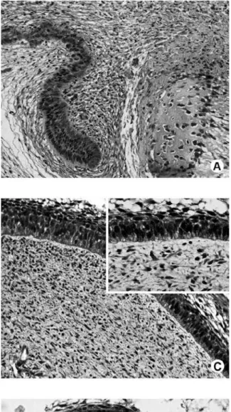
In the dental lamina, the most peripheral cells facing the developing tooth showed a positive immu-nostaining for DMP1. On the other hand, the opposite side was negative (Fig. 1A).
During the bell stage, we observed in the enamel organ a conspicuous labeling of the cytoplasm and nucleus of the cells of the outer enamel epithelium, stellate reticulum and stratum intermedium (Fig. 1-B, C). However, some cells did not label for DMP1 in the internal dental epithelium (Fig. 1C) and cervical loop region (future root area) (Fig. 1D).
When dentinogenesis begins and the amelogen-esis process occurs (crown stage), we have observed a strong immunolabeling for DMP1 in the ameloblast layer (Fig. 1E). Intense immunoreaction for DMP1 has also
also been observed in the dentinal tubules of coronal dentin (Fig. 1E). No staining was found in the enamel in the crown stage (Fig. 1E).
In the dental papilla, in the bell and crown stages, DMP1 was present in the nucleus and/or cytoplasm of some cells and also in the extracellular matrix (Fig. 1C).
In the erupted teeth with complete tooth forma-tion, DMP1 was immunoexpressed only in the dentinal tubules mainly nearer to the dental pulp (Fig. 1F). No staining was observed in the dentin, predentin, or extra-cellular matrix of the dental pulp (Fig. 1F). The enamel was not present due to the demineralization process.
DISCUSSION
The present results demonstrated a varying ex-pression pattern of DMP1 in developing human teeth comparing with complete tooth formation erupted teeth.
DMP1 is the major acidic noncollagenous matrix protein identified from mineralized matrix of dentin and bone (4,6) due to its highly acidic property and the capacity to bind Ca+2 ions playing a regulatory role in the nucleation of hydroxyapatite within the collagenous matrix of bone and dentin (4).
In the present study, DMP1 was present in all developing dental structures (dental lamina, enamel organ, dental papilla) implying a direct role for this protein in tooth formation and matrix mineralization.
In the dental lamina, the most peripheral cells facing of the developing tooth showed a positive im-munostaining for DMP1. On the other hand, the opposite side was negative (Fig. 1A). These results suggest that these positive cells might be involved in tooth forma-tion and cell differentiaforma-tion. It is suggested that DMP1 may be involved in the maintenance of the mineralized tissue microenvironment (7).
DMP1 is a key regulator of odontoblast differen-tiation, the formation of the dentin tubular system and mineralization (10).
during the crown stage (Fig. 1E). However, no staining for DMP1 was detected in the enamel in the examined specimen.
Several studies have demonstrated that DMP1 is not dentin specific, but it is expressed in bone (11), in several mineralized tissues (12), and even in non-mineralized tissues (5). DMP1 has not been detected in ameloblasts in the rat studies (7,8), but our positive results agree with those of Kim et al. (13), who showed the immunolocalization of DMP1 in ameloblasts of porcine developing teeth.
Intense immunoreaction for DMP1 has also been found in the nucleus and cytoplasm of odontoblasts in both coronal and root regions (Fig. 1E). Massa et al. (9) also detected DMP1 in the nucleus of odontoblasts cells. Narayanan et al. (14) have already demonstrated that DMP1 resides in the nucleus, cytoplasm and extracellular matrix of osteoblasts depending on their differentiation state suggesting a bifunctional role of this protein, as a transcriptional regulator of specific genes that controls osteoblast differentiation before its exports into the extracellular matrix.
In the dental papilla, in the bell and crown stages, DMP1 was present in the nucleus and/or cytoplasm of some cells and also in the extracellular matrix. The present findings may suggest that these positive cells are committed to differentiate into odontoblast lineage cells and the negatively stained cells represent mesenchymal cells or stem cells present in dental papilla (15).
In the present study, positive immunoreactions for DMP1 in the dentinal tubules of coronal dentin in both developing and completed tooth formation erupted teeth were observed, but no staining was found in the dentin. Toyosawa et al. (8) demonstrated the expression of DMP1 along the dentinal tubules is related to their mineralization. Moreover it is suggested that DMP1 is degraded after its secretion in dentinal tubules. Futher-more, some proteases, such as matrix metalloproteinase 8, were reported to be expressed in odontoblasts (16) suggesting that DMP1 in dentin is degraded by proteases.
In the erupted teeth with complete tooth forma-tion, no staining was observed in the predentin, or extracellular matrix of the dental pulp. No enamel is observed in the specimen which was demineralized. Nevertheless, it is possible to infer, based on the results in the developing human teeth that the enamel in the
Previous studies have shown that DMP1 is crucial for normal dentinogenesis (17) and mineralization in vivo
(18). The absence of DMP1 expression in the predentin and dentin indicates that this protein is required for the mineralization process. It is suggested that DMP1 plays a role in dentin formation (19) and also in the mainte-nance of the dentin tubular space (20). Moreover, when detected in nonmineralized tissues, DMP1 may have an important role in various cell activities (5). In the erupted teeth with complete tooth formation, the enamel is almost completely composed by hydroxyapatite crystals (96%) and is lost during the demineralization process.
In conclusion, the present findings showed that DMP1 is present in all developing dental structures (dental lamina, enamel organ, dental papilla) with few immunoexpression variations, with no staining in mineralized enamel and dentin in the complete tooth formation erupted teeth.
RESUMO
A proteína da matriz dentinária 1 (DMP1) é uma fosfoproteína ácida que tem sido relacionada diretamente ao processo de mineralização dos tecidos em formação sendo iniciadora do processo de nucleação e modulação da fase mineral. O objetivo desse trabalho foi avaliar a imunoexpressão da DMP1 em germes dentários em diferentes fases da odontogênese, obtidos de 7 fetos humanos em diversos estágios gestacionais (14, 16, 19, 20, 21, 23 e 24 semanas), comparando-se com dentes com rizogênese completa. Os resultados mostraram que a DMP1 esteve expressa na lâmina dentária, bem como, nas células do epitélio externo, retículo estrelado e estrato intermediário do órgão do esmalte. Diferentemente, no epitélio interno do órgão do esmalte, alça cervical e papila dentária algumas células não apresentaram a DMP1. Nas fases de coroa, os ameloblastos e odontoblastos apre-sentaram marcação positiva para a DMP1, bem como os túbulos dentinários da dentina coronária próximos à região odontoblástica. Os dentes com rizogênese completa exibiram marcação para a DMP1 apenas nos túbulos dentinários principalmente próximos à polpa dentária. Nenhuma marcação foi observada na matriz de esmalte ou pré-dentina, nem na polpa dentária. Concluímos que a DMP1 está presente em todas as fases da odontogênese, tanto na lâmina dentária, órgão do esmalte, bem como na papila dentária, com pequenas variações de nuances de expressão, estando ausente na dentina e esmalte mineralizados.
ACKNOWLEDGEMENTS
REfERENCES
1. Thesleff I, Vaahtokari A, Kettunen P, Aberg T. Epithelial-mes-enchymal signaling during tooth development. Conn Tissue Res 1995;32:9-15.
2. Robey PG. Vertebrate mineralized matrix proteins: structure and function. Connect Tissue Res 1996;35:131-136.
3. Papagerakis P, Berdal A, Mesbah M, Peuchmaur M, Malaval L, Nydegger J, et al.. Investigation of osteocalcin, osteonectin, and dentin sialophosphoprotein in developing human teeth. Bone 2002;30:377-385.
4. George A, Sabsay B, Simonian PA, Veis A. Characterization of a novel dentin matrix acidic phosphoprotein. J Biol Chem 1993;268:12624-12630.
5. Terasawa M, Shimokawa R, Terashima T, Ohya K, Takagi Y, Shimokawa H. Expression of dentin matrix protein 1 (DMP1) in nonmineralized tissues. J Bone Miner Metab 2004;22:430-438. 6. MacDougall M, Gu TT, Luan X, Simmons D, Chen J.
Identifica-tion of a novel isoform of mouse dentin matrix protein 1: spatial expression in mineralized tisues. J Bone Miner Res 1998;13:422-431.
7. Baba O, Qin C, Brunn JC, Wygant JN, Mcintyre BW, Butler WT. Colocalization of dentin matrix protein 1 and dentin sialoprotein at late stages of rat molar development. Matrix Biol 2004;23:371-379.
8. Toyosawa S, Okabayashi K, Komori T, Ijuhin N. mRNA expres-sion and protein localization of dentin matrix protein 1 during dental root formation. Bone 2004;34:124-133.
9. Massa LF, Ramachandran A, George A, Arana-Chavez VE. De-velopmental appearance of dentin matrix protein 1 during the early dentinogenesis in rat molars as identified by high-resolution immunocytochemistry. Histochem Cell Biol 2005;124:197-205. 10. Lu Y, Ye L, Yu S, Zhang S, Xie Y, McKee MD, et al.. Rescue of
odontogenesis in DMP1-deficient mice by targeted re-expression of DMP1 reveals roles for DMP1 in erly odontogenesis and dentin apposition in vivo. Dev Biol 2007;303:191-201.
11. D’Souza RN, Cavender A, Sunavala G, Alvarez j, Ohshima T, Kulkarni AB, et al.. Gene expression patterns of murine dentin matrix protein 1 (DMP1) and dentin sialophosphoprotein (DSPP) suggest distinct developmental functions in vivo. J Bone Miner Res 1997;12:2040-2049.
12. Feng JQ, Huang H, Lu Y, Ye L, Xie Y, Tsutsui TW, et al.. The dentin matrix protein 1 (DMP1) is specifically expressed in mineralized, but not soft tissues during development. J Dent Res 2003:82:776-780.
13. Kim J-W, Yamakoshi Y, Iwata T, Hu YY, Zhang H, Hu JC-C, et al.. Porcine dentin matrix protein 1: gene structure, cDNA sequence, and expression in teeth. Eur J Oral Sci 2006;114:33-41.
14. Narayanan R, Ramachandran A, Hao J, He G, Park KW, Cho M, et al.. Dual functional roles of dentin matrix protein 1. Implica-tions in biomineralization and gene transcription by activation of intracellular Ca2+ store. J Biol Chem 2003;278:17500-17508. 15. Gronthos S, Mankani M, Brahim J, Robey PG, Shi S. Postnatal
human dental pulp stem cells (DPSCs) in vitro and in vivo. Proc Natl Acad Sci USA 2000;97:13625-13630.
16. Palosaari H, Wahlgren J, Larmas M, Rönkä H, Sorsa T, Salo T, et al.. The expression of MMP-8 in human odontoblasts and dental pulp cells is down-regulated by TGF-beta1. J Dent Res 2000;79:77-84.
17. Ye L, Mishina Y, Chen D, Huang H, Dallas SL, Dallas MR, ET al.. Dentin matrix protein 1 (DMP1) deficient mice display severe defects in cartilage formation responsible for a chondrodysplasia-like phenotype. J Biol Chem 2005;280:6197-6203.
18. Ling Y, Rios HF, Myers ER, Lu Y, Feng JQ, Boskey AL. DMP1 depletion decreases bone mineralization in vivo: an FTIR imaging analysis. J Bone Miner Res 2005;20:2169-2177.
19. Hao J, Ramachandran A, George A. Temporal and spatial localiza-tion of the dentin matrix proteins during dentin biomeniralizalocaliza-tion. J Histochem Cytochem 2008;57:227-237.
20. Orsini G, Ruggeri A, Mazzoni A, Nato F, Falconi M, Putignano A, et al.. Immunohistochemical localization of dentin matrix protein 1 in human dentin. Eur J Histochem 2008;52:215-220.