210 Radiol Bras. 2012 Jul/Ago;45(4):210–214
Evaluation of medical radiation exposure in pediatric
interventional radiology procedures
*
Avaliação de exposições médicas em procedimentos pediátricos de radiologia intervencionista
Valéria Coêlho Costa Navarro1, Marcus Vinícius Teixeira Navarro2, Ana Figueiredo Maia3, Adriano Dias Dourado Oliveira4, Aline da Silva Pacheco Oliveira5
Objective: To evaluate pediatric radiation exposure in procedures of interventional radiology in two hospitals in the Bahia state, aiming at contributing to delineate the scenario at the state and national levels. The knowledge of exposure levels will allow an evaluation of the necessity of doses optimization, considering that peculiarities of radiology and pediatrics become even more significant in interventional radiology procedures which involve exposure to higher radiation doses. Materials and Methods: A total of 32 procedures were evaluated in four rooms of the two main hospitals performing pediatric interventional radiology procedures in the Bahia state. Air kerma rate and kerma-area product were evaluated in 27 interventional cardiac and 5 interventional brain procedures. Results: Maximum values for air kerma rate and kerma-area product and air kerma obtained in cardiac procedures were, respectively, 129.9 Gy.cm2
and 947.0 mGy; and, for brain procedures were 83.3 Gy.cm2
and 961.0 mGy. Conclusion: The present study results showed exposure values up to 14 times higher than those found in other foreign studies, and approximating those found for procedures in adults. Such results demonstrate excessive exposure to radiation, indicating the need for constant procedures optimization and evaluation of exposure rates.
Keywords: Radiation protection; Interventional radiology; Pediatrics.
Objetivo: Avaliar as exposições pediátricas de radiologia intervencionista em dois hospitais do Estado da Bahia, visando contribuir para a construção de um cenário estadual e nacional, possibilitando o conhecimento das exposições e da necessidade de sua otimização, visto que as peculiaridades que envolvem a radiologia e a pediatria se potencializam quando se trata de procedimentos de radiologia intervencionista, em razão das doses elevadas de radiação. Materiais e Métodos: Foram avaliados 32 procedimentos em quatro salas nos dois principais hospitais que realizam procedi-mentos de radiologia intervencionista pediátrica na Bahia. Foram avaliados os valores de kerma no ar incidente e o produto kerma-área no ar de 27 procedimentos cardiológicos e 5 procedimentos cerebrais. Resultados: Os valores máximos de produto kerma-área e kerma obtidos para procedimentos cardiológicos foram, respectivamente, 129,9 Gy.cm2
e 947,0 mGy, e para procedimentos cerebrais, 83,3 Gy.cm2
e 961,0 mGy. Conclusão: Os resultados deste estudo mostraram valores de exposições superiores em até 14 vezes os obtidos em estudos realizados em outros países, chegando próximos de resultados obtidos para procedimentos em adultos. Isto revela quão elevadas podem ser as expo-sições pediátricas, indicando a necessidade de constante otimização dos procedimentos e avaliação das expoexpo-sições.
Unitermos: Proteção radiológica; Radiologia intervencionista; Pediatria.
Abstract
Resumo
* Study developed in the Postgraduate Programme in Physics at Universidade Federal de Sergipe (UFS), Aracaju, SE, Brazil.
1. Master, Fellow PhD degree, Postgraduate Programme in Physics, Universidade Federal de Sergipe (UFS), Professor at Ins-tituto Federal de Educação, Ciência e Tecnologia da Bahia (IFBA), Salvador, BA, Brazil.
2. PhD, Professor at Instituto Federal de Educação, Ciência e Tecnologia da Bahia (IFBA), Salvador, BA, Brazil.
3. PhD, Professor at Universidade Federal de Sergipe (UFS), Aracaju, SE, Brazil.
4. MD, Hemodynamicist, Director for Quality at Sociedade Bra-sileira de Hemodinâmica e Cardiologia Intervencionista, Salva-dor, BA, Brazil.
5. Radiology Technologist, Scientific Initiation Student, Insti-tuto Federal de Educação, Ciência e Tecnologia da Bahia (IFBA), Salvador, BA, Brazil.
Mailing Address: Valéria Coêlho Costa Navarro. Rua Apoema, 240, Condomínio Aldeia Jaguaribe, Piatã. Salvador, BA, Brazil, 41613-044. E-mail: [email protected]
Navarro VCC, Navarro MVT, Maia AF, Oliveira ADD, Oliveira ASP. Evaluation of medical radiation exposure in pediatric interventional radiology procedures. Radiol Bras. 2012 Jul/Ago;45(4):210–214.
cal specialties such as cardiology, hepa-tology and neurology have utilized this tool. Additionally, there is a strong trend towards the utilization of such technique in pediatrics considering that this is a group of patients at high surgical risk.
Until not long ago, it was not possible to think about correction of complex cardio-pathies, pulmonary venous drainage, cor-rection of defects such as pulmonary and aortic stenosis, correction of urological disorders such as urolithiasis, among other indications, without a surgical intervention, a method that poses high mortality risk(2). INTRODUCTION
Interventional radiology comprises pro-cedures with predominantly therapeutic ob-jectives, utilizing a percutaneous approach and imaging guidance usually with fluoros-copy, but also computed tomography or magnetic resonance imaging(1). The num-ber of invasive procedures utilizing inter-ventional radiology in pediatrics has been increasing in recent years, as other
Thus, interventional radiology is one of the applications of radiation in medicine whose benefits should exceed the risks, in compliance with the first and most funda-mental principle of radiological protection,
the principle of justification, even being the
radiological practice with the highest lev-els of medical and occupational exposures, sometimes reaching the levels of determin-istic effects.
In such a context of possible high expo-sure levels, pediatric interventional radiol-ogy demands special attention, as children are more radiosensitive than adults because of high cell multiplication and longer life expectancy, with a risk for lethal cancer/ dose unit two to four times higher than for adults(3).
As regards deterministic effects, there is a possibility of occurrence of severe radio-induced lesions, erythema, epilation, ne-crosis, sterility and cataract(4–8).
Because of the above mentioned risks, studies have pointed towards the need for adopting specific approaches to optimize pediatric procedures by means of imaging quality assessment simultaneously with the investigation of radiation doses, with the purpose of maintaining them as low as possible(9–13).
In the Brazilian State of Bahia, five he-modynamics centers perform pediatric procedures; but a demand of approxi-mately 80% of such procedures is concen-trated in the two hospitals involved in the present study, as the other three services only perform pediatric procedures in emer-gency situations. In order to ensure the rep-resentativeness of the sample, the informa-tion required for the present study was col-lected in these two services over a three-month period. The low rate of pediatric procedures in the state is a consequence of the limited number of professionals quali-fied for the performance of such proce-dures.
Thus, the present study was aimed at evaluating the pediatric exposure to radia-tion in intervenradia-tional radiology procedures performed in two hospitals in the State of Bahia, Brazil, with a view of contributing to the construction of a scenario both at state and national levels, allowing for the knowledge of exposure levels and their optimization.
MATERIALS AND METHODS
The present study was undertaken in co-operation with two hospitals which per-form pediatric procedures of interventional radiology in the city of Salvador, in the state of Bahia, Brazil. Each one of the A and B institutions have two rooms (1 and 2) avail-able for pediatric procedures. The follow-ing apparatuses were utilized: Angio Diag-nost 5 (Philips Medical Systems) in hospi-tal A, and Innova 3100 (GE Healthcare) in hospital B. In the present study, the levels of radiation exposure in pediatric proce-dures performed between April and June 2011 were evaluated. The identity of both patients and institutions was not disclosed. It is important to highlight that both hos-pitals perform interventional procedures in adults and in children, and both face limi-tations in the number of professionals who perform pediatric procedures. Thus, their capacity to perform pediatric procedures is still limited, a fact which explains the small amount of data in the period. The proce-dures were categorized according to the medical specialty: 27 cardiological proce-dures and five brain proceproce-dures were stud-ied, in a total of 32 procedures. Age and sex were the biometric data collected.
In the statistical analysis, values of mean, median and range of air kerma dis-tribution and air kerma area-product (PKA) were calculated. And, purposes of compari-son adult reference level, the third quartile of the air kerma and PKA were calculated for pediatric procedures.
The evaluations were performed by uti-lizing the indications from the gauges on the apparatuses themselves. In spite of the fact that all the apparatuses were equipped with transmission chambers with the pos-sibility of indicating kerma and PKA, the in-dication of these two data was available only in rooms 1 and 2 of the institution B, while in the institution A, room 1, only the PKA indication was enabled, and in room 2, only the kerma indication was enabled.
For the PKA assessment, the correction factor was initially calculated, taking as ref-erence an external PKA meter manufactured by Iba Dosimetry. The meter was placed at the beam exit, over the equipment’s head, and the results were corrected with basis on the obtained calibration factors. Thus, the
PKA measurements in the present study are those indicated on the apparatuses, ad-justed by the correction factor.
The kerma values on the reference point indicated by the apparatuses, were cor-rected with basis on the values indicated by the Rapidose (Radcal Corporation) multi-meter. Such multimeter was placed under the phantom CIRS NEMA XR21 config-ured as recommended by the manufacturer and in compliance with the IEC 601-2-43 item 203.5.2.4.5.101 (d). The meters uti-lized in the present study had calibration certificates valid until June 2012.
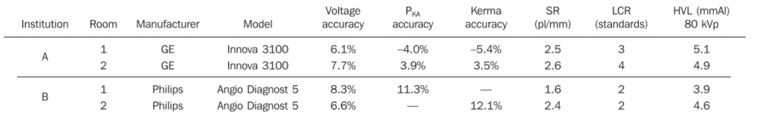
The apparatuses of both institutions had their performance evaluated and, besides air kerma and PKA, the following param-eters were verified: tube voltage, exposure time, half-value layer, filtration, spatial resolution, high contrast resolution, low contrast resolution, dynamic range and field size.
As in only one service there was indi-cation of scopy time and numbers of im-ages, such parameters were not considered in the present study.
RESULTS
In the present study, 32 pediatric medi-cal exposures were evaluated and catego-rized according to procedure, institution and room as shown on Table 1.
The mean air kerma rate and PKA for each medical specialty are presented on Table 2.
A summary of the parameters evaluated in the apparatuses is presented on Table 3. As regards the patients’ age, the study sample comprised patients in the age range between 0 and 14 years. The age distribu-tion is shown on Figure 1.
In all age groups there were patients who were submitted to cardiological pro-cedures, while no brain procedure was per-formed in patients in the 1 to 10 years age group. The age distribution by institution and by room is presented on Figure 2.
As regards sex, 41.9% of the patients were girls and 58.1% were boys.
catheterization and transluminal coronary angioplasty represented 83.9% of the pro-cedures.
DISCUSSION
In the present study, the evaluation of pediatric medical exposures for
interven-Figure 2. Age distribution by institution and by room. Age distribution (years)
N
u
m
b
e
r
o
f
p
a
ti
e
n
ts
Figure 1. Distribution by age in the procedures categorized as cardiological and cerebral.
N
u
m
b
e
r
o
f
pa
ti
e
n
ts
Age distribution (years)
Table 1 Mean, median and range of PKA, air kerma rate and number of patients for the rooms in institutions A and B.
Institution
A
B
Room
1
2
1
2
Procedures
RA CC
CA CC PTCA
CC
CE CC
n
1 11
4 6 3
4
1 1
PKA (Gy.cm2) Kerma(mGy)
Minimum
34.1 10.1
— — —
27.1
83.3 64.2
Maximum
34.1 129.9
— — —
80.8
83.3 64.2
Mean
34.1 57.8
— — —
54.9
83.3 64.2
3rd quartile
34.1 86.2
— — —
83.8
83.3 64.2
Minimum
— —
248.5 28.3 145.9
229.0
961.0 947.0
Maximum
— —
527.0 492.3 153.3
675.0
961.0 947.0
Mean
— —
415.3 399.3 149.6
460.3
961.0 947.0
3rd quartile
— —
498.6 486.6 151.5
575.0
961.0 947.0
n, number of patients; RA, radiofrequency ablation; CC, cardiac catheterization; CA, cerebral angiography; PTCA, percutaneous transluminal coronary angiography; CE, cerebral embolization.
Table 2 Mean, median and 3rd quartile values of PKA and air kerma rate for cardiological and cerebral procedures in the present study.
Cardiological procedures Cerebral procedures
Mean Median 3rd quartile
PKA (Gy.cm2)
38.7 27.9 45.9
Kerma (mGy)
393.3 454.9 491.8
PKA (Gy.cm2)
42.3 30.9 59.2
Kerma (mGy)
551.7 498.7 635.5
Table 3 Characterization of the apparatuses evaluated in the present study.
Institution
A
B
Room
1 2
1 2
Manufacturer
GE GE
Philips Philips
Model
Innova 3100 Innova 3100
Angio Diagnost 5 Angio Diagnost 5
Voltage accuracy
6.1% 7.7%
8.3% 6.6%
PKA
accuracy
–4.0% 3.9%
11.3% —
Kerma accuracy
–5.4% 3.5%
— 12.1%
SR (pl/mm)
2.5 2.6
1.6 2.4
LCR (standards)
3 4
2 2
HVL (mmAl) 80 kVp
5.1 4.9
3.9 4.6
tional cardiological and cerebral proce-dures, as compared with results of similar studies, demonstrated that the method uti-lized for PKA calculation was similar to that utilized by Bacher et al.(14) and Tsapaki et al.(8). In the evaluation of kerma, the au-thors utilized a method similar to that uti-lized for PKA, since other methods such as dosimetric films or thermoluminescent dosimetry were not available.
The present results demonstrated PKA values between 57.8 and 64.2 Gy.cm2, while the maximum and minimum values were, respectively, 10.1 and 129.9 Gy.cm2. On the other hand, the mean PKA values ob-tained by Tsapaki et al.(8) for cardiac cath-eterization in a study involving 20 coun-tries in Africa, Asia and Europe, with a pediatric population representing approxi-mately 2% of the study sample, were be-tween 0.1 and 36.7 Gy.cm2. Also, accord-ing to the study developed by Bacher et al.(14) to estimate the dose in a sample in-cluding 60 pediatric patients submitted to cardiac catheterization, the mean PKA value was 4.5 Gy.cm2, with maximum and mini-mum values of 0.4 and 20.4 Gy.cm2, re-spectively. As the above mentioned results are compared, the mean PKA values are be-tween two and fourteen times higher and the maximum PKA value is approximately six times higher.
As compared with the results found by Padovani & Quai(6) and by TECDOC-1641(15) for procedures in adults, the P
KA for cardiac catheterization was in the range of 12.0–67.0 Gy.cm2 and 5.1–221.0 Gy.cm2, respectively, which demonstrates that the results in the present study are similar to those found for adults and reported in the literature.
It is important to highlight that such equivalence between the PKA ranges – pe-diatric and adult – may reflect the complex-ity of the studied pediatric procedures, as well as the need for optimization of the pro-cedures in the mentioned institutions.
As regards the kerma for cardiac cath-eterization, the mean results in the present study are between 399.3 and 947.0 mGy, while the minimum and maximum values were, respectively, 28.3 and 947.0 mGy. As such results are compared with those ob-tained by Bacher et al.(14) who obtained a mean value for cardiac catheterism of 23.9
mGy and interval between minimum and maximum values of 1.49 – 297.5 mGy, one observes that the results in the present study are much higher than those reported by the above mentioned authors, with the mini-mum value in the present study being above the mean value obtained by those authors. The mean kerma value obtained in the present study was 961.0 mGy for cerebral procedures, while the minimum and maxi-mum values were respectively 248.3 mGy and 961.0 mGy. For PKA, the mean value was 83.3 Gy.cm2. In a similar study, Raelson et al.(16) obtained mean values be-tween 103.8 and 340.3 Gy.cm2 for P
KA and a mean value of 580.0 mGy for kerma. Rampado et al.(17), in a study on neuroin-terventional procedures in 18 pediatric pa-tients, have obtained intervals between minimum and maximum values for kerma and PKA corresponding to, respectively, 88.0–1710.0 mGy and 16.7–343.0 Gy.cm2. As the results from the present study are compared with those reported by Raelson et al.(16) and Rampado et al.(17), one ob-serves that the mean value for PKA in the present study is within the range of values reported by the mentioned authors, while, in the present study, the maximum kerma value is higher than that obtained by Raelson et al.(16) and lower than that ob-tained by Rampado et al.(17), a fact that may reflect the complexity of the cases under analysis.
The cerebral techniques evaluated by Miller et al.(18) revealed third quartile values for cerebral embolization in adults in the range of 339.5–403.2 Gy.cm2 for P
KA, well above the value found in the present study, which was 83.3 Gy.cm2, and third quartile values in the range of 4169.0–4441.0 mGy for kerma, also above the value in the present study, which was 961.0 mGy.
As the analysis of dose by institution in the present study is considered, one ob-serves that, for institution A, the maximum air kerma rate for cerebral procedures was 527.0 mGy, and for cardiac catheterization the maximum PKA value was 129.9 Gy.cm
2.
For the institution B, the maximum kerma and PKA values for cerebral procedures were 961.0 mGy e 83.3 Gy.cm2, respec-tively, while the results for cardiac catheter-ization were 675.0 mGy and 80.8 Gy.cm2 for kerma and PKA, respectively.
In the present study, the groups were classified according to age and sex. As re-gards age, there was a prevalence of con-genital cardiopathies in the neonatal (0–30 days) and in the infant (31 days–2 years) groups, indicating a predominance of car-diological procedures in these age groups(19). It is important to highlight that the body mass index cannot be disregarded as a routine information in the hemody-namics centers, as this is a contributing factor to be considered in the presence of high radiation doses, since the beam attenu-ation is related to the increase in the patient’s body mass index(18). As the body mass index is related to the weight and height of the patient, such biometric data was not evaluated in the present study for not being available in the institutions.
The differences observed between the institutions, as well as those between other studies, may be related to several other fac-tors such as the complexity of each proce-dure, patients’ weight and age, the level of the clinical staff or the utilized techniques. However, such parameters and correlations were not included as objectives of the present study, and, as the authors continue their investigations on pediatric interven-tional radiology, they will be evaluated.
As regards the characterization of the ap-paratuses, in spite of the absence of specific Brazilian regulations on the assessment and performance of interventional radiology equipment, the Ordinance SVS/MS 453/ 98(20) establishes that interventional radiol-ogy is under its regulatory scope, with the fluoroscopy requirements being applicable to interventional radiology.
All apparatuses included in the present study presented deviations < 10% in tube voltage indication, in compliance with the Ordinance SVS/MS 453/98(18). The devia-tions of the PKA and kerma indications were > 10% in two apparatuses.
The visualization of the low contrast patterns should be capable of resolving, at least, three of the four patterns present in the phantom. Two of the four apparatuses were below the minimum level of visual-ization.
Thus, based on the Brazilian regula-tions, it was possible to consider that all the apparatuses were compliant with respect to parameters of accuracy in the indication of tube voltage and half-value layer.
However, as far as spatial resolution is concerned, all manufacturers indicate in their manuals, values between 4 and 5 pl/ mm. In the present study, the highest value was 2.6 pl/mm, and the lowest, 1.6 pl/mm, three times lower than the value informed by the manufacturer. The spatial resolution is an important parameter in the evaluation of imaging quality, which, in association with low contrast resolution, strongly influ-ences the imaging quality; and their low performance values may increase examina-tion time and, consequently, the medical exposures, on account of difficulties in the images visualization.
CONCLUSION
The present study results demonstrated exposure values up to 14 times those ob-tained in studies developed in other coun-tries, with values similar to the results ob-tained for procedures in adults. Thus, it is possible to conclude that the high pediat-ric exposures indicate the need for constant optimization of procedures and assessment of exposures.
The practice of not recording data on pa-tients and procedures, such as biometric data, exposure time, kerma and PKA, hin-ders the performance of studies and the possibility of follow-up in those cases where the exposure levels are close to the deterministic effects threshold.
Also, as the indication of scopy time and number of images were available in only one service, such parameters were not
taken into consideration, which represented a limitation in the present study. The results of the present study are the first in the State of Bahia, and constitute a contribution to-wards delineating a scenario at state and national levels, with the prospect of con-tributing to the development of public poli-cies, including specific regulations regard-ing interventional and pediatric radiology.
Acknowledgements
The authors wish to thank Instituto Fed-eral de Educação, Ciência e Tecnologia da Bahia (IFBA), Universidade Federal de Sergipe (UFS), Instituto Nacional de Ciência e Tecnologia (INCT) em Metro-logia das Radiações em Medicina and Instituto Nacional de Ciência Inovação e Tecnologia em Saúde (INCT-CITECS) for the technical and financial support.
REFERENCES
1. World Health Organization. Efficacy and radia-tion safety in intervenradia-tional radiology. Geneva, Switzerland: WHO; 2000.
2. Atik E. Cateterismo cardíaco intervencionista na cardiologia pediátrica. O posicionamento médico quanto às aplicações atuais e perspectivas. [Edi-torial]. Arq Bras Cardiol. 2002;79:443–5.
3. Pettersson HB, Fälth-Magnusson K, Persliden J, et al. Radiation risk and cost-benefit analysis of a paediatric radiology procedure: results from a national study. Br J Radiol. 2005;78:34–8.
4. Canevaro L. Aspectos físicos e técnicos da radio-logia intervencionista. Rev Bras Fís Med. 2009; 3:101–15.
5. Vano E, Sanchez R, Fernandez JM, et al. Patient dose reference levels for interventional radiology: a national approach. Cardiovasc Intervent Radiol. 2009;32:19–24.
6. Padovani R, Quai E. Patient dosimetry approaches in interventional cardiology and literature dose data review. Radiat Prot Dosimetry. 2005;117: 217–21.
7. Rösch J, Keller FS, Kaufman JA. The birth, early years, and future of interventional radiology. J Vasc Interv Radiol. 2003;14:841–53.
8. Tsapaki V, Kottou S, Korniotis S, et al. Radiation doses in paediatric interventional cardiology pro-cedures. Radiat Prot Dosimetry. 2008;132:390– 4.
9. Aroua A, Besançon A, Buchillier-Decka I, et al. Adult reference levels in diagnostic and interven-tional radiology for temporary use in Switzerland. Radiat Prot Dosimetry. 2004;111:289–95. 10. Ubeda C, Vano E, Miranda P, et al. Radiation dose
and image quality for paediatric interventional cardiology systems. A national survey in Chile. Radiat Prot Dosimetry. 2011;147:429–38.
11. Vano E, Ubeda C, Leyton F, et al. Radiation dose and image quality for paediatric interventional cardiology. Phys Med Biol. 2008;53:4049–62. 12. Silva MSR, Khoury HJ, Borrás C, et al.
Dosime-tria de pacientes e médicos em intervenções co-ronárias percutâneas em Recife, Pernambuco, Brasil. Radiol Bras. 2011;44:90–6.
13. Soares FAP, Pereira AG, Flôr RC. Utilização de vestimentas de proteção radiológica para redução de dose absorvida: uma revisão integrativa da li-teratura. Radiol Bras. 2011;44:97–103. 14. Bacher K, Bogaert E, Lapere R, et al.
Patient-specific dose and radiation risk estimation in pe-diatric cardiac catheterization. Circulation. 2005; 111:83–9.
15. International Atomic Energy Agency. Patient dose optimization in fluoroscopically guided inter-ventional procedures. Final report of a coordi-nated research project. IAEA-TECDOC-1641. Vienna, Austria: IAEA; 2010.
16. Raelson CA, Kanal KM, Vavilala MS, et al. Ra-diation dose and excess risk of cancer in children undergoing neuroangiography. AJR Am J Roentgenol. 2009;193:1621–8.
17. Rampado O, Ropolo R. Entrances skin dose dis-tribution maps for interventional neuroradio-logical procedures: a preliminary study. Radiat Prot Dosimetry. 2005;117:256–9.
18. Miller DL, Kwon D, Bonavia GH. Reference lev-els for patient radiation doses in interventional radiology: proposed initial values for U. S. prac-tice. Radiology. 2009;253:753–64.
19. Miyague NI, Cardoso SM, Meyer F, et al. Estudo epidemiológico de cardiopatias congênitas na infância e adolescência. Análise em 4538 casos. Arq Bras Cardiol. 2003;80:269–73.