241
Vascular resistance index in breast nodules
Radiol Bras. 2009 Jul/Ago;42(4):241–244 Original Article • Artigo Original
Utilization of vascular resistance index
in the differentiation between benign and malignant
breast nodules*
Utilização do índice de resistência vascular na diferenciação entre nódulos mamários benignos e malignos
Joel Schmillevitch1, Hélio Antonio Guimarães Filho2, Harley De Nicola3, Ana Cheila Gorski1
OBJECTIVE: The present study was aimed at evaluating the role played by spectral Doppler as a method to measure the vascular resistance index in the differentiation between benign and malignant breast lesions. MATERIALS AND METHODS: Nineteen malignant and 18 benign lesions histologically diagnosed had their vascularization previously analyzed through the resistance index. RESULTS: A statistically significant difference (p < 0.001) was observed between the mean values of resistance index for benign and malignant lesions (respectively 0.62×0.80) in nodular breast lesions greater than 1 cm. A resistance index ≥≥≥≥≥ 0.69 was highly associated with malignant lesions, with 84.2% sensitivity, 88.9% specificity, false-positive rate of 11.1%, and false-negative rate of 15.8%. CONCLUSION: The analysis of vascular resistance index combined with findings on grayscale sonographic images can be of great assistance in the assessment of nodular breast lesions greater than 1 cm, with high sensitivity and specificity.
Keywords: Breast; Breast lesions; Ultrasonography; Doppler; Resistance index.
OBJETIVO: O objetivo deste estudo foi avaliar o significado do Doppler espectral por meio da obtenção do índice de resistência vascular na diferenciação entre lesões mamárias benignas e malignas. MATERIAIS E MÉTODOS: Dezenove lesões malignas e 18 benignas, diagnosticadas por estudo histológico, foram subme-tidas previamente a análise de sua vascularização por meio do Doppler espectral para se obter o índice de resistência vascular. RESULTADOS: Observou-se diferença estatisticamente significante (p < 0,001) entre os valores médios do índice de resistência para os resultados benigno e maligno (0,62×0,80, respectiva-mente), em nódulos maiores que 1 cm. Um índice de resistência ≥≥≥≥≥ 0,69 foi altamente associado a lesões malignas, com sensibilidade de 84,2%, especificidade de 88,9%, taxa de falso-positivo de 11,1% e taxa de falso-negativo de 15,8%. CONCLUSÃO: A análise do índice de resistência vascular pode fornecer grande auxílio na avaliação das lesões nodulares da mama maiores que 1 cm, em conjunto com as informações obtidas por meio da escala de cinzas, com elevada sensibilidade e especificidade.
Unitermos: Mama; Lesões mamárias; Ultra-sonografia; Doppler; Índice de resistência. Abstract
Resumo
* Study developed at Centro Diagnóstico Schmillevitch, São Paulo, SP, Brazil.
1. Specialists in Radiology and Imaging Diagnosis, Directors for Centro Diagnóstico Schmillevitch, São Paulo, SP, Brazil.
2. PhD, Professor at Centro de Treinamento em Imaginologia (Cetrim), João Pessoa, PB, Brazil.
3. PhD, MD, Centro Diagnóstico Schmillevitch, São Paulo, SP, Brazil.
Mailing address: Dr. Hélio Antonio Guimarães Filho. Rua Rei-naldo Tavares de Melo, 142, ap. 901, Manaíra. João Pessoa, PB, Brazil, 58038-300. E-mail: [email protected] .
Received October 13, 2008. Accepted after revision June 23, 2009.
utilization of Doppler in the diagnosis of breast diseases. Several studies have adopted qualitative and quantitative crite-ria in an attempt to distinguish malignant from benign breast lesions and also to pre-dict the disease prognosis(6–15). The results achieved by these studies are unlike each other and, for this reason the Doppler util-ity in the diagnosis of breast cancer is still to be defined.
For the purpose of aiding in the defini-tion of the role of Doppler in the diagnosis of breast lesions, the authors present the re-sults of a study aimed at evaluating the util-ity of vascular resistance índex values in the differentiation between malignant and benign breast nodules.
Schmillevitch J, Guimarães Filho HA, De Nicola H, Gorski AC. Utilization of vascular resistance index in the differentiation between benign and malignant breast nodules. Radiol Bras. 2009;42(4):241–244.
circumscribed lesions visible at mammog-raphy, evaluation of palpable nodules with-out mammographic expression, as an aid in the diagnosis of focal asymmetries, and as a method of screening patients with in-creased breast density in the search for occult lesions. Additionally, it represents a relevant adjuvant method in invasive diag-nostic procedures, such as biopsies and preoperative punctures(1–4).
Gray scale characteristics have been widely accepted by the international litera-ture as a sonographic criterion for differen-tiating malignant from benign breast le-sions, since the publication of the study developed by Stavros et al.(5). However, a consensus is still to be reached about the
0100-3984 © Colégio Brasileiro de Radiologia e Diagnóstico por Imagem
INTRODUCTION
242
Schmillevitch J et al.
Radiol Bras. 2009 Jul/Ago;42(4):241–244 MATERIALS AND METHODS
The present prospective study involved 37 female patients evaluated in the period between January and July 2006. All the patients participating in the study have signed a term of free and informed consent. The inclusion criterion was the presence of breast nodules > 1 cm sonographically vi-sualized and classified into BI-RADS®
categories III, IV or V for ultrasonography. These patients were submitted core needle biopsy for histological diagnosis, resulting in 37 biopsied nodules. The patients’ ages ranged between 20 and 93 years.
All the echographic examinations were performed previously to the invasive pro-cedure. Both procedures were performed by a single, experienced investigator. The lesions were evaluated through the grayscale and subsequently by amplitude Doppler ultrasound. The color box was ad-justed to include the lesion and also a small margin of the adjacent healthy breast tissue. The Doppler study was considered as posi-tive in cases where at least one vessel was detected within or adjacent to the lesion, demonstrating an arterial flow pattern at the spectral analysis. In this case, pulsed Dop-pler was performed to evaluate the flow velocity, invariably in the most calibrous vessel, with subsequent calculation of the vascular resistance index (RI). Adjustments in pulse repetition frequency, gain, wall filter, and sample volume depth were made as necessary to improve de image, mini-mizing artifacts.
The specimens for anatomopathological study were obtained through ultrasonogra-phy-guided core needle biopsy performed in all of the nodules, by the same investi-gator, in all the cases evaluated in the present study. Three to six fragments were collected from each solid nodule identified, with 14 or 16 Gauge needle and an auto-matic pistol device. The histological samples were fixed in buffer formaldehyde (10%) and subsequently sent to two pa-thologists specialized in breast diseases, who performed a joint analysis of all the cases. All of the nodules included in the present study were greater than 1.0 cm in diameter.
For the purposes of statistical analysis, the RI values for the 37 patients were
re-corded on an Excel 2003 worksheet and exported to the statistical software package SPSS-13.0. Initially, the quantitative “RI” study variable was coded (ranging between 0 and 1), and “nodule”, as the categorical variable, was classified as malignant or be-nign. The t-test was applied for determin-ing the presence of statistically significant difference between two independent samples at a significance level of α = 0.05. The normality of both populations was analyzed by the Kolmogorov-Smirnov nor-mality test, while the Levene’s test was utilized to evaluate the homocedasticity between both results (benign and malignant lesions). It is important to highlight that both statistical tests correspond to manda-tory assumptions for the application of the
t-test for comparison of mean values for both populations.
RESULTS
Among the 37 biopsied nodules, 19 were diagnosed as malignant, and 18 as benign (Tables 1 to 3).
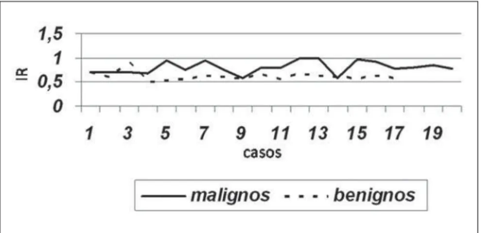
The mean RI was 0.80 for malignant nodules, and 0.61 for benign nodules (Table 4). Figure 1 shows the distribution of the RI values in benign and malignant nodules.
The t-test applied for comparison of two independent samples presented the follow-ing result: t = 5.435, with degree of free-dom ≅ 32 and p-value = 0.000, indicating that, at a significance level corresponding to α = 0.05 the results demonstrated a sta-tistically significant difference pr RI in the malignant nodules as compared with the
Table 3 Histological results for 19 malignant nodules.
Histological diagnosis
Lobular carcinoma
Ductal carcinoma in situ
Invasive ductal carcinoma
n
2
1
16
n, no. of nodules.
Table 2 Histological results for 18 benign nodules.
Histological diagnosis
Lipoma
Adenosis
Typical ductal hyperplasia
Fibroadenoma
Fibrosis
n
2
3
3
9
1
n, no. of nodules.
Table 1 Distribution of frequencies of nodules classification by Doppler.
Nodule
Benign
Malignant
Total
Quantity
18
19
37
Percentage
48.6%
51.4%
100.0%
Figure 1. Distribution of resistance index values for benign and malignant nodules.
benign nodules. Considering the variable “nodule type”, the cut-off value (RImedian =
243
Vascular resistance index in breast nodules
Radiol Bras. 2009 Jul/Ago;42(4):241–244 DISCUSSION
Ultrasonography plays a relevant role in the breast imaging evaluation. The techno-logical development can be noticed as a relevant component in the images analysis and processing. In this context, doppler-fluxometry has benefited from the continu-ous improvement in the temporal resolu-tion of modern ultrasonography equipment. Thus, both the color Doppler signal gener-ated in small vessels and their spectral analysis have demonstrated a significant improvement in the characterization of the blood flow in the breast tissue, allowing a better investigation of the vascularization pattern(16,17).
In the present study, the authors evalu-ated vascular RI of breast nodules greater than 1.0 cm in diameter. Later, the RI data were crossed with the histopathological result for each nodule. A statistically sig-nificant difference could be observed for the RI results in relation to the nodules clas-sification (benign or malignant), with the malignant results demonstrating a signifi-cantly higher vascular RI as compared with the benign results (0.80 versus 0.61, re-spectively, with p < 0.001). Similar results have already been observed in some stud-ies utilizing similar methods(18–22).
Several studies have analyzed the vas-cular RI within breast nodules in an attempt to differentiate malignant from benign le-sions(18–22). Choi et al. have observed that the RI exceeded 0.70 in more than 80% of
patients with malignant nodules with 80.9% sensitivity and 89.1% specificity(20). Peters-Engl et al. have also observed a RI of 0.70 as the best cut-off value to be uti-lized as an aid in the identification of ma-lignant nodules, with 82% sensitivity, 81% specificity, 70% positive predictive value and 89% negative predictive value(21). In the present study, the method sensitivity for malignant nodules identification was of 84.2%, with 88.9% specificity, 11.1% false-positive rate, and 15.8% false-negative rate for a RI cut-off value ≥ 0.69, a value prac-tically identical to the ones observed by the above mentioned studies.
Almost one decade after the publication of results from relevant studies about the role played by the vascular RI in the evalu-ation of breast nodules, the authors ob-served that the present study results were similar to those results, in spite of the sig-nificant technological development ob-served in ultrasonography equipment along this period of time. Consequently, it is un-derstood that such results can be consid-ered as duly validated and that seemingly they are not subjected to the variations re-sulting from the improvements in both hardware and software directly related to the Doppler function (temporal resolution) in currently available ultrasonography units. Finally, the Doppler technique prob-ably plays a role as an adjuvant to the grayscale in the evaluation of suspicious nodules. It is important to note that this method is not a diagnostic study.
Table 4 Descriptive resistance index measurements for both groups of nodules.
Resistance index Nodule Benign Malignant n 18 19 Mean 0.8047 0.6178 Standard deviation 0.1212 0.0859
Mean standard deviation
0.0278
0.0202
n, no. of nodules.
Table 5 Vascular resistance index versus nodule classification.
Vascular resistance index
Resistance index ≤ 0.69
Resistance index > 0.69
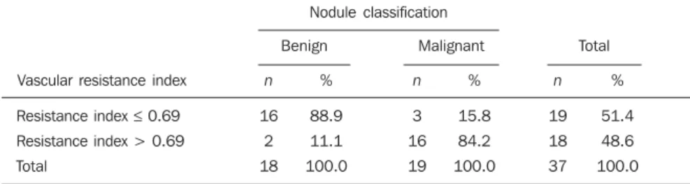
Total Nodule classification Total n 19 18 37 % 51.4 48.6 100.0 n 3 16 19 Malignant % 15.8 84.2 100.0 Benign n 16 2 18 % 88.9 11.1 100.0
n, no. of nodules.
CONCLUSIONS
The results of the present study demon-strate that a RI ≥ 0.69 in a nodule > 1 cm suggests a high risk for malignancy, and may represent additional information to be taken into consideration in the selection of lesions eligible for histopathological study.
REFERENCES
1. Souza LRMF, De Nicola H, De Nicola ALA, et al. Nódulos mamários: correlação entre caracte-rísticas ultra-sonográficas e achados histológicos em 433 nódulos biopsiados. Rev Imagem. 2005; 27:225–30.
2. Chala LF, Barros N. Avaliação das mamas com métodos de imagem. Radiol Bras. 2007;40(1):iv– vi.
3. Roveda Jr D, Piato S, Oliveira VM, et al. Valores preditivos das categorias 3, 4 e 5 do sistema BI-RADS em lesões mamárias nodulares não-palpá-veis avaliadas por mamografia, ultra-sonografia e ressonância magnética. Radiol Bras. 2007;40: 93–8.
4. Fleury EFC, Rinaldi JF, Piato S, et al. Apresenta-ção das lesões mamárias císticas à ultra-sonogra-fia utilizando a elastograultra-sonogra-fia. Radiol Bras. 2008; 41:167–72.
5. Stavros AT, Thickman D, Rapp CL, et al. Solid breast nodules: use of sonography to distinguish between benign and malignant lesions. Radiol-ogy. 1995;196:123–34.
6. Cosgrove DO, Kedar RP, Bamber JC, et al. Breast diseases: color Doppler US in differential diag-nosis. Radiology. 1993;189:99–104.
7. Raza S, Baum JK. Solid breast lesions: evalua-tion with power Doppler US. Radiology. 1997; 203:164–8.
8. Kook SH, Park HW, Lee YR, et al. Evaluation of solid breast lesions with power Doppler sono-graphy. J Clin Ultrasound. 1999;27:231–7.
9. Mehta TS, Raza S. Power Doppler sonography of breast cancer: does vascularity correlate with node status or lymphatic vascular invasion? AJR Am J Roentgenol. 1999;173:303–7.
10. Holcombe C, Pugh N, Lyons K, et al. Blood flow in breast cancer and fibroadenoma estimated by colour Doppler ultrasonography. Br J Surg. 1995; 82:787–8.
11. Yang WT, Metreweli C, Lam PKW, et al. Benign and malignant breast masses and axillary nodes: evaluation with echo-enhanced color power Dop-pler US. Radiology. 2001;220:795–802. 12. Birdwell RL, Ikeda DM, Jeffrey SS, et al.
Prelimi-nary experience with power Doppler imaging of solid breast masses. AJR Am J Roentgenol. 1997; 169:703–7.
13. Kubek KA, Chan L, Frazier TG. Color Doppler flow as an indicator of nodal metastasis in solid breast masses. J Ultrasound Med. 1996;15:835–41. 14. McNicholas MM, Mercer PM, Miller JC, et al. Color Doppler sonography in the evaluation of palpable breast masses. AJR Am J Roentgenol. 1993;161:765–71.
244
Schmillevitch J et al.
Radiol Bras. 2009 Jul/Ago;42(4):241–244 16. Mehta TS, Raza S, Baum JK. Use of Doppler
ul-trasound in the evaluation of breast carcinoma. Semin Ultrasound CT MR. 2000;21:297–307.
17. Tozaki M, Toi M, Miyamoto Y, et al. Power Dop-pler sonography of breast masses: correlation of Doppler spectral parameters with tumor angio-genesis and histologic growth pattern. J Ultra-sound Med. 2000;19:593–600.
18. Youssefzadeh S, Eibenberger K, Helbich T, et al.
Use of resistance index for the diagnosis of breast tumours. Clin Radiol. 1996;51:418–20.
19. Blohmer JU, Oellinger H, Schmidt C, et al. Com-parison of various imaging methods with particu-lar evaluation of color Doppler sonography for planning surgery for breast tumors. Arch Gynecol Obstet. 1999;262:159–71.
20. Choi HY, Kim HY, Baek SY, et al. Significance of resistive index in color Doppler ultrasonogram:
differentiation between benign and malignant breast masses. Clin Imaging. 1999;23:284–8.
21. Peters-Engl C, Medl M, Leodolter S. The use of colour-coded and spectral Doppler ultrasound in the differentiation of benign and malignant breast lesions. Br J Cancer. 1995;71:137–9. 22. Chao TC, Lo YF, Chen SC, et al. Color Doppler