90 Radiol Bras. 2011 Mar/Abr;44(2):90–96
Radiation dosimetry in patients and physicians during
percutaneous coronary angioplasty in Recife, Pernambuco,
Brazil
*
Dosimetria de pacientes e médicos em intervenções coronárias percutâneas em Recife, Pernambuco, Brasil
Maria do Socorro Rocha da Silva1, Helen Jamil Khoury2, Cari Borrás3, André Ferrão Oliveira4, Hugor Ferreira de Paiva Vianna5, Flavio Roberto Azevedo de Oliveira6, Flavio Adolpho Aranha Japyassú6, Flávio Braga Mota6
Objective: The present study was aimed at estimating the values of radiation doses received by physicians and patients during interventional cardiology procedures performed at a public hospital in the city of Recife, Pernambuco, Brazil.
Materials and Methods: Measurements were made in two cardiologists with more than ten years of experience and in 31 adult patients, with 22 of them being clinically followed-up after the procedure. The individual irradiation parameters were documented. Results: The values for maximum patients skin dose ranged between 612 and 8,642 mGy, achieving more than 2,000 mGy in 53% of the patients; such dose values may cause deterministic effects. As regards the physicians, the mean effective dose per procedure was 11 µSv, and the highest mean equivalent doses in the limbs were 923 µSv in the left foot, 514 µSv in the right foot, 382 µSv in the left hand, and 150 µSv in the left eye. Depending on the number of procedures, the doses received by the physician may exceed the dose limits established by the Brazilian and international standards. Conclusion: The obtained results indicate the necessity of adopting strategies for optimizing the radiological protection for both patients and physicians.
Keywords: Dosimetry in physicians and patients; Interventional cardiology; Dosimetry in interventional cardiology.
Objetivo: Este trabalho teve como objetivo estimar os valores de doses de radiação recebidas por médicos e pacien-tes em procedimentos intervencionistas cardíacos realizados em um hospital público na cidade de Recife, Pernambuco.
Materiais e Métodos: As medidas foram determinadas em 31 pacientes adultos, dos quais 22 tiveram acompanha-mento clínico após o procediacompanha-mento, e em dois cardiologistas com mais de dez anos de experiência. Parâmetros de irradiação para cada procedimento foram registrados. Resultados: Os valores obtidos para a dose absorvida máxima na pele do paciente variaram entre 612 e 8.642 mGy, sendo que 53% foram maiores que 2.000 mGy, valores estes que podem causar efeitos determinísticos. Com relação aos médicos, a dose efetiva média por procedimento foi de 11 µSv e os valores médios do equivalente de dose nas extremidades, mais altos, foram: 923 µSv no pé esquerdo, 514 µSv no pé direito, 382 µSv na mão esquerda e 150 µSv no olho esquerdo. Dependendo do número de procedi-mentos, as doses recebidas pelos médicos podem exceder os valores limites de doses estabelecidos pelas normas nacionais e internacionais. Conclusão: Os resultados obtidos sinalizam a necessidade de adoção de estratégias para otimização da proteção radiológica tanto de pacientes quanto de médicos.
Unitermos: Dosimetria de pacientes e médicos; Cardiologia intervencionista; Dosimetria em cardiologia intervencio-nista.
Abstract
Resumo
* Study developed at the Department of Nuclear Energy – Universidade Federal de Pernambuco (UFPE), Dosimetry and Nuclear Instrumentation Group, and Instituto de Medicina Inte-gral Professor Fernando Figueira (IMIP), Recife, PE, Brazil.
1. Master in Biomedical Engineering, Professor at Instituto Federal de Ciências, Educação e Tecnologia de Pernambuco, Recife, PE, Brazil.
2. PhD of Physics, Titular Professor at Department of Nuclear Energy, Universidade Federal de Pernambuco (UFPE), Recife, PE, Brazil.
3. PhD of Sciences, Visiting Professor at Department of Nuclear Energy, Universidade Federal de Pernambuco (UFPE), Recife, PE, Brazil.
Silva MSR, Khoury HJ, Borrás C, Oliveira AF, Vianna HFP, Oliveira FRA, Japyassú FAA, Mota FB. Radiation dosimetry in patients and physicians during percutaneous coronary angioplasty in Recife, Pernambuco, Brazil. Radiol Bras. 2011 Mar/Abr;44(2):90–96.
INTRODUCTION
Technological developments in the field of imaging methods utilizing ionizing ra-diations have allowed the performance of minimally invasive surgeries in different areas of medicine, particularly interven-tional cardiology. The safety, credibility and effectiveness of such invasive tech-niques have allowed the performance of 4. MD, Resident, Department of Surgery, Hospital das
Clíni-cas de Pernambuco, Recife, PE, Brazil.
5. MD, Resident, Department of Surgery, Hospital da Aeronáu-tica de Pernambuco, Recife, PE, Brazil.
6. MDs, Interventional Cardiologists, Department of Hemody-namics, Instituto de Medicina Integral Professor Fernando Fi-gueira (IMIP), Recife, PE, Brazil.
Mailing Address: Maria do Socorro Rocha da Silva. Grupo GDOIN-DEN, Universidade Federal de Pernambuco. Avenida Pro-fessor Luiz Freire, 1000, Cidade Universitária. Recife, PE, Bra-zil, 50740-540. E-mail: [email protected]
increasingly complex and sophisticated interventions, with the advantage of being less aggressive to patients yielding clinical benefits comparable to those of conven-tional surgery(1).
However, such procedures expose both the patient and the medical team to high ra-diation doses. Several studies have demon-strated high skin entrance doses in patients, causing erythema and, in some cases, skin necrosis(2,3).
The absorbed dose on the patient’s skin surface can be determined by means of appropriate dosimeters, or estimated from dose indicators parameters displayed by the angiographic system during the clinical procedure, such as “cumulative dose” (Ka,r)
and air kerma area product (PKA) (4,5). This
cumulative dose, internationally denomi-nated as cumulative reference point air kerma, is calculated from the irradiation parameters selected by the fluoroscopic angiographic C arm during the clinical pro-cedure. The reference for such calculation is a point denominated interventional ref-erence point (IRP), defined by the IEC 601-2-43 standard, and is located on the central axis, at 15 cm from the isocenter towards the focal point. The IRP is defined as the location that represents the beam entrance into the patient’s skin(5,6). The skin
ab-sorbed dose in the most irradiated region and the Ka,r are considered as the
appropri-ate magnitudes for estimating the risk for occurrence of deterministic effects (such as erythema, alopecia and sterility), while PKA
is utilized for estimating the occurrence of stochastic effects (such as cancer)(5,6).
The International Commission on Ra-diological Protection (ICRP) recommends that, in interventional procedures, the val-ues and location of maximum cumulative skin dose (MSD) be registered on the hos-pital files or on the patient’s records, when-ever such dose exceeds 1,000 mGy in pro-cedures which may need to be repeated, such as in cardiology, in case of restenosis (reoccurrence of obstruction of an artery previously treated by means of angio-plasty). The ICRP and several guidelines also recommends clinical follow-up and recording of such data in case of any inter-ventional procedure in which MSD ex-ceeds 3,000 mGy, Ka,r exceeds 5,000 mGy
or PKA exceeds 500 Gy.cm 2(5,7).
Besides the patient who is directly ex-posed to the primary radiation beam, pro-fessionals, particularly physicians, also re-ceive high doses as they are exposed to the scattered radiation, because they need to be in close proximity with the patients during the procedures. The radiation dose values may also be high as a function of the num-ber of procedures performed by the physi-cian. Studies developed by the American Society of Cardiovascular and Interven-tional Radiology demonstrated that high ra-diation doses have been observed even in cases where modern apparatuses are uti-lized by qualified professionals(8,9).
Accord-ing to Filippova, cardiologists are the pro-fessionals who receive the highest doses(10).
The present study was aimed at evaluat-ing the radiation dose levels in interventional cardiology procedures such as transluminal coronary angioplasty at a hospital where such procedures are routinely performed. The relevance of the present study is to pro-vide data for physicians, contributing to the optimization of the radiation dose reduction both for patients and for the medical team.
MATERIALS AND METHODS
In the present study, 34 procedures of percutaneous transluminal coronary artery angioplasty, with and without placement of stents, were analyzed. All the procedures were performed in a public hospital in the city of Recife, PE, Brazil, between April of 2007 and September of 2009.
The equipment utilized for such proce-dures was an Integris Allura 12 monoplanar angiography system (Philips Medical Sys-tems; Best, The Netherlands), equipped with a 3-phase X-ray generator and a micropro-cessor high frequency converter, nominal voltage 40–150 kV and maximum current of 1 A. The X-ray tube had focal points at 0.5 and 0.8 mm and was equipped with two collimators, one of them of the iris type (au-tomatic) and the other of the rectangular type (manual). The X-ray beam filtration was done with 2.5 mmAl at 100 kV, besides added filtration with aluminum and copper selected by software. The image intensifier had a diameter of 30 cm, with four fields of view (30/22/17/12 cm), selectable by the physician. Neither the leaded drape nor a lead glass window was installed in the
equipment. Such devices aid in the reduc-tion of the incidence of scattered radiareduc-tion on the physician(11).
All the procedures were performed with the normal pulsed fluoroscopy mode (30 pulses per second) and pulsed cinegraphy mode (15 images per second), with normal images matrix (512 × 512). In the fluoros-copy mode, the images are viewed in real time and are not recorded, while at the cinegraphy mode the images are recorded in a cinegraphic sequence (filming) to be viewed after the procedure. Fluoroscopy and cinegraphy modes may be pulsed or continuous, according to the X-rays pro-duced by the generator.
The randomly selected patients sample included 31 adults, with no restriction re-garding gender, age or weight, and was based on the elective scheduling of the hos-pital hemodynamics unit. Three of the 31 pa-tients underwent two procedures over the study period, and 21 patients were clinically followed-up after the procedure, with the purpose of checking for skin lesions. Nine of such patients had their backs photo-graphed and were evaluated by the physi-cians both before and 14 days after the pro-cedure, in compliance with international recommendations(8).
The present study was approved by the Committee for Ethics in Research of the hospital. The patients or their guardians who agreed to participate in the study were appropriately informed on the risks and benefits of the research and signed a term of free and informed consent in compliance with the requirements defined by the Reso-lution CNS 196/96 that regulates research involving human beings in Brazil(12).
The physicians sample included two professionals specialized in interventional cardiology, from the hospital medical staff. The dosimetric monitoring was performed on the physician who performed the pro-cedure. The physician assistant, when present, was not monitored.
Radiation on patients
The patients’ dosimetry was approached in two manners as follows:
1. Evaluation of “cumulative dose” (Ka,r)
and PKA
proce-dures, the following data were recorded: level (normal/high/low) and mode (continu-ous/pulsed) of fluoroscopy and cinegraphy; total number of images acquired in the cinegraphy mode; and fluoroscopy time (min). Also, the angle and rotation of the C-arm (X-ray tube and image intensifier) were recorded for each projection of the radiation fields resulting in the different magnifications of the images intensifier; the distances between the focal point and the intensifier (source to image detector distance – SID) and between the focal point and the patient (source to skin distance – SSD); and irradiation parameters such as voltage (kV), pulse width (ms) and current (mA) applied to the X-ray tube.
Based on the irradiation parameters (kV, mA and ms) which are automatically se-lected by the equipment according to the utilized projections, the patient character-istics and radiographic contrast, the angio-graphic equipment calculates the Ka,r Such
calculation is performed with reference to a (IRP) point, without considering back-scattering, as defined by the IEC 601-2-43 Standard(6). From the value of K
a,r it was
possible to calculate the incident air kerma Ka,i (mGy) at the point corresponding to the
patient skin surface (at the level of the table surface), taking into consideration the variation of radiation intensity with the inverse square distance. Considering that Ka,r does not account for the contribution
of the radiation scattered by the patient, for determining the patient’s skin entrance surface air kerma (Ka,e) the Ka,i was
multi-plied by a backscattering factor (1.3). Such backscattering factor represents the contri-bution of radiation that is scattered from the central beam due to the presence of the sur-gical table, the mattress and the patient himself.
With the distance from the intensifier to the X-ray tube and the diameter of the ir-radiation field, the irradiated area was cal-culated for each angle/rotation at the level of the surgical table, by utilizing the appro-priate geometrical factors. With such val-ues, PKA was calculated by multiplying Ka,i
by the obtained area. Total PKA was
calcu-lated by summing up the partial PKA at the
different projections utilized in the proce-dure.
2. Radiochromic film dosimetry
In order to evaluate the radiation distri-bution and location of the maximum ab-sorbed dose on the backs of nine patients, radiochromic films of the GafChromic XR V2 (International Specialty Products; New Jersey, USA) type were utilized. Such films are available in the 35 × 43 cm format and are developed for uses in dosimetry in pa-tients during fluoroscopy and radiotherapy procedures. The films were positioned over the surgical table at the level of the poste-rior chest area, with the white face of the film positioned towards the entrance of the X-ray beam, according to the film manu-facturer’s usage instructions(13). The film
was wrapped in a black plastic envelope to protect it from light as well as from liquid products utilized in the asepsis of the pa-tient.
The radiation changes the colors of the radiochromic film in such a manner that the most darkened areas correspond to the most irradiated areas. The entrance surface air kerma – Ka,e (mGy) – was estimated from
the readings of the optical density on these darkened areas, 24 hours after the proce-dure, in order to assure the stability of the film’s optical absorbance, as recommended by the manufacturer, and by the application of calibration factors established at the Laboratory of Metrology of Ionizing Ra-diations, Department of Nuclear Energy of
Universidade Federal de Pernambuco (LMRI/DEN/UFPE)(13,14).
In order to establish a relationship be-tween the location of the most irradiated areas on the films and the regions on the backs of the patients, the film was divided into nine regions as follows: upper right side (UR), middle right side (MR) and lower right side (LR), upper middle (UM), central middle (CM), lower middle (LM), and upper left side (UL), middle left side (ML) and lower left side (LL), as shown on Figure 1.
The maximum skin dose (MSD) on the patient’s skin surface was calculated by multiplying the highest value of Ka,e,
de-termined by the optical density on the sur-face of the film, by an energy absorption coefficient (1.054) that relates the absorp-tion of radiaabsorp-tion on human tissue with the absorption of radiation in the air, as in the case of Ka,e.
Radiation on physicians
On the physicians, the dosimetry was performed with 38 TL dosimeters (TLD-100) distributed in pairs as shown on Fig-ure 2.
The effective dose E (mSv) was esti-mated from the measurements obtained with the TL dosimeters placed at the level of the neck, over the thyroid protector, and another at the level of the waist, under the
Figure 1. Regions of the radiochromic film. Upper right
(UR)
Middle right (MR)
Lower left (LL) Lower right
(LR)
Upper middle (UM)
Central middle (CM)
Lower middle (LM)
Upper left (UL)
Figure 2. Location of the TL dosimeters over the apron and on the body of the physicians: face – fore-head (1) right eye region (2) and left eye region (3); upper limbs –hands, at the wrist level, inside the procedure glove [right hand (4) and left hand (5)]; thyroid region – on the external surface of the thyroid shield (8) and inside the thyroid shield (9); torso – at the level of the shoulders, over the apron, right side (6) and left side (7); thorax - lower abdomen – at waist level: on the apron, right side (10), left side (11), at the middle, over the apron (12) at the middle, underneath the apron (13); at the knee level, over the apron, right side (14) and left side (15), at the middle, over the external surface (16) and inside the apron (17); lower limbs – feet, anterior region, right foot (18) and left foot (19).
protective apron (TL 8 and 13, respectively, shown on Figure 2), and utilizing the Niklason algorithm(15), as follows:
E = 0.02 (HO – HU) + HU
where: HO corresponds to the value of
HP(0.07) (mSv) for the TL (8) dosimeter
mea-surement and HU is the value of HP(10)
(mSv) obtained by the TL (13) dosimeter. The readings from the TL dosimeters (8 and 13) were corrected by means of the coefficients defined at ICRP 74(16) for the
conversion of such measurements into the corresponding personal dose equivalents HP(0.07) and HP(10),respectively.
RESULTS
Radiation on patients
The 31 evaluated adult patients (39% women and 61% men) were between 36 and 78 years old (60 ± 11 years), weighted between 49 and 116 kg (73 ± 14 kg) and were between 1.50 e 1.80 m tall (1.63 ± 0.10 m). The values in parenthesis repre-sent the mean standard deviation (M ± 1s). The results of the dosimetric evaluation by the described methods were the follow-ing:
1.Evaluation of “cumulative dose”(Ka,r)
and PKA
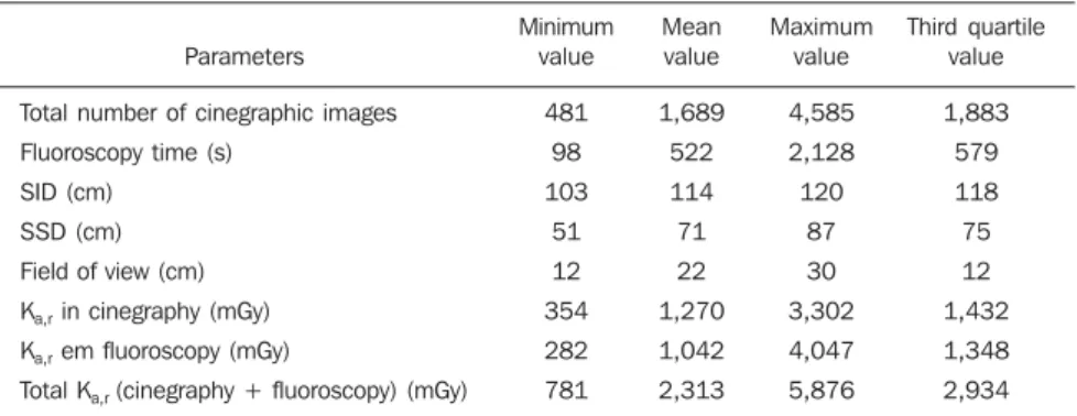
Minimum, mean, maximum and third quartile values of the parameters recorded by the equipment during the procedures are shown on Table1.
On Table 1, one observes that the Ka,r
values, both at fluoroscopy and cinegraphy, are practically the same, in spite of the dif-ferences between patients and procedures. On Table 2, the values for Ka,r, Ka,e, PKA
are expressed at the level of skin entrance surface. As regards PKA values, one
ob-serves that none of the patients exceed 500 Gy.cm2 in a single procedure.
2. Radiochromic film dosimetry
The results for absorbed dose distribu-tion by utilizing the radiochromic film in nine patients are summarized on Table 3.
The results for localization of the maxi-mum absorbed dose indicated higher doses at the middle right side (MR) and lower middle side (LM) of the film, correspond-ing to the right lower middle and lower
Table 1 Data recorded by the angiography equipment during the monitored procedures.
Parameters
Total number of cinegraphic images Fluoroscopy time (s)
SID (cm) SSD (cm) Field of view (cm) Ka,r in cinegraphy (mGy) Ka,r em fluoroscopy (mGy)
Total Ka,r (cinegraphy + fluoroscopy) (mGy)
Minimum value
481 98 103
51 12 354 282 781
Mean value
1,689 522 114 71 22 1,270 1,042 2,313
Maximum value
4,585 2,128 120
87 30 3,302 4,047 5,876
Third quartile value
1,883 579 118 75 12 1,432 1,348 2,934
SID, source to image detector distance; SSD, source to skin distance.
Note: The Ka,r values are expressed at the IRP reference point, as presented on the angiography equipment,
without correction for the backscattering factor and geometry for the table surface.
Protection goggles
Thyroid shield
LEFT SIDE RIGHT SIDE
Protective apron
TL dosimeters over the apron
Table 3 Minimum, mean, maximum and third quartile values for the location of maximum absorbed dose on skin surface, estimated by region of the radiochromic film, in nine patients.
Region Back (right side) Back (middle) Back (left side) Location Upper (UR) Middle (MR) Lower (LR) Upper (UM) Middle (CM) Lower (LM) Upper (UL) Middle (ML) Lower (LL) MSD (mGy) Minimum value 328.0 328.0 268.0 297.0 112.0 214.0 214.0 127.0 248.0 Mean value 1,255.0 2,454.0 1,011.0 947.0 466.0 1,681.0 362.0 912.0 555.0 Maximum value 3,230.0 3,967.0 2,277.0 3,230.0 1,351.0 4,180.0 649.0 1,960.0 1,218.0 Third quartile value 2,285.0 3,924.0 1,383.0 636.0 721.0 2,398.0 396.0 1,363.0 768.0
middle regions of the patients’ backs. Such locations are consistent with clinical stud-ies, as 65% of the patients presented lesions in the left coronary vessels, which causes the most irradiated areas to be located be-tween the middle and the right side of the backs.
The images with the dose distribution on the backs of the two patients with the highest doses are shown on Figure 3. At the first image (Figure 3A) one observes that in spite of the utilization of different angles, the MSD is located in a region with fields overlapping; at the second image (Figure 3B), the MSD is a result of the projection most utilized in the procedure.
The maximum absorbed dose on skin surface, determined with radiochromic film (MSDFILM), was higher than 2,000 mGy in
63% of the patients. In spite of the possi-bility of the occurrence of erythema after exposure levels > 2,000 mGy, such reaction was not observed on the skin of any of the patients. The results obtained for both the localization of maximum dose, as well as for determining the maximum dose value have demonstrated the superiority and practicity in utilizing the radiochromic film.
The analysis of results obtained for the patients monitored with the radiochromic film allowed observing that Ka,r maximum, i.e.,
the Ka,r in the projection most utilized
dur-ing the procedure and the maximum ab-sorbed dose measured with the film, pre-sented a significant correlation (r2 = 0.808).
Based on such relation (Ka,r maximum and
MSDFILM), it was possible to estimate the
maximum absorbed dose for the other pa-tients in the study (MSDKa,r max). The
indi-vidual results for maximum absorbed dose measured with the radiochromic films (MSDFILM) and estimated from de relation
with Ka,r maximum (MSDKa,r max) are shown on
Table 2.
The maximum absorbed dose on pa-tients’ skin surface was higher than 1,000 mGy in 94% (32 out of 34) of the evalu-ated procedures, and higher than 3,000 mGy in 29% (10 out of 34).
The patients that presented maximum absorbed dose higher than 3,000 mGy or cumulative air kerma higher than 5,000 mGy were contacted and did not report the occurrence of lesions.
Table 2 Ka,r, Ka,e, PKA and MSD measured and estimated by means of radiochromic films, on patients’ skin entrance surface.
ID F1 F2 F3 F4 F5 F6 F7 F8 F9 F10 F11 F12 F13 M1 M2 M3 M4 M5 M6 M7 M8 M9 M10 M11 M12 M13 M14 M15 M16 M17 M18 M19 M20 M21 Ka,r (mGy) 3,961.7 1,378.2 3,852.1 2,993.5 2,989.8 1,665.2 1,412.8 805.0 1,158.2 5,876.1 2,101.3 1,068.2 1,593.5 3,503.0 1,643.5 1,832.6 2,765.4 1,459.3 2,508.3 2,558.2 1,661.9 1,626.1 2,237.7 5,835.6 1,760.4 1,425.4 3,130.5 2,133.7 1,232.6 2,032.8 781.2 2,561.0 3,388.2 1,693.2 Ka,e (mGy) 3,976.0 1,383.2 3,866.0 3,004.3 3,000.6 1,671.2 1,417.9 807.9 1,162.4 5,897.3 2,108.9 1,072.0 1,599.2 3,515.6 1,649.4 1,839.2 2,775.4 1,464.6 2,517.3 2,567.4 1,667.9 1,632.0 2,245.8 5,856.6 1,766.7 1,430.5 3,141.8 2,141.4 1,237.0 2,040.1 784.0 2,570.2 3,400.4 1,699.3 PKA (Gy.cm2)
135 43 131 89 92 52 46 28 73 228 82 32 55 143 62 72 84 45 85 84 79 77 74 240 60 61 166 70 73 87 28 99 113 57 MSDFILM (mGy) — — — — 2,779.9 3,228.5 1,350.8 1,013.6 — — — — — — — — — — 3,965.6 4,180.0 2,306.9 — — — — — — — — — — 648.6 — — MSD(Ka,r max) (mGy) 2,368.0 1,301.3 2,985.8 4,232.4 3,949.0 1,689.2 1,587.7 1,288.8 1,185.8 8,642.3 2,692.5 1,545.0 2,012.6 1,962.6 1,420.4 2,231.9 1,833,4 611.6 3,825.7 3,845.1 2,090.8 1,804.2 2,670.9 6,828.2 1,345.0 2,725.9 1,241.8 4,729.7 3,276.9 1,242.7 1,551.6 710.3 4,365.0 2,169.1
DISCUSSION
As regards dose parameters, the com-parison of the results from the present study with those published in the literature (see Table 5) shows that the recorded values are comparable with those reported by Mavri-kou et al.(17) and Tsapaki et al.(18) and with
the diagnostic reference levels (DRL) sug-gested by the IAEA(19) (TF = 22 min, P
KA
= 125 Gy.cm2) and by the European
DI-MOND III project(20).
Based on the results obtained in the present study, where 94% of the patients presented maximum absorbed dose > 1,000 mGy and, considering that restenosis oc-curs in 40% of patients submitted to per-cutaneous coronary interventions within the first six months following the proce-dure(21,22), it is recommended that the
he-modynamics department of the hospital records the doses and perform the clinical follow-up of all patients submitted to angioplasty procedures (transluminal coro-nary angioplasty), to evaluate both the oc-currence of immediate as well as late ef-fects (cancer).
As regards dosimetry on physicians, the doses on the legs were very high and could be reduced by means of an accessory such as a lower lead curtain placed on the lower part of the table, according to recommen-dations on Ordinance 453 of the Brazilian Ministry of Health(23). The mean dose per
procedure on the left eye of the physicians was 69 µSv. Such value could be signifi-cantly reduced in case the upper protective Figure 3. Image demonstrating the dose distribution (Ka,e) on the film’s surface of two patients who presented the highest doses.
A B
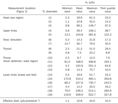
Table 4 Air kerma measured in different regions of the apron (inside and outside) and on physicians’ skin surface.
Measurement locations (Figure 2)
Head (eye region)
Upper limbs
Torso (shoulder)
Thyroid
Thorax
(lower abdomen: waist region)
Lower limbs (knees and feet)
Effective dose (µSv.procedure–1)
HT (µSv)
Minimum value
2.3 1.1 9.8
3.6 13.2
0.3 14.7
2.5 0.8
2.5 61.0
4.2 0.8
5.5 170.8
66.2 4.4 79.0 119.6
1.1
Mean value
19.9 10.8 69.2
39.4 104.8
14.3 30.7
21.3 7.5
6.7 168.0 104.6 13.3
19.6 310.2 227.6 14.3 236.2 309.9
10.8
Maximum value
61.3 42.0 149.7
106.1 381.6
31.8 76.0
51.0 20.2
12.0 406.6 551.4 70.7
51.7 465.1 730.7 35.5 514.1 922.7
44.0
Third quartile value
23.3 14.3 97.0
38.7 122.3
17.3 35.5
29.4 6.8
7.9 164.1
64.6 10.0
24.2 350.8 243.5 16.2 265.9 327.8
10.4 TL dosimeter
(1) (2) (3)
(4) (5)
(6) (7)
(8) (9)
(10) (11) (12) (13)
(14) (15) (16) (17) (18) (19)
Radiation on physicians
The mean effective dose per procedure on the physicians was 10.8 ìSv. The mini-mum, mean, maximum and third quartile values of the dose equivalents obtained by the TL dosimeters on the bodies of the phy-sicians are shown on Table 4.
The mean values for the dose equiva-lents on the extremities, which were the highest ones, were the following: 923 ìSv
shield of the equipment or protection goggles were utilized. In general, physi-cians do not wear such goggles during the procedures either because they are not com-fortable or because they are not available at the unit. However, several studies have demonstrated the occurrence of injuries to the crystalline lens of interventional phy-sicians as a consequence of the scattered radiation received during such proce-dures(24).
Considering the limit values for the dose equivalents established by both na-tional and internana-tional standards (CNEN, ICRP), corresponding to 500 mSv/year for extremities and 150 mSv/year for the crys-talline lens, it is estimated that such limits would be exceeded with 20 to 32 weekly transluminal coronary angioplasty proce-dures.
CONCLUSION
The results of the present study indicate the necessity of adopting strategies to op-timize radiological protection both for pa-tients and physicians. The first steps in that direction could be the adoption of measures to control the cumulative dose, clinical follow-up of patients, and utilization of protective accessories by physician (lower lead curtains and protection goggles, among others).
REFERENCES
1. Atik E. Cateterismo cardíaco intervencionista na cardiologia pediátrica. O posicionamento médico quanto às aplicações atuais e perspectivas. Arq Bras Cardiol. 2002;79:443–5.
2. United Nations Scientific Committee on the Ef-fects of Atomic Radiation. Sources and efEf-fects of ionizing radiation. UNSCEAR 2000 Report to the General Assembly, with scientific annexes –
an-nexes D and E. [acessado em 3 de março de 2008]. Disponível em: http://www.unscear.org/ docs/reports/annexd.pdf
3. Miller DL, Balter S, Wagner LK, et al. Quality im-provement guidelines for recording patient radia-tion dose in the medical record. J Vasc Interv Radiol. 2004;15:423–9.
4. Miller DL, Balter S, Cole PE, et al. Radiation
doses in interventional radiology procedures: the RAD-IR study. Part I: overall measures of dose. J Vasc Interv Radiol. 2003;14:711–27.
5. Stecker MS, Balter S, Towbin RB, et al. Guide-lines for patient radiation dose management. J Vasc Interv Radiol. 2009;20(7 Suppl):S263–73.
6. International Electrotechnical Commission. IEC report 60601. Medical electrical equipment – Part 2-43: particular requirements for the safety of x-ray equipment for interventional procedures. Geneva, Switzerland: International Electrotech-nical Commission; 2000.
7. International Commission on Radiological Pro-tection. ICRP Publication 103: The 2007 Recom-mendations of the International Commission on Radiological Protection. Annals of the ICRP (v. 37/2-4). New York, NY: Elsevier; 2007.
8. International Commission on Radiological Pro-tection. ICRP Publication 85. Avoidance of radia-tion injuries from medical intervenradia-tional proce-dures. Ann ICRP 30(7-67). Oxford/New York: Pergamon Press; 2000.
9. Vano E. Radiation exposure to cardiologists: how it could be reduced. Heart. 2003;89:1123–4. 10. Filippova I. Patient and staff doses in radiology
and cardiology in Estonia. Radiat Prot Dosimetry.
2005;117:59–61.
11. Philips Medical System. Integris Allura 12 & 15: technical handbook. Netherland: Philips Elec-tronics N.V.; 2003.
12. Brasil. Ministério da Saúde. Conselho Nacional de Saúde. Diretrizes e normas regulamentadoras de psquisa envolvendo seres humanos. Resolu-ção CNS nº 196, de 10 de outubro de 1996. [aces-sado em 15 de setembro de 2009]. Disponível em: http://conselho.saude.gov.br/resolucoes/1996/ Reso196.doc
13. International Specialty Products. GAFCHROMIC®
XR-RV2 dosimetry film. Characteristic perfor-mance data. [acessado em 10 de março de 2009]. Disponível em: http://online1.ispcorp.com/_lay-outs/Gafchromic/
14. Silva MSR, Khoury HJ, Borrás C, et al.
Calibra-ção do filme radiocrômico GAFCHROMIC XR-RV2 para radiologia. Rev Bras Fís Méd. 2010; 4:45–8.
15. Kim KP, Miller DL, Balter S, et al. Occupational radiation doses to operators performing cardiac catheterization procedures. Health Phys. 2008; 94:211–27.
16. International Commission on Radiological Pro-tection. ICRP Publication 74: Conversion coeffi-cients for use in radiological protection against external radiation. Annals of the ICRP (v. 37/2-4). New York, NY: Pergamon; 1997.
17. Mavrikou I, Kottou S, Tsapaki V, et al. High pa-tient doses in interventional cardiology due to physicians’ negligence: how can they be pre-vented? Radiat Prot Dosimetry. 2008;129:67–70.
18. Tsapaki V, Patsilinakos S, Voudris V, et al. Level of patient and operator dose in the largest cardiac
centre in Greece. Radiat Prot Dosimetry. 2008;
129:71–3.
19. International Atomic Energy Agency. Establish-ing guidance levels in X ray guided medical interventional procedures: a pilot study. Safety Reports Series nº 59. Vienna: IAEA; 2009.
20. DIMOND website. Patient and staff dosimetry database/Interventional cardiology/Diagnostic reference levels for CA and PTCA. [acessado em 10 de janeiro de 2010]. Disponível em: http:// www.dimond3.org/WEB_DIMOND3/home.htm 21. Martinez EE, Ribeiro EE. Hemodinâmica e car-diologia intervencionista: abordagem clínica. Barueri, SP: Manole; 2008.
22. Unimed. Central de Serviços Auxiliares. Câmara Técnica de Medicina Baseada em Evidências. Avaliação das evidências comparativas entre os
stents coronarianos recobertos por drogas: Siro-limus (Cypher®) x Paclitaxel (Taxus®). [acessado
em 10 de janeiro de 2010]. Disponível em: http:// www.unimedvtrp.com.br/autoriza/evidencias_ aprovadas/avaliacao_stent_coronariano– recoberto.pdf
23. Brasil. Ministério da Saúde. Secretaria de Vigi-lância Sanitária. Diretrizes de proteção radioló-gica em radiodiagnóstico médico e odontológico.
Portaria/MS/SVS no 453, de 1º de junho de 1998.
Brasília, DF: Diário Oficial da União, 2 de junho de 1998.
24. Lie qq, Paulsen GU, Wrhni T. Assessment of
effective dose and dose to the lens of eye for the interventional cardiologist. Radiat Prot Dosim-etry. 2008;132:313–8.
Table 5 Values observed in the present study and in other published studies.
Parameters
Number of procedures FT(min)
Number of images Ka,r (mGy) PKA (Gy.cm2)
Mavrikou et al.(17) (2008)
Mean
214 21.4 1,493 2,100 158
Tsapaki et al.(18) (2008)
Mean / (min–max)
203 10.4 / (3.2–53) 1,257 / (398–5,940)
— 62 / (8–861)
Present study (2009)
Mean / (min–max) / third quartile
34 11 / (2.6–37.2) / 12 1,682 / (481–4,585) / 1,883 2,313 / (781.2–5,876.1) / 2,934
174 / (60.2–503.6) / 199
DRL(20) (2010)
Third quartile
— 20 1,700
— 110