Revista Brasileira de Anestesiologia 545 Vol. 59, No 5, Setembro-Outubro, 2009
RESUMO
Gimenez S, Teixeira ML, Myashiro R, Carmona MJC, Auler Jr JOC, Malbouisson LMS - Avaliação Pulmonar em Crianças Portadoras de Cardiopatia Congênita Acianótica e Hiperfluxo Pulmonar Através de Tomografia Computadorizada.
JUSTIFICATIVA E OBJETIVOS: Disfunção respiratória é frequente em crianças com cardiopatias congênitas acianóticas com hiperfluxo pul-monar (CCAHP), porém pouco é conhecido sobre a estrutura pulmo-nar destes pacientes. O objetivo deste estudo foi quantificar os volumes de gás e tecido e a distribuição da aeração pulmonar nes-ta população.
MÉTODOS: Após aprovação do Comitê de Ética institucional e obten-ção do consentimento escrito pós-informado, foram obtidas tomografias computadorizadas torácicas em sete crianças com CCAHF. As imagens pulmonares direita e esquerda foram contornadas em todas as imagens e os volumes e pesos pulmonares foram computados a partir dos da-dos volumétricos. As comparações entre esquerda e direita foram ana-lisadas usando teste t de Student pareado e as correlações através de regressão exponencial.
RESULTADOS: A idade mediana foi 20 meses e o peso foi de 9,9 kg.
Volume pulmonar total (VPT) foi de 66,7 ± 23,1 mL.kg-1, o de tecido
33,5 ± 15,7 mL.kg-1 e o de gás 33,1 ± 8,3 mL.kg-1. O pulmão direito
re-presentou 57,9% e o pulmão esquerdo 42,1% do VPT (p < 0,001). O volume pulmonar de gás à direita foi 60,5% do volume de gás total (p
< 0,001) e a quantidade de parênquima pulmonar normalmente aerado foi significativamente menor à esquerda (27,6 ± 6,8 vs. 18,1 ± 8% p < 0,001).
CONCLUSÕES: As crianças portadoras de CCAHP apresentaram au-mento no volume de tecido pulmonar maior que o esperado, possivel-mente por edema intersticial. A aeração pulmonar está reduzida no pulmão esquerdo pela compressão imposta pelo coração ao pulmão subjacente.
Unitermos: COMPLICAÇÕES: edema pulmonar; DOENÇAS, Congênita: cardiopatia; EXAMES COMPLEMENTARES: tomografia computadori-zada espiral.
SUMMARY
Gimenez S, Teixeira ML, Myashiro R, Carmona MJC, Auler Jr JOC, Malbouisson LMS – Computed Tomography in Pulmonary Evaluation of Children with Acyanotic Congenital Heart Defect and Pulmonary Hyperflow.
BACKGROUND AND OBJECTIVES: Respiratory dysfunction is common in children with acyanotic congenital heart defects (ACHD) with pulmonary hyperflow; however, little is known about the pulmonary structure of those patients. The objective of this study was to quantify the volumes of air and tissue, as well as the distribution of pulmonary aeration in this population.
METHODS: After approval by the Ethics Committee of the institution and signing of an informed consent, seven children with ACHD with pulmonary hyperflow underwent computed tomographies of the chest. All images included the left and right pulmonary contour, and pulmonary volumes and weight were calculated using volumetric data. Paired Student t test was used to compare left and right, and exponential regression was used for correlations.
RESULTS: Patients had a mean age of 20 months and weight of 9.9
kg. Total pulmonary volume (TPV) was 66.7 ± 23.1 mL.kg-1, tissue
vo-lume of 33.5 ± 15.7 mL.kg-1, and air volume of 33.1 ± 8.3 mL.kg-1. The
right lung represented 57.9% of TPV and the left, 42.1% (p < 0.001). The pulmonary volume of air on the right was 60.5% of the total air volume (p < 0.001), and the volume of pulmonary parenchyma normally aerated was significantly lower on the left (27.6 ± 6.8 vs. 18.1 ± 8%, p < 0.001).
CONCLUSIONS: The volume of lung tissue was greater than expected in children with ACHD with pulmonary hyperflow, possibly due to interstitial edema. Pulmonary aeration is reduced in the left lung due to the compression of the lung by the heart.
Keywords: COMPLEMENTARY EXAMS: spiral computed tomography; COMPLICATIONS: pulmonary edema; DISEASES, Congenital: car-diopathy.
Avaliação Pulmonar em Crianças Portadoras de Cardiopatia
Congênita Acianótica e Hiperfluxo Pulmonar Através de
Tomografia Computadorizada *
Computed Tomography in Pulmonary Evaluation of Children with
Acyanotic Congenital Heart Defect and Pulmonary Hyperflow*
Solange Gimenez 1, Mariana Limeira Teixeira 2, Rodrigo Myashiro 3, Maria José Carvalho Carmona, TSA 4,
José Otávio Costa Auler Jr, TSA 5, Luiz Marcelo Sá Malbouisson, TSA 6
* Recebido do Instituto do Coração (InCor) do Hospital das Clínicas da Facul-dade de Medicina da UniversiFacul-dade de São Paulo (HC/FMUSP), São Paulo, SP
1. Doutora em Ciências pela USP; Médica Assistente do Serviço de Anestesiologia e Terapia Intensiva do InCor do HC/FMUSP
2. Médica Residente da Disciplina de Anestesiologia da FMUSP 3. Acadêmico de Medicina da FMUSP
4. Professor Associada da Disciplina de Anestesiologia da FMUSP; Especia-lista em Terapia Intensiva – AMIB; Diretora do Serviço de Anestesiologia e Terapia Intensiva Cirúrgica do HC/FMUSP
5. Professor Titular da Disciplina de Anestesiologia da FMUSP; Diretor do Ser-viço de Anestesiologia e Terapia Intensiva Cirúrgica do InCor do HC/FMUSP; Diretor Clínico do HC/FMUSP
6. Doutor em Ciências - Médico Supervisor da UTI-Anestesia do Instituto Cen-tral do HC/FMUSP
Apresentado (Submitted) em 30 de janeiro de 2009 Aceito (Accepted) para publicação em 19 de maio de 2009
Endereço para correspondência (Correspondence to): Dr. Luiz Marcelo Sá Malbouisson
Instituto Central
Hospital das Clínicas da Faculdade de Medicina da USP Av. Enéas de Carvalho Aguiar, 255
Computed Tomography In Pulmonary
Evaluation Of Children With Acyanotic
Congenital Heart Defect And Pulmonary
Hyperflow
Solange Gimenez, M.D. 1; Mariana Limeira Teixeira, M.D. 2; Rodrigo Myashiro 3; Maria José Carvalho Carmona, TSA, M.D. 4; José Otávio Costa Auler Jr, TSA, M.D. 5; Luiz Marcelo Sá Malbouisson, TSA, M.D. 6
INTRODUCTION
Acyanotic congenital heart defects with pulmonary hyperflow represent a group of congenital cardiopathies characterized by the presence of intracardiac or large vessels malformation that leads arterialized blood after passing through the pul-monary circulation to flow from the left to the right chambers of the heart or pulmonary artery. Those anomalies in the formation of the heart occur during intra-uterine life and include a large variety of cardiocirculatory malformations, ranging from patent ductus arteriosus to absence of interatrial and interventricular septi. Physiologically, the development of clinical manifestations depends on the mag-nitude of the flow through the right-left communication and it is essentially translated by the presence of pulmonary congestion of varying degrees and cardiomegaly1. A conside-rable percentage of this population undergoes surgical correction of cardiac defects in the first two years of life to avoid the harmful consequences of persistent hyperflow on the pulmonary circulation.
Despite advances on the knowledge of the physiology of pulmonary circulation in this population2,3, little is known on the structure and distribution of air in the pulmonary pa-renchyma of children with acyanotic congenital cardiopathy with pulmonary hyperflow. Several reasons hinder the in vivo investigation of the structure and function of the respiratory system in this age group, including the availability of few accurate non-invasive methods, incapacity of patients to cooperate with exams such as spirometry, the need of seda-tion to perform exams in small children, and the low incidence of this group of disorders in the general population.
In adult patients in different clinical conditions and in experimentation animals helical computed tomography allows the quantitative and qualitative evaluation of pulmo-nary changes using volumes and X-ray attenuation by the pulmonary parenchyma4,5.
METHODS
After approval by the Ethics Commission of the institution and signing of the informed consent by the legal guardian, seven children with acyanotic congenital cardiopathy with pul-monary hyperflow admitted to the Instituto do Coração (InCor) of the Hospital das Clínicas of the Medical School of Universidade de São Paulo for surgical correction were investigated. This study is part of a research project that investigates the impact of cardiac surgery and mechanical ventilation on the pulmonary physiology of children with acyanotic congenital cardiopathy with pulmonary hyperflow and discusses aspects related to preoperative pulmonary changes.
Inclusion criteria were as follows: 1) diagnosis of acyanotic congenital cardiopathy by transthoracic echocardiogram or cardiac catheterization; 2) hemodynamic stability charac-terized by the absence of the need for vasoactive drugs; 3) pulse oximetry of at least 95% in room air; and 4) ages 6 to 24 months. Exclusion criteria were: 1) untreated respiratory infections; 2) pulmonary edema with clinical manifestations; 3) need of supplemental oxygen; and 4) partial or total surgical or percutaneous correction of the congenital cardiopathy.
On the day before the surgery, after the evaluation of inclusion and exclusion criteria, patients were transported by two physicians to the department of imaging diagnosis for a computed tomography. Since patients were unable to follow commands for apnea, the test was done during sponta-neous breathing after the children get used to the environ-ment. To prevent accidents, patients were immobilized on the tomography table with Velcro straps over their head, on the hips and lower limbs with the necessary tension to avoid movements and a physician properly protected against radiation remained in the tomography room during the exam. This restriction minimized artifacts caused by movements during the 10 seconds necessary for image acquisition. During transportation and the exam, patients were monitored with continuous cardioscopy, pulse oximetry, and non-in-vasive blood pressure using a transport Philips M3 monitor (Philips, Eindhoven, Netherlands).
A helical Toshiba Aquilon 16 CT scanner (Toshiba Medical Division, Japan) was used for acquisition of spiral volumetric tomographies of the chest. Exposures were done at 120 kV and 250 mAs, using a one-second rotation time, 10-mm collimation, and pitch of one. Continuous axial images were reconstructed from the volumetric data using the recons-truction algorithm of the CT equipment, with 5-mm width. Images were recorded in the pulmonary parenchyma window (window width = 1.400 HU and window center = -400HU). Reconstructed images were recorded in the image storage system of the hospital and retrieved on a Windows® compatible (Microsoft, São Paulo, Brazil) PC for further ana-lysis, which was undertaken with a program conceived to
measure volumes and X-rays attenuation coefficients from reconstructed volumetric images (Osiris 4.19, Geneva Uni-versity Hospital, Switzerland).
The pulmonary volume was computed adding the total number of voxels (elemental volume unit of computed to-mography) whose dimensions were known in all areas of pulmonary delineation in different contiguous images. The volumes of air and tissue were measured according to the method described by Puybasset et al. 6 based on the linear correlation between the X-rays attenuation coefficient through tissues and the physical density4,7. The attenuation coefficient (CT coefficient) of each voxel is defined as the attenuation coefficient of X-rays crossing the study material minus the water attenuation coefficient divided by the water attenua-tion coefficient and expressed in Hounsfield units (HU). By convention the water CT coefficient is 0 HU and the air is considered as -1,000 HU. One pulmonary voxel with a CT coefficient of -500 HU is accounted as composed by 50% air and 50% tissue. One voxel with a CT coefficient of -200 HU is considered to be formed by 20% air and 80% tissue. Using this analysis, it is possible to compute the volume of air and tissue present in the lung. Initially, voxels with ra-diological density between -1,000 HU and +200 HU are distributed in 1,200 compartments with 1 HU interval. For each compartment with known number of voxels the total vo-lume, volume of air, tissue vovo-lume, and the weight of the pulmonary parenchyma were computed using the following formulas:
1) Voxel volume = (pixel area) x section width, in which the pixel area is provided for each tomographic study; 2) Total compartment volume = number of voxels x voxel
vo-lume for each range of radiological density;
3) Air volume = (CT coefficient/1,000) x total compartment volume, if the compartment has a CT coefficient between -1,000 HU and 0 HU, (3’) air volume = 0, if the compart-ment has a CT coefficient greater than 0 HU, and (3’’) air volume = total compartment volume, if the CT coefficient is -1,000 HU;
4) Tissue volume = (1 + CT coefficient/1,000) x total com-partment volume, if the comcom-partment has a CT coefficient between -1,000 HU and 0 HU, (4’) tissue volume = number of voxels x voxel volume, if the compartment has a CT coefficient greater than 0 HU, and (4’’) tissue volu-me = 0, if the compartvolu-ment has a CT coefficient lower than -1,000 HU;
The tissue volume measured by the CT represents the sum-mation of the volumes of the pulmonary parenchyma, blood and its cellular components, and pulmonary extravascular water.
Parallel to the determination of the volumes of air and pul-monary tissue, computed tomography allows the study of the pulmonary parenchyma as a function of the degree of aeration. In this analysis, the pulmonary parenchyma is classified according to the CT coefficient in:
1) over-aerated pulmonary parenchyma: CT coefficient between -1,000 HU and -900 HU.
2) well-aerated pulmonary parenchyma: CT coefficient between -900 HU and -500 HU.
3) poorly aerated pulmonary parenchyma: CT coefficient between -500 HU and -100 HU.
4) non-aerated pulmonary parenchyma: CT coefficient between -100 HU and +100 HU.
Those limits indicate that the “over-aerated area” contains more than 90% air, the “well-aerated area” between 50% and 90% of air, the “poorly aerated area” between 50% and 10%, and the “non-aerated area” less than 10% of air.
Anthropometric data and those related to the total results of both lungs were expressed descriptively. Since the weight of the patients varied the data on the volumes of pulmonary air and tissue and the weight of pulmonary compartments according to the degree of aeration were presented as a fraction of the total volume and weight, respectively. The nor-mal distribution of all parameters measured in this study was tested by the Kolmogorov-Smirnov test. Paired Student
t test or Wilcoxon test were used to compare the left and right lungs. Regression curves were adjusted using an expo-nential model. The Aabel 2.4.2 (Gigawitz) software was used for the statistical analysis with a level of significance of 0.05. Results were expressed as mean ± standard deviation, unless otherwise specified.
RESULTS
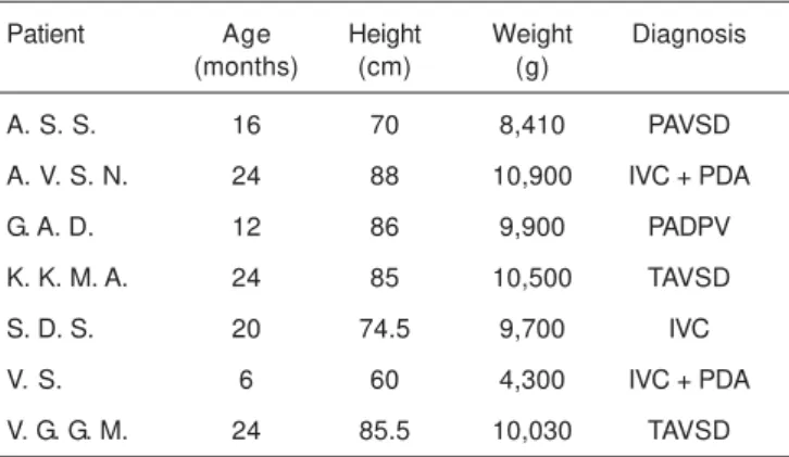
Seven children with acyanotic congenital cardiopathy with pulmonary hyperflow with mean age of 20 months (ranging from 6 to 24 months), and mean weight 9.9 kg (from 4.3 to 10.9 kg) participated in this study. Table I shows individual anthropometric data and the diagnosis of the patients in-cluded in the study. All CT scans were done without inter-currences. Figure 1 shows a representative CT scan of the chest of a child with congenital cardiopathy with pulmonary hyperflow.
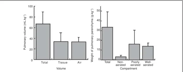
As can be seen on the left panel of Figure 2, the mean total lung volume was 66.7 ± 23.1 mL.kg-1, tissue volume 33.5 ± 15.7 mL.kg-1, and air volume 33.1 ± 8.3 mL.kg-1. When tissue and air were considered as a fraction of the total lung volu-me, it was observed that they represented 49.3 ± 6.2% and 50.7 ± 6.2%, respectively. The mean weight of the pulmonary parenchyma was 33.6 ± 15.7 g.kg-1. When the pulmonary parenchyma was analyzed in relation to the distribution of
aeration, it was observed that the non-aerated pulmonary parenchyma represented 9.7 ± 3% of the total pulmonary weight, poorly aerated parenchyma 44.6 ± 14.8%, and well-aerated pulmonary parenchyma represented 45.7 ± 14.4% (right panel of Figure 2). A significant correlation was obser-ved between the weight of the patients and total lung volume (r = 0.97), air volume (r = 0.86) and the weight of the pulmo-nary parenchyma (r = 0.97), as can be seen in figure 3. Figure 4 shows the volume and weight of the right and left lungs in relation to the total values of both lungs. On the left panel, one observes that the right lung represented 57.9 ± 2.3% of the total lung volume, while the left lung represented 42.1 ± 2.3% (p < 0.001). As for the total tissue volume, the right lung represented 55.4 ± 2.3% and the left 44.6 ± 2.3% of the total with a significant difference of 10.8% (p < 0.001). Table I: Anthropometric Data and Diagnosis of the Patients
Patient Age Height Weight Diagnosis
(months) (cm) (g)
A. S. S. 16 70 8,410 PAVSD
A. V. S. N. 24 88 10,900 IVC + PDA
G. A. D. 12 86 9,900 PADPV
K. K. M. A. 24 85 10,500 TAVSD
S. D. S. 20 74.5 9,700 IVC
V. S. 6 60 4,300 IVC + PDA
V. G. G. M. 24 85.5 10,030 TAVSD
PAVSD – partial atrioventricular septal defect; IVC – interventricular communication; PDA – patent ductus arteriosus; PADPV – partial anomalous drainage of the pulmonary veins; TAVSD – total atrioventricular septal defect.
As for air distribution, the amount of air on the right is 21% greater than on the left (60.5 ± 3.1% vs. 39.5 ± 3.1%, p < 0.001). The right panel of figure 4 shows that the fraction of pulmonary parenchyma classified according to the degree of aeration in relation to the total weight of the parenchyma are compared between the right and left lungs. Significant
differences were not observed between non-aerated (5 ± 2.4% vs. 4.6 ± 1.3%, p = NS) and poorly aerated (22.7 ± 7.9%
vs 21.9 ± 7.1%, p = NS) in the right and left lungs, respectively; however, a considerable higher fraction of well-aerated pulmonary parenchyma is in the right lung (27.6 ± 6.8 vs 18.1 ± 8%, p = NS).
Figure 2 – Distribution of Total, Tissue, and Air Volumes (left panel) and Distribution of Pulmonary Parenchyma According to Aeration (right panel). Data presented as mean ± standard deviation.
Figure 3 – Exponential Regression Curves Demonstrating the Correlation between the Weight of the Patient and Total Pulmonary Volume, Air Volume, and Weight of the Pulmonary Parenchyma.
Pulmonary volume (mL.kg
-1)
100
80
60
40
20
0
50
40
30
20
10
0 Total Tissue Air
Volume
Total Non- Poorly
Well-aerated aerated aerated
Compartment
Weight of pulmonary parenchyma (g.kg
-1)
150
100
50
0
2 4 6 8 10 12 2 4 6 8 10 12
Air volume (mL.kg
-1)
80
70
60
50
40
30
20
10
0 Total pulmonary volume = 400 x Weight;
r = 0.97 Air volume = 400 x Weight; r = 0.86
T
o
tal pulmonary volume (mL.kg
-1)
100
80
60
40
20
0
Weight of the Pulmonary Parenchyma (g.kg
-1)
2 4 6 8 10 12
DISCUSSION
Children with acyanotic congenital cardiopathy and pul-monary hyperflow have left-to-right shunt with mixture of ar-terial blood from the systemic circulation with the venous blood from the pulmonary circulation. Understanding this clinical situation depends on the knowledge of the fetal circulation and that of the postnatal transition. In intrauterine life the presence of the foramen ovale and ductus arteriosus associated to the elevated pressure in the pulmonary vas-cular bed and the low systemic resistance due to the pla-centa favors the distribution of the blood flow through the systemic circulation and only a small fraction of blood flows through the pulmonary arteries to the left atrium. In general, one third of the total blood volume of a child flows to the left atrium through the foramen ovale, while the remaining two thirds flow to the pulmonary artery. However, only 10% of this volume goes to the pulmonary circulation, and the remaining is redirected through the ductus arteriosus. In normal conditions, after birth with the occlusion of the umbilical cord and pulmonary expansion, vasodilation and reduction in vascular resistance is seen in the pulmonary circulation with the consequent increase in pulmonary blood flow and in venous pulmonary pressure. The increase in the volume of blood returning to the left atrium and consequent increase in the pressure in this chamber leads to the functional closure of the foramen ovale a few hours after birth. The increase in partial pressure of oxygen leads to vasoconstriction of the ductus arteriosus and eventual closure in the first three to four weeks of life. In children with acyanotic congenital cardiopathy the foramen ovale and ductus arteriosus remain patent, or the defects in the interatrial septum, interventricular septum, or atrioventricular septum are not closed perpetuating the fetal circulation described3,8. The pathophysiological changes
depend on the size of the shunt frequently causing respira-tory complications related to interstitial-alveolar edema. The increase in the volume of water in the extravascular space of the lungs is secondary to the increase in pulmonary blood flow associated with varying degrees of congestive heart failure due to the interdependence of both ventricles9. In the present study, the volume of tissue in the pulmonary parenchyma (volume of pulmonary parenchyma, volume of intrapulmonary blood and its components, and pulmonary extravascular water) was approximately 50% of the total lung volume, and the volume of air was responsible for the re-mainder 50%. Studies in healthy children evaluating accura-tely by computed tomography the proportion of tissue and air in the pulmonary parenchyma do not exist; however, several studies with adults with normal lungs show that the air/tis-sue relationship is around 70%/30%. Puybasset et al. stu-dying the helical chest computed tomography of 11 healthy volunteers observed that the volume of air was 2,085 ± 537 mL and that of tissue was 943 ± 143 mL, representing 70% and 30% of the total lung volume respectively10. Using a combination of chest CT and helium dilution technique, Gattinoni et al. found similar results11. Those results indicate that the increase in the volume of pulmonary tissue is secon-dary to an increase in the volume of blood in the pulmonary circulation and pulmonary extravascular water of approxi-mately 30% to 40% of the total weight of pulmonary paren-chyma. Unfortunately, it is impossible to separate the blood and pulmonary extravascular water components.
When the pulmonary parenchyma of the children was eva-luated according to the degree of aeration, it was observed that non-aerated pulmonary parenchyma represented 9.7 ± 3% of the total weight of the pulmonary parenchyma, poorly aerated parenchyma 44.6 ± 14.8%, and well-aerated paren-chyma 45.7 ± 14.4%. Under physiological conditions, it is
Figure 4: Distribution of Total, Tissue, and Air Volumes (left panel) and Distribution of the Pulmonary Parenchyma According to Aeration (right panel) in the right (black bars) and left (gray bars) Lungs. Data presented as Mean ± Standard Deviation.
Fraction of total pulmonary volume (%)
70
60
50
40
30
20
10
0
65
55
45
35
25
15
5 Right lung
Left lung
Right lung Left lung
Fraction of the weight of pulmonary
parenchyma (%)
Total Tissue Air Volume
Total Non- Poorly
Well-aerated aerated aerated
Compartment P < 0,001
P = 0,001 P < 0,001
P < 0,001
expected that the aeration of the pulmonary parenchyma is distributed in such a way that at least 90% of the lungs are normally aerated according to tomographic criteria. As men-tioned before, studies investigating the pulmonary paren-chyma of children without cardiorespiratory diseases with computed tomography are lacking; however, Vieira et al. observed in chest CTs obtained under conditions of FRC in six healthy adult volunteers that 92 ± 3% of the total lung vo-lume was well-aerated, 7 ± 3% was poorly aerated, and less than 1% was non-aerated12. Due to the increase in circulating blood inside the lungs and consequent increase in the cali-ber of the pulmonary vessels secondary to pulmonary hyper-flow, an increase in non-aerated pulmonary parenchyma is expected, since voxels that characterize blood, liquid ele-ments, and vascular structures have a CT coefficient close to zero. On the other hand, none of the children had peri-pheral saturation of oxygen below 95% during the study, and small hyperdense images were observed in dependent regions of the lungs below the heart on dorsal decubitus, which is against the notion that the presence of large areas of atelectasis would explain the non-aerated fraction of pulmonary parenchyma observed. On the other hand, des-pite normal peripheral saturation of hemoglobin, a consi-derable fraction of the pulmonary parenchyma was poorly aerated. This compartment of the pulmonary parenchyma is probably increased due to interstitial edema and increase in the volume of blood in pulmonary capillaries, but maintaining the aeration of the alveoli and small airways.
When the right and left lungs were compared, it was obser-ved that the mass of lung tissue on the right represented 55% of the total lung volume, while the left represented 45%. Those results are similar to those observed in other po-pulations of patients and they are justified by the position of mediastinal structures that are located, mainly, in the left hemithorax. However, it was observed that the ratio air/tissue on the left was significantly higher on the right and the mass of tissue with normal aeration was considered smaller on the left, demonstrating that the heart and other mediastinal structures cause effective compression of areas of the left lung under the heart, causing a reduction in regional venti-lation. In some patients with acute respiratory distress syndrome, Malbouisson et al. observed an increase in the pressure exerted by the edematous heart on the left lower lobe when compared to healthy volunteers13. Several authors described the compression of the left lower lobe and pul-monary artery induced by cardiomegaly14-17. This phenome-non, associated with the muscular relaxation caused by anesthetic agents, is responsible for the frequent atelectasis formation in the left lower lobe in children undergoing surgeries to correct congenital cardiopathies18,19.
This study has some limitations. Technically, it should be emphasized that it was not possible to control the moment of the respiratory cycle when tomographic images were acquired because children of this age group cannot follow complex verbal commands. Thus, it was not possible to
com-pare the measurements of specific respiratory parameters such as functional residual capacity (FRC) with levels pre-dicted by formulas, like those proposed by Stokes and Quanjer20 and determine the impact of the cardiopathy on FRC. The absence of a control group is another important factor, since one cannot justify performing CT scans in chil-dren without cardiopulmonary disorders.
One should not forget that the growth and developmental pro-cesses of the pulmonary parenchyma continue until the age of 8 years, and during this period an important increase in the number of alveoli is seen21-23. Some studies demons-trated that the number of alveoli at birth is 1/3 to half of the total number seen in adults and that in the first two years of life the architecture of gas exchange structures is immature and, in extrauterine life, they develop into respiratory bron-chioles, ducts, and alveolar sacs by septation23-25. This would explain the proportion of pulmonary volume in relation to the body weight, such as the correlations reported here, since the efficiency of gas exchange in this organ increases in the first years of life allowing the reduction of its mass in relation to the body weight.
To conclude, children with acyanotic congenital cardiopathy with pulmonary hyperflow have an increase in the volume of pulmonary tissue greater than expected in normal condi-tions. It also has been accurately recorded that the volume of air in the left lung is proportionally smaller than in the right lung due to the compression exerted by the heart, whose size is increased, and other mediastinal structures.
REFERÊNCIAS – REFERENCES
01. Nichols DG, Cameron DE, Greeley WJ et al. - Critical Heart Di-sease in Infants and Children. Saint Louis, Mosby - Year Book, 1995. 02. Rosenthal M, Redington A, Bush A - Cardiopulmonary physiology after surgical closure of asymptomatic secundum atrial septal defects in childhood. Exercise performance is unaffected by age at repair. Eur Heart J, 1997;18:1816-22.
03. Teitel DF, Iwamoto HS, Rudolph AM - Changes in the pulmonary circulation during birth-related events. Pediatr Res, 1990;27:372-378
04. Malbouisson LM, Preteux F, Puybasset L et al. - Validation of a software designed for computed tomographic (CT) measurement of lung water. Intensive Care Med, 2001;27:602-608
05. Malbouisson LM, Muller JC, Constantin JM et al. - Computed to-mography assessment of positive end-expiratory pressure-indu-ced alveolar recruitment in patients with acute respiratory distress syndrome. Am J Respir Crit Care Med, 2001;163:1444-1450. 06. Puybasset L, Cluzel P, Chao N et al. - A computed tomography
scan assessment of regional lung volume in acute lung injury. The CT Scan ARDS Study Group. Am J Respir Crit Care Med, 1998;158:1644-1655.
07. Mull RT - Mass estimates by computed tomography: physical density from CT numbers. AJR Am J Roentgenol 1984;143:1101-1104.
10. Puybasset L, Cluzel P, Gusman P et al. - Regional distribution of gas and tissue in acute respiratory distress syndrome. I. Consequences for lung morphology. CT Scan ARDS Study Group. Intensive Care Med, 2000;26:857-869
11. Gattinoni L, Pesenti A, Avalli L et al. - Pressure-volume curve of total respiratory system in acute respiratory failure. Computed tomographic scan study. Am Rev Resp Dis, 1987;136:730-736. 12. Vieira SR, Puybasset L, Richecoeur J et al. - A lung computed
tomographic assessment of positive end-expiratory pressure-induced lung overdistension. Am J Respir Crit Care Med, 1998;158:1571-1577.
13. Malbouisson LM, Busch CJ, Puybasset L et al. - Role of the heart in the loss of aeration characterizing lower lobes in acute respiratory distress syndrome. CT Scan ARDS Study Group. Am J Respir Crit Care Med, 2000;161:2005-2012.
14. Alexander MS, Arnot RN, Lavender JP - Left lower lobe ventilation and its relation to cardiomegaly and posture. BMJ, 1989;299:94.
15. Alexander MS, Peters AM, Cleland JP et al. - Impaired left lower lobe ventilation in patients with cardiomegaly. An isotope study of mechanisms. Chest, 1992;101:1189-1193.
16. Wiener CM, McKenna WJ, Myers MJ et al. - Left lower lobe ven-tilation is reduced in patients with cardiomegaly in the supine but not the prone position. Am Rev Respir Dis, 1990;141:150-155. 17. Beckmann CF, Levin DC, Ulreich S - Cardiomegaly as a cause of nonuniform pulmonary artery perfusion. AJR Am J Roentgenol, 1977;129:661-666.
18. Benjamin JJ, Cascade PN, Rubenfire M et al. - Left lower lobe atelectasis and consolidation following cardiac surgery: the effect of topical cooling on the phrenic nerve. Radiology, 1982; 142:11-14.
19. Emhardt JD, Moorthy SS, Brown JW et al. - Chest radiograph changes after cardiopulmonary bypass in children. J Cardiovasc Surg (Torino), 1991;32:314-317.
20. Stocks J, Quanjer PH - Reference values for residual volume, functional residual capacity and total lung capacity. ATS Work-shop on Lung Volume Measurements. Official Statement of The European Respiratory Society. Eur Respir J, 1995;8:492-506. 21. Thurlbeck WM - Lung growth and alveolar multiplication.
Patho-biol Annu, 1975;5:1-34
22. Wigglesworth JS, Desai R, Aber V - Quantitative aspects of perinatal lung growth. Early Hum Dev, 1987;15:203-212. 23. Hislop AA, Wigglesworth JS, Desai R - Alveolar development in
the human fetus and infant. Early Hum Dev, 1986;13:1-11. 24. Zeltner TB, Bertacchini M, Messerli A et al. - Morphometric
estimation of regional differences in the rat lung. Exp Lung Res, 1990;16:145-158.
25. Thurlbeck WM - Postnatal human lung growth. Thorax, 1982;37: 564-571.
RESUMEN
Gimenez S, Teixeira ML, Myashiro R, Carmona MJC, Auler Jr JOC, Malbouisson LMS - Evaluación Pulmonar en Niños Portadores de Cardiopatía Congénita Acianótica e Hiperflujo Pulmonar a través de Tomografía Computadorizada.
JUSTIFICATIVA Y OBJETIVOS: La disfunción respiratoria es frecuente en niños con cardiopatías congénitas acianóticas con hiperflujo pulmonar (CCAHP), sin embargo, se conoce muy poco sobre la estructura pulmonar de esos pacientes. El objetivo de este estudio, fue cuantificar los volúmenes de gas y tejido y la distri-bución de la aeración pulmonar en esa población.
MÉTODOS: Después de la aprobación por parte del Comité de Éti-ca Institucional y de la obtención del consentimiento escrito infor-mado, se obtuvieron tomografías computadorizadas torácicas en siete niños con CCAHF. Las imágenes pulmonares derecha e izquierda fueron perfiladas en todas las imágenes, y los volúmenes y los pesos pulmonares fueron computados a partir de los datos volumétricos. Las comparaciones entre izquierda y derecha fueron analizadas usando el test t de Student pareado y las correlaciones a través de regresión exponencial.
RESULTADOS: La edad mediana fue de 20 meses y el peso fue de 9,9 kg. El volumen pulmonar total (VPT) 66,7 ± 23,1 mL.kg-1, y del tejido 33,5 ± 15,7 mL.kg-1 y el del gas 33,1 ± 8,3 mL.kg-1. El pulmón derecho representó un 57,9% y el pulmón izquierdo 42,1% del VPT (p < 0,001). El volumen pulmonar de gas a la derecha fue de un 60,5% del volumen de gas total (p < 0,001) y la cantidad de parénquima pulmonar normalmente aerado fue significativamente menor a la izquierda (27,6 ± 6,8 vs. 18,1 ± 8%, p < 0,001).