Article
ISSN 0102-695X
doi: 10.1590/S0102-695X2011005000050 Received 26 Mar 2010
Accepted 7 Nov 2010 Available online 1 Apr 2011
Stachytarpheta cayennensis
by scavenger of
mitochondrial reactive oxygen species
Pierre André de Souza,
*,1Clara Rodrigues,
2Ana Paula S. A.
Santiago,
3Nanci Camara de Lucas,
1Gilda Guimaraes Leitao,
4Antonio Galina Filho
21Instituto de Química, Departamento de Química Orgânica, Laboratório de Fotoquímica, Universidade Federal do Rio de Janeiro, Brazil,
2Instituto de Biofísica Carlos Chagas Filho, Programa de Biologia Celular
e Parasitologia, Universidade Federal do Rio de Janeiro, Brazil,
3Instituto de Bioquímica Médica, Programa de Biofísica e Bioquímica
Celular e Programa de Biologia Molecular e Biotecnologia, Universidade Federal do Rio de Janeiro, Brazil,
4Núcleo de Pesquisa de Produtos Naturais, Universidade Federal do Rio de
Janeiro, Brazil.
Abstract: The etiology of a range of diseases is associated with an excessive generation of reactive oxygen species (ROS). Exacerbated ROS production leads to mitochondrial dysfunction, cellular damage and apoptosis. Nowadays, many strategies are being developed for the targeted delivery of antioxidants compounds to mitochondria, trying to minimize the damages. Production of ROS was investigated by the molecular probes CM-H2DCFDA and Amplex Red. The purpose of this work is to evaluate the antioxidant capacity of verbascoside, martinoside, betulinic acid from the Stachytarpheta cayennensis
and quercetin by an in vitro assay with isolated mitochondria from mice’s brain The results showed that all compounds tested exhibited a scavenger effect on the ROS generated by the isolated mitochondria, which displayed a dependent dose increase.
Keywords:
Satchytarpheta cayennensis
antioxidants mitochondria
reactive oxygen species fluorescent probes
Introduction
Antioxidants react differently with the diverse free radicals and oxidant sources (Huang et al., 2005). During the last decade, natural antioxidant compounds from medicinal plants have aroused much attention, what resulted on increasing efforts to search for plant-derived antioxidants against free radicals like reactive oxygen (ROS) and nitrogen species (RNS) (Huang et al., 2005; Pulido et al., 2005; Roginsky et al., 1996). Mitochondrial electron transport system (ETS) is considered to be one of the major sources of cellular ROS. The formation of ROS is inevitable during normal oxidative metabolism, and it is well established
that some of the oxygen (O2) consumed by the ETS
generate ROS, mostly at complexes I and III (Cadenas et al., 1977, Cadenas & Davies, 2000; Chen et al., 2003; Boveris et al., 1976; Boveris & Chance, 1973; Turrens & Boveris, 1980). The partial reduction of molecular oxygen, during oxidative phosphorylation,
leads to a constant flux of superoxide anion (O2-), the
precursor of hydrogen peroxide (H2O2) radicals (Liu,
2002; Oliveira & Kowaltowski, 2004). It has been described in the literature that in pathological conditions ROS production can be exacerbated, resulting in an imbalance between ROS generation and the intracellular levels of antioxidant defenses (Brand et al., 2004; Turrens 2003). It is involved in the pathology of many neurodegenerative diseases like Parkinson’s disease, Alzheimer’s disease, Huntington’s disease, epilepsy and atherosclerosis (Freitas et al., 2005; Schapira, 2006). Others studies showed important role of antioxidant drugs such as ascorbic acid and α-tocopherol can be used as scavengers of free radicals, reducing mice’s brain injury (Tomé et al., 2010a; 2010b; Xavier et al., 2007).
Penido et al., 2006; Schapoval et al., 1998). The most frequent ethnomedical usages of roots and leaves of
S. cayennensis are as inflammations, ulcers, fever and diuretics (Penido et al., 2006; Schapoval et al., 1998).
The anti-inflammatory activity of S. cayennensis has
been demonstrated in rats by and Penido et al. (2006). The aim of this study was to demonstrate the antioxidant properties of verbascoside, martinoside and
betulinic acid compounds isolated from the roots of S.
cayennensis. The antioxidant capacity was evaluated
by an in vitro assay using mitochondria isolated from
mice’s brains like ROS source. For that, we used two
fluorescent probes, Amplex Red and CM-H2DCFDA,
that are highly sensitive and stables markers for
mitochondrial generated ROS with selectivity to H2O2
and free radicals, respectively (Valeur, 2001; Votyakova & Reynolds, 2004; Zhou et al., 1997).
Materials and Methods
Collection and extraction of plant material
Roots of Stachytarpheta cayennesis (Rich.)
Vahl, Verbenaceae, were collected at Conceição lake, Florianópolis, Santa Catarina, Brazil. Botanical identification was done by Inês M. Oliveira da Silva from the Department of Botany at Universidade Federal Rural do Rio de Janeiro, Brazil, where a voucher specimen is deposited (RBR 5979). The air-dried and powdered roots (367.8 g) were placed in a Soxhlet apparatus and exhaustively extracted with
n-hexane followed by ethanol, yielding 5.0 g and 48.0
g of crude hexane and ethanol extracts, respectively. The dried ethanol extract was suspended in MeOH/
H2O 3:2 and extracted with hexane, chloroform, ethyl
acetate and butanol, in this order. High-speed counter-current chromatography (HSCCC) was applied to the preparative separation of the compounds verbascoside
(2) (300.0 mg) and martinoside (3) (54.0 mg) from
the ethyl acetate fraction and is described elsewhere
(Leitao et al., 2005). Betulinic acid (4) (45.0 mg) was
obtained from the chloroform fraction from the ethanol extract by HSCCC.
Selection of two-phase solvent system by the tube partitioning test
A small quantity of the chloroform fraction
from the ethanol extract of the roots of S. cayennensis
was dissolved in a series of test tubes containing the
solvent systems CH2Cl2:EtOH:H2O (1:1:1, 1.5:1:1,
2:1:1 v/v) and hexane:CH2Cl2:EtOH:H2O (1:0.5:1:1,
1:1:1:1, 1:2:1:1 v/v). The test tubes were shaken and the compounds allowed to partition between the two phases. Equal aliquots of each phase were spotted
beside each other separately on silica gel TLC plates
and developed with the solvent system CHCl3:ethyl
acetate 3:2. The results were visualized under UV light and by spraying with vanillin-sulfuric acid (1%) solution followed by heating. The solvent system was
defined as hexane: CH2Cl2:EtOH:H2O (1:2:1:1 v/v).
HSCCC separation procedure
The solvents of the selected solvent system were thoroughly equilibrated in a separation funnel at room temperature. The upper and lower phases were separated and degassed in an ultrasonic bath for 20 min before use. The sample was prepared by dissolving
400 mg of the chloroform fraction from S. cayennesis
in 2.5 mL of each phase of the solvent system and loaded into an injection valve (Rheodyne model 5010 A) equipped with a 5 mL loop. A P.C Inc counter-current chromatograph equipped with a multi-layer coil equilibrated by a counterweight was used. The 80 mL coil was initially filled with the stationary upper phase of the solvent system. Then, rotation started (850 rpm) and the lower mobile phase was pumped into the coil in the head to tail (H→T) direction at a flow rate of 2.0 mL/min until hydrodynamic equilibrium was achieved. Under these conditions, the retention of the stationary
phase (SF) in the coil was 82.5 % (Vm=14.0 mL). CCC
rotation was interrupted in tube 60 and the coil content (organic and aqueous phases) was collected (“wash-off”), giving a total of seventy fractions, of 5.0 mL each. The whole separation experiment was conducted at 27 °C. All fractions obtained by CCC were analyzed by TLC using dichloromethane: acetone: methanol (2:1:0.5) as eluent. The major compound, betulinic acid, was obtained in fractions (FC 38-40) and identified by
1H and 13C NMR spectroscopy and the data compared
with those from the literature (Mahato 1994).
Animals and reagents
Male swiss mice, two month old, were maintained on a 12 h light/dark cycle (lights on at 7 a.m.), with free access to tap water and standard laboratory chow. The experimental protocols using animals were approved by the Committee for Ethics in Animal Research of the Universidade College for Animal Experimentation. The reagents were purchased from Sigma (USA), Amersham Biosciences (USA), Invitrogen (USA) and Merck (Germany) and Molecular Probes (Eugene, OR).
N-acetyl-3,7-dihydroxyphenoxazine (Amplex-Red)
and 5-(-6)-chloromethyl-2`,7`-dichlorohydroluorescein
diacetate (CM-H2DCFDA) probes were obtained from
used as mitochondrial source for oxidative stress assay. The experiments using Amplex Red and CM-H2DCFDA probes with isolated mitochondria were carried out at 37 °C. Quercetin was used as antioxidant standard, obtained from Sigma Chemicals and used in all experiments, due to its well-established antioxidant activity. All organic solvents used for High-Speed Countercurrent Chromatography (HSCCC) were of analytical grade and purchased from Tedia Brazil.
Mitochondrial isolation
Mitochondria from forebrains were isolated as described by Rosenshtraukh et al. (1978) with minor modifications, from two months old swiss mice. Their brains were rapidly removed (within 1 min) and put into 10 mL of ice-cold isolation buffer containing 300 mM mannitol, 1 mM K+-EGTA, 0.1% bovine serum
albumin (BSA; free fatty acid), 10 mM K+-HEPES
pH 7.2. The tissues were cut into small pieces using surgical scissors and extensively washed. Then, they were ressuspend in a proportion of 1/10 (w/v) with the cold isolation buffer, and manually homogenized during two cycles of 10 s in a Teflon glass potter. The
homogenates were centrifuged at 2.000 x g for 3 min
in a Hitachi Himac SCR20B RPR 20-2 rotor. After centrifugation, the supernatant was centrifuged for 8
min at 12.000 x g. The pellet was suspended in 5 mL
of isolation buffer containing 20 µL of 10% digitonin and centrifuged again for 8 min at 12.000 x g. The supernatant was discarded and the final pellet gently washed and suspended in cold isolation buffer at an approximate protein concentration of 10 mg/mL. The protein concentration in the samples was determined by the Folin-Lowry method using bovine serum albumin as standard (Lowry et al., 1951). All of the experiments with isolated mitochondria were carried out at 37 °C with continuous stirring in a respiration buffer containing 10 mM Tris-HCl, pH 7.4, 0.32 M mannitol,
8 mM inorganic phosphate, 4 mM MgCl2, 0.08 mM
EDTA, 1 mM EGTA and 0.2 mg/mL fatty acid-free bovine serum albumin.
Spectrofluorometric measurements of mitochondrial ROS generation
Mitochondrial release of reactive oxygen species (ROS) was assessed by two methods using 10-acetyl-3,7-dihydroxyphenoxazine (Amplex Red)
to evaluate H2O2 or
5-(-6)-chloromethyl-2`,7`-dichlorohydrofluorescein diacetate (CM-H2DCFDA) to
access whole ROS release from mitochondria (Cadenas & Davies 2000; Cadenas et al., 1977; Chen et al., 2003; Turrens 2003; Turrens & Boveris 1980; Valeur 2001; Votyakova & Reynolds 2004). Briefly, for evaluation
of H2O2 release from mitochondria, 0.5 mg protein/
mL of isolated mitochondria were incubated in the standard respiration buffer supplemented with 1.5 µM Amplex Red and 10 units/mL horseradish peroxidase. (HRP). Amplex Red, is a nonfluorescent molecule that when oxidized by hydrogen peroxide in the presence of HRP originates resorufin, a highly fluorescent product. Fluorescence was monitored at excitation and emission wavelengths of 563 nm and 587 nm, respectively, using a multi-mode microplate reader spectrofluoremeter
Spectra Max® M5 (Molecular Devices). For evaluation
of whole mitochondrial ROS release, CM-H2DCFDA
fluorescence, 0.5 mg protein/mL of isolated
mitochondria were incubated in the standard respiration
buffer supplemented with 2 µM of CM-H2DCFDA.
Fluorescence was monitored at excitation and emission wavelengths of 485 nm and 535 nm, respectively, using a multi-mode microplate reader spectrofluoremeter
Spectra Max® M5 (Molecular Devices). The acetate
group of CM-H2DCFDA is hydrolysed by esterases
when it enters the cell and the molecule is trapped inside as the non-fluorescent
5-(-6)-chloromethyl-2`,7`-dichlorodihydrofluorescein (CM-H2DCFH). Oxidation
of CM-H2DCFH by ROS yields the fluorescent product
carboxymethyldichlorofluorescein (CM-DCF), and
indirectly measures mitochondrially produced O2- that
has dismutated to H2O2 through the action of superoxide
dismutase. Calibrations were performed by the addition
of known quantities of H2O2. Each experiment was
repeated at least three times with different mitochondrial preparations with increasing concentrations (1-45µM)
of quercetin (1), verbascoside (2), martinoside (3) and
betunilic acid (4) compounds. In all experiments, we
detected small changes in the rate of H2O2 formation
among different preparations, but the overall pattern of response to different modulators was not affected. The
maximal rate (100%) of mitochondrial H2O2 or ROS
formation was assumed to be the difference between
the rate of H2O2 or ROS formation in the absence or in
the presence of succinate.
Statistical analysis
Statistical analyses were performed using a Tukey’s test and ANOVA. Differences were considered
statistically significant for p<0.05. All results are
expressed as means±S.E.M. for n independent
experiments.
Results and Discussion
The excess of ROS induce lipid peroxidation, especially in brain, since it is inadequately equipped with scavenger antioxidant defense mechanisms and which has easily peroxidizable fatty acid, high content of iron and ascorbate. These features turn brain tissue very vulnerable to free radical and ROS injury (Chen et al., 2003; Halliwell & Gutteridge, 2002; Halliwell, 2006; Liu, 2002; Sheu et al., 2006). Mitochondria is the most important cellular source of the free radical
superoxide anion (O2•‾), which was established as the
stoichiometric precursor of mitochondrial H2O2 (Chen
et al., 2003; Sheu et al., 2006; Halliwell & Gutteridge, 2002).
Little information is available on the levels of natural antioxidant compounds in brain (Halliwell & Gutteridge, 2002). On the other hand, there is an evidence, based on animal studies, that some phenolics antioxidants compounds (feluric acid and flavonoids such as naringenin and hesperitin) can cross the blood-brain barrier (Halliwell & Gutteridge, 2002; Sheu et al., 2006, Spencer et al., 2004). Some of the pharmacological properties of medicinal plant like anti-inflammatory property may be linked to their antioxidant potential, antioxidant compounds, which can be due to their ability to scavenge free radicals and
ROS, such as H2O2 and other very reactive non-radical
species (Amaral et al., 2009).
In this context we tested the ROS production using two fluorescent probes, Amplex Red (AR) and
CMH2DCDFA, in the experiments using isolated
mitochondria from mice’s brain, in order to investigate the antioxidant potential of the natural compounds
from S. cayennensis. The chemiluminescence of
CM-H2DCFDA and Amplex Red (AR) catalyzed by
horseradish peroxidase (HRP) enzymatic coupled
assays have been employed in several in vitro studies
for the quantification of low concentrations of ROS and
H2O2 (Chen et al., 2003; Sheu et al., 2006; Valeur, 2001).
The addition of H2O2 to a sample consisting of AR and
HRP. HRP in pH 7.4, buffer immediately generated a strong absorption band with maximum at 587 nm, indicating conversion of AR to resorufin. Increasing
H2O2 produced more resorufin, the concentration of
which was proportional to the concentrated H2O2 used
(Chen et al., 2003; Sheu et al., 2006). The acetate
group of CM-H2DCFDA is hydrolysed by esterases
when it enters the cell and the molecule is trapped
inside as the non-fluorescent CM-H2DCFH.
CM-H2DCFH was chosen because, unlike underivatized
dichlorodihydrofluorescein, it is well retained in cells and, in this case, in the mitochondrial matrix (Valeur, 2001; Votyakova & Reynolds, 2004).
The verbascoside (2), martinoside (3) and
betulinic acid (4) resulting from the roots of ethanol
extract of S.cayennensis has been previously assayed
by our group for antioxidant activities (DPPH
free-radical assay and Saccharomyces cerevisiae cells,
an eukaryotic cell model) (De Souza et al., 2010). All compounds afforded the best results in the two assays and were therefore, chosen for this study. The remarkable capacity of these natural compounds to
function in vitro as antioxidants has been demonstrated
in this study.
O OH O OH HO
OH OH
1
O O
OH O
O R
OH O
O HO HO HO
R
HOH OH
2 R = OH
3 R = OCH3 H
HO
H HH
CO2H
4
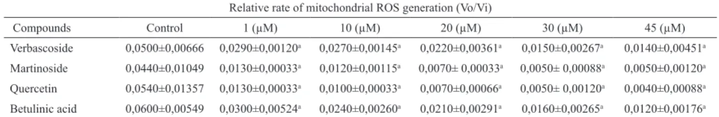
We induced the isolated mitochondria to produce ROS using the mitochondrial substrate succinate and the mitochondrial complex III inhibitor antimycin A. Each experiment was repeated at least three times with different mitochondrial preparations. The compounds verbascoside and martinoside presented the best antioxidant effects, according to their IC50 for ROS scavenger (Table 1). Table 2 and Table 3 show representative experiments. The differences between the antioxidant efficacy of verbascoside, martinoside and betunillic acid comparing to quercetin became clear in the concentration range of 1 to 20 µM (Table 2) and of 1 to 10 µM (Table 3).
Table 1. Effect of different compounds with antioxidant activities.
IC50 (uM) values for ROS scavenger Compounds H2O2 (Amplex red)
IC50
ROS (CMH2DCFDA) IC50
Betunilic acid 28.11 2.07
Martinoside 19.34 0.54
Quercetin - 7.27
Verbascoside 5.25 0.54
Table 1 shows for the Amplex red probe, after the titulation curve with H2O2, with the IC50 (inhibitory concentration, 50%) of 19,000 pmol of
H2O2 consumed by each compound. For the CMH2DCFDA probe, the
IC50 was 29,325 pmol of H2O2 consumed by each compound.
On the other hand, the current study demonstrates that AR (Table 3) is more sensitive probe than
CMH2DCFDA (Table 2) when the assay is conducted at pH
7.4. Perhaps, the hydrolysis of CM-H2DCFDA in buffer at
several minutes, and where buffer capacity is low and pH is variable. In addition, the oxidized product of Amplex Red has excitation and emission maxima at 563 and 587
nm, in comparison with those of CMH2DCFDA at 485
and 535 nm. These superior spectral properties maybe are important for avoiding interference from autoluorescence in assays involving biological samples. Finally, there are a few factors affecting the sensitivity of resorufin for
detecting H2O2 (Valeur, 2001).
The number of phenolic hydroxyl groups may play an important role in their scavenging activity (Justino et al., 2004; Van Acker et al., 1993). The literature reports that compounds with hydroxyl in the ortho-aromatic position, like quercetin, have more antioxidant activity against ROS (Justino et al., 2004;
Soobratte et al., 2005). In the presence of H2O2, for
example, phenolic compounds such as ortho-quinol suffer a reduction reaction generating ortho-quinones and water as products (Dewick, 2002; Justino et al., 2004; Soobratte et al., 2005; Van Acker et al., 1993). The
verbascoside (2) and the flavonoid quercetin (1) have
four hydroxyls in their structures capable of suffering oxidation reactions in the presence of peroxides, such
as H2O2 (Dewick, 2002; Zhou & Sadik, 2008).
Table 3 shows the antioxidant activity of the
tested compounds in H2O2. Again, the verbascoside
presented the better antioxidant capacity among the compounds tested with about 10 µM of concentration.
Both betulinic acid (4) and martinoside (3) showed
similar antioxidant effect around the concentration of 10 µM (Table 3). A possible explanation for this would be that the carboxylic acid group presents a
greater oxidation potencial comparing to the phenolic compound (Dewick, 2002; Justino et al., 2004). This property could give to betulinic acid a similar antioxidant capacity when compared with martinoside, which has only a phenolic hydroxyl group on each of its two aromatic rings. Surprisingly, both verbascoside and martinoside showed more antioxidant activity than quercetin that was used as standard, due to its well-established antioxidant activity (Table 1 and 3) (Belinha et al., 2007; Ho et al., 2004; Santos et al., 2008; Oken et al., 1998; Soobrattee et al., 2005; Spencer et al., 2004; Zhou & Sadik, 2008).
Nevertheless, only verbascoside has four hydroxyls on ortho position, in its two aromatic rings. This structural information could justify its high antioxidant activity when compared to the other compounds tested (Zhou & Sadik, 2008). Perhaps, the weak antioxidant activity of quercetin reported here may be related to the solubility and ability of quercetin to be distributed to a less or greater extent of its diffusion speed, between the lipid-water interface, or the distribution in the hydrophobic core of the mitochondrial membrane (Justino et al., 2004; Oliveira & Kowaltowski, 2004; Pulido et al., 2005; Spencer et al., 2004).
In summary, present results show that verbascoside, martinoside and betulinic acid are useful antioxidants for preventing oxidative stress-related mitochondrial dysfunction, and as such may find use in the management of a range of acute and chronic disease processes. However, very little is known about the
Table 2. ROS generation rate using the luorescent probe CM-H2DCFDA, during state 2 respiration induced by 10 mM succinate (suc), with increasing compounds concentrations (1-45 µM) of verbascoside, quercetin, betunilic acid and martinoside.
Relative rate of mitochondrial ROS generation (Vo/Vi)
Compounds Control 1 (µM) 10 (µM) 20 (µM) 30 (µM) 45 (µM)
Verbascoside 0,0500±0,00666 0,0290±0,00120a 0,0270±0,00145a 0,0220±0,00361a 0,0150±0,00267a 0,0140±0,00451a
Martinoside 0,0440±0,01049 0,0130±0,00033a 0,0120±0,00115a 0,0070± 0,00033a 0,0050± 0,00088a 0,0050±0,00120a
Quercetin 0,0540±0,01357 0,0130±0,00033a 0,0100±0,00033a 0,0070±0,00066a 0,0050± 0,00120a 0,0040±0,00088a
Betulinic acid 0,0600±0,00549 0,0300±0,00524a 0,0240±0,00260a 0,0210±0,00291a 0,0160±0,00265a 0,0120±0,00176a ap<0.05 relative to 1, 10,20,30 and 45 µM. The values are mean±SE from at least three independent mitochondrial preparations for each assay
conditiona: control vs 1,10,20,30,45.
Table 3. ROS (H2O2) generation rate using the luorescent probe Amplex Red, during state 2 respiration induced by 10 mM succinate (suc), with increasing compounds concentrations (1-45 µM) of verbascoside, quercetin, betunilic acid, and martinoside.
Relative rate of mitochondrial ROS generation (Vo/Vi)
Compounds Control 1 (µM) 10 (µM) 20 (µM) 30 (µM) 45 (µM)
Verbascoside 6,911±0,08170 1,363±0,06059a 0,4367±0,01618a,b 0,2557±0,04156a,b 0,2800±0,05029a,b 0,2833±0,06164a,b
Martinoside 6,651±0,09650 1,381±0,06351 a 1,015±0,06784a,b 0,6773±0,07518 a,b 0,6100±0,08685a,b,c 0,5267±0,08225a,b,c
Quercetin 6,943±0,10513 1,362±0,6529 a 1,477±0,3324 a 1,107±0,2052 a 0,7627±0,1621 0,8267±0,1470a
Betulinic acid 6,873±0,06754 1,428±0,1159 a 1,053±0,1195a 0,9297±0,09556a,b 0,6543±0,07911a,b 0,5670±0,07365a,b,c ap<0.05 relative to control, bp<0.05 relative to 1 µM, cp<0.05 relative to 10 µM. The values are mean±SE from at least 3 independent mitochondrial
effects of natural compounds isolated from plants in the cellular metabolism of humans, especially after cross the blood-brain barrier and interaction with brain cells (Justino et al., 2004; Spencer et al., 2004). Preclinical
studies in vivo models are needed to evaluate the
effectiveness and toxicity of mitochondrially targeted antioxidants before recommending specific doses for human treatment.
Acknowledgments
This research was supported by grants from CAPES and CNPq, Brazil. We thank Dr. Inês M. Oliveira (UFRRJ) for her help in identifying the voucher specimen.
References
Amaral S, Mira L, Nogueira JMF, da Silva AP, Florencio MH 2009. Plant extracts with anti-inflammatory properties - a new approach for characterization of their bioactive compounds and establishment of structure–antioxidant activity relationships. Bioorg Med Chemi 17: 1876-1883.
Belinha I, Amorim MA, Rodrigues P, De Freitas V, Moradas-Ferreira P, Mateus N, Costa V 2007. Quercetin increases oxidative stress resistance and longevity in
S. cerevisiae. J Agric Food Chem 55: 2446-2451. Boveris A, Cadenas E, Stoppani AO 1976. Role of ubiquinone
in the mitochondrial generation of hydrogen peroxide.
Biochem J 156: 435e444.
Boveris A, Chance B 1973. The mitochondrial generation of hydrogen peroxide: general properties and effect of hyperbaric oxygen. Biochem J 134: 707-716.
Brand DM, Affourtit CE, Green K, Lambert JA, Miwa S, Pakay LJ, Parker N 2004. Serial Review: The powerhouse takes control of the cell: the role of mitochondria in signal transduction. Free Rad Biol Med 37: 755-767. Cadenas E, Boveris A, Ragan CI, Stopani AO 1977.
Production of superoxide radicals and hydrogen peroxide by NADH-ubiquinone reductase and ubiquinol-cytochrome c reductase from beef-heart mitochondria. Arch Biochem Biophys 180: 248-257. Cadenas E, Davies KJ 2000. Mitochondrial free radical
generation, oxidative stress, and aging. Free Radic Biol Med 29: 222-230.
Chen Q, Vazquez EJ, Moghaddas S, Hoppel CL, Lesnefsky EJ 2003. Production of reactive oxygen species by mitochondria: central role of complex III. J Biol Chem 278: 36027-36031.
De Souza PA 2004. Estudo fitoquímico e farmacológico P. bahienenis e S .cayennensis. Rio de Janeiro, 139p. Dissertação de Mestrado, Programa de Pós-graduação em Química Orgânica, Universidade Federal Rural do Rio de Janeiro.
De Souza PA, Silva CG, Machado BRP, Lucas NC, Leitão GG, Eleutherio ECA, Dellamora Ortiz GM, Benchetrit LC 2010. Evaluation of antimicrobial, antioxidant and phototoxic activities of extracts and isolated compounds from Stachytarpheta cayennesis (Rich.) Vahl. Rev Bras Farmacogn 20: 922-928.
Dewick PM 2002. Medicinal Natural Products. A Biosynthetic Approach. 2nd ed.John Wiley & Sons Ltd. England.
Freitas RM, Vasconcelos SMM, Souza FCF, Viana GSB, Fonteles MMF 2005. Oxidative stress in the hippocampus after pilocarpine-induced status epilepticus in Wistar rats. FEBS J 272: 1307-1312. Halliwell B 2006. Oxidative stress and neurodegeneration?
Where are we now. J Neurochem 97: 1634-1658. Halliwell B, Gutteridge JMC 2002. Free Radicals in Biology
and Medicine. Oxford University Press.
Ho J, Heo HJ, Lee CY 2004. Protective effects of quercetin and vitamin C against oxidative stress-induced neurodegeneration. J Agric Food Chem 52: 7514-751.
Huang D, Boxin O, Prior LR 2005. The chemistry behind antioxidant capacity assays. J Agric Food Chem 53: 1841-185.
Justino CG, Santosa MR, Canarioa S, Borgesa C, Florencio MH, Miraa L 2004. Plasma quercetin metabolites: structure-antioxidant activity relationships. Arch Biochem Biophys 432: 109-121.
Leitão GG, De Souza PA, BrowL, Moraes AA 2005. Step-gradient CCC separation of phenylpropanoid and iridoid glycosides from roots of Stachytarpheta cayennesis (Rich.) Vahl. J Liq Chromatogr Relat Technol 28: 2053-2060.
Liu Y 2002. Generation of reactive oxygen species by the mitochondrial electron transport chain. J Neurochem 80: 780-787.
Lowry OH, Rosebrough NJ, Farr AL, Randall RJ 1951. Protein measurement with the Folin phenol reagent. J Biol Chem 193: 265-275.
Mahato BS 1994. 13C NMR Spectra of pentacyclic
triterpenoids - a compilation and some salient features.
Phytochemistry 37: 1517-1575.
Oken BS, Storzbach DM, Kaye JA 1998. The efficacy of
Ginkgo biloba on cognitive function in Alzheimer disease. Archiv Neurol 55: 1409-1415.
Oliveira GA, Kowaltowski AJ 2004. Phosphate increases mitochondrial reactive oxygen species release. Free Radic Res 38: 1113-1118.
Penido C, Costa KA, Futuro DO, Paiva SR, Kaplan MAC, Figueiredo MR, Henriques MGMO 2006. Anti-inflammatory and anti-ulcerogenic properties of
Stachytarpheta cayennensis (L.C. Rich) Vahl. J Ethnopharmacol 104: 225-233.
Roginsky VA, Barsukova TK, Remorova AA 1996. Moderate antioxidative efficiencies of flavonoids during peroxidation of methyl linoleate in homogenous and micellar solutions. J Am Oil Chem Soc 73: 777-786. Rosenshtraukh LV, Saks VA, Undrovinas AI, Chazov EI,
Smirnov VN, Sharov VG 1978. Studies of energy transport in heart cells. The effect of creatine phosphate on the frog ventricular contractile force and action potential duration. Biochem Med 19: 148-164.
Santos MR, Rodrıguez-Gomez MJ, Justino GC, Charro
N, Florencio MH, Mira L 2008. Influence of the metabolic profile on the in vivo antioxidant activity of quercetin under a low dosage oral regimen in rats. Br J Pharmacol 153: 1750-1761.
Schapira AH 2006. Etiology of Parkinson’s disease. Neurology 66: S10-S23.
Schapoval EES, Winter de Vargas MR, Chaves CG, Bridi R, Zuanazzi JA, Henriques AT 1998. Antiinflammatory and antinociceptive activities of extracts and isolated compounds from Stachytarpheta cayennensis. J Ethnopharmacol 60: 53-59.
Sheu SS, Nauduri D, Anders MW 2006.Targeting antioxidants to mitochondria: A new therapeutic direction. Biochim Biophys Acta 1762: 256 -265.
Soobrattee MA, Neergheen VS, Luximon-Ramma A, Aruoma OI, Bahorun T 2005. Phenolics as potential antioxidant therapeutic agents: Mechanism and actions. Mutat Res 579: 200-213.
Spencer JPE, El Mohsen MMA, Rice-Evans C 2004. Cellular uptake and metabolism of flavonoids and their metabolites: implications for their bioactivity. Arch Biochem Biophys 423: 148-161.
Tomé AR, Feng D, Freitas RM 2010a.The effects of alpha-tocopherol on hippocampal oxidative stress prior to in pilocarpine-induced seizures. Neurochem Res 35: 580-587.
Tomé AR, Ferreira PMP, Freitas RM 2010b. Inhibitory action
of antioxidants (ascorbic acid or α-tocopherol) on
seizures and brain damage induced by pilocarpine in
rats. Arq Neuropsiquiatr 68: 355-361.
Turrens JF 2003. Mitochondrial formation of reactive oxygen species. J Physiol 552: 335-344.
Turrens JF, Boveris A 1980. Generation of superoxide anion by the NADH dehydrogenase of bovine heart mitochondria. Biochem J 191: 421-427.
Valeur B 2001. Molecular Fluorescence: Principles and Applications. Wiley-VCH Verlag GmbH Press. Van Acker SA, Koymans LM, Bast A 1993. Molecular
pharmacology of vitamin E: structural aspects of antioxidant activity. Free Rad Biol Med 15: 311-328. Votyakova TV, Reynolds IJ 2004. Detection of hydrogen
peroxide with Amplex Red: interference by NADH and reduced glutathione auto-oxidation. Arch Biochem Biophys 431: 138-44.
Xavier SM, Barbosa CO, Barros DO, Silva RF, Oliveira AA, Freitas RM 2007. Vitamin C antioxidant effects in hippocampus of adult Wistar rats after seizures and status epilepticus induced by pilocarpine. Neurosci Lett 1: 76-79.
Zhou A, Sadik AO 2008. Comparative analysis of quercetin oxidation by electrochemical, enzymatic, autoxidation, and free radical generation techniques: A mechanistic study. J Agric Food Chem 56: 12081-12091.
Zhou M, Diwu Z, Panchuk-Voloshina N, Haugland RP 1997. A stable nonfluorescent derivative of resorufin for the fluorometric determination of trace hydrogen peroxide: applications in detecting the activity of phagocyte NADPH oxidase and other axidases. Anal Biochem 253: 162-168.
*Correspondence
Pierre André de Souza
Instituto de Química, Departamento de Química Orgânica, Laboratório de Fotoquímica
Universidade Federal do Rio de Janeiro, 21949-900 Rio de Janeiro-RJ, Brazil