ABSTRACT
Inluence of light-exposure methods and depths
of cavity on the microhardness of dual-cured core
build-up resin composites
Keiichi YOSHIDA1, Xiangfeng MENG2
1- Clinic of Fixed Prosthodontics, Nagasaki University Hospital, Nagasaki, Japan.
2- Department of Prosthodontics, The Stomatological Hospital Afiliated Medical School, Nanjing University, Nanjing, China.
Corresponding address: Keiichi Yoshida - Clinic of Fixed Prosthodontics - Nagasaki University Hospital, 1-7-1 - Sakamoto, - Nagasaki - 852-8588 - Japan - Phone: +81-95-819-7688 - Fax: +81-95-819-7689 - e-mail: [email protected]
Submitted: May 26, 2013 - Modiication: August 19, 2013 - Accepted: September 18, 2013
O
bjective: The purpose of this study was to evaluate the Knoop hardness number(KHN) of dual-cured core build-up resin composites (DCBRCs) at 6 depths of cavity after 3 post-irradiation times by 4 light-exposure methods. Material and Methods: Five
specimens each of DCBRCs (Clearil DC Core Plus [DCP] and Uniil Core EM [UCE]) were illed in acrylic resin blocks with a semi-cylindrical cavity and light-cured using an LED
light unit (power density: 1,000 mW/cm2) at the top surface by irradiation for 20 seconds
(20 s), 40 seconds (40 s), bonding agent plus 20 seconds (B+20 s), or 40 seconds plus light irradiation of both sides of each acrylic resin block for 40 seconds each (120 s). KHN was measured at depths of 0.5, 2.0, 4.0, 6.0, 8.0, and 10.0 mm at 0.5 hours, 24 hours, and 7 days post-irradiation. Statistical analysis was performed using repeated measures
ANOVA and Tukey’s compromise post-hoc test with a signiicance level of p<0.05. Results:
For both DCBRCs, at 0.5 hours post-irradiation, the 20 s and 40 s methods showed the
highest KHN at depth of 0.5 mm. The 40 s method showed signiicantly higher KHN than
the 20 s method at all depths of cavity and post-irradiation times, except UCe at depth
of 0.5 mm (p<0.05). The 120 s method did not result in signiicantly different KHN at all
depths of cavity and post-irradiation times (p>0.05). In DCP, and not UCe, at 24 hours
and 7 days post-irradiation, the B+20 s method showed signiicantly higher KHN at all
depths of cavity, except the depth of 0.5 mm (p<0.05). Conclusion: KHN depends on the light-exposure method, use of bonding agent, depth of cavity, post-irradiation time, and material brand. Based on the microhardness behavior, DCBRCs are preferably prepared by the effective exposure method, when used for a greater depth of cavity.
Keywords: Composite resins. exposure time. Hardness.
INTRODUCTION
Core build-up materials are often required to reconstruct and provide an ideal morphology to severely damaged teeth prior to their preparation for indirect foundation restorations. Despite substantial documented evidence of the long-term success of large amalgam restorations26,40, resin composites,
since the early days of self-cured materials, have also been used for this purpose. More recently, light-cured core build-up materials that are more convenient to use than chemically cured composites
however, have their disadvantages12,29. While
chemically cured materials do not allow clinicians to adjust the setting time individually, light-cured resin composites do not ensure adequate polymerization in areas with limited access to the curing light.
However, resin-based composites are associated with polymerization shrinkage, causing stress
development under conined conditions36. Several
restrict clinicians from using elaborate multi-layering techniques. Therefore, dual-cured resin composites, in which polymerization is chemically initiated in the deeper portions of the canal or preparations, have been developed for use as core build-up materials; this has allowed clinicians to build extended foundation restorations quickly, and in bulk.
At the coronal region, dual-cured core build-up resin composites are mainly polymerized through photo-initiated reactions, whereas, in the apical region, polymerization is chemically initiated. However, the incorporation of self- and light-curing modes in the same material does not ensure maximal curing of the material. Due to incomplete
compensation for deicient light activation, lower
hardness values of dual-cured core build-up resin composites have been observed with increased depth of cavity1. It has been speculated that a delay
in light activation would be beneicial in enhancing
the degree of conversion of dual-cured resin cements, since immediate exposure to light could interfere with the chemical-curing mechanism24. On
the other hand, it has been reported that time delay and duration of light exposure does not increase microhardness at different depths of a dual-cured core build-up resin composite 2 weeks after light irradiation39. Moreover, light-activation delayed by
5 minutes after seating the iber-reinforced post did
not affect the microhardness of dual-cured resin cements at 3 regions of the root after 3 months of storage in water28. Although dual-cured core
build-up resin composites have been recently used to prepare prefabricated posts and core or coronal-radicular build-ups, their hardness behavior at greater depths of cavity is unknown. Moreover, no information is available in the literature regarding the effect of applying a bonding agent on the cavity wall on polymerization of the dual-cured core build-up resin composite.
Based on these considerations, the purpose of
the present study was to investigate the inluence
of light-exposure durations (20, 40, and 120 seconds) and application of a bonding agent on the extent of polymerization by measuring the microhardness of 2 dual-cured core build-up resin composites at different depths of cavity without prefabricated posts and various post-irradiation times. The following null hypotheses were tested: (1) an increase in light-exposure time results in no difference in hardness, regardless of the depth of cavity and type of dual-cured resin composite; (2) hardness is not affected by depth of cavity and post-irradiation time; and (3) application of the bonding agent does not improve hardness.
MATERIAL AND METhODS
Specimen preparation
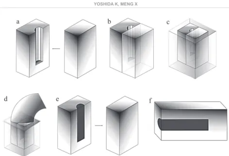
Forty semi-cylindrical cavities with a diameter of 3 mm and a depth of 11 mm were prepared in 5x10x16 mm acrylic resin blocks (Figure 1). Two acrylic resin blocks, with or without a semi-cylindrical cavity, were placed in a silicon impression material mold (15x15x20 mm). either 1 of 2
dual-cured core build-up resin composite pastes [Clearil DC Core Plus (DCP) or Uniil Core EM (UCE) (Figure 2)] was illed directly in the cavities using
auto-mixing tips, being sure to avoid entrapment of air, according to the manufacturer’s instructions. The upper surface in the resin composite material was covered with a plastic strip and pressed with a thin cover glass to remove any excess resin. Light-irradiation was provided by placing the tip of the LeD light unit (power density: 1,000 mW/ cm2;Pencure, J. Morita MFG Corp., Kyoto, Japan)
on the plastic strip. Power output was veriied with
a curing radiometer (Cure Lite; Dentsply Caulk, Milford, CT, USA) immediately before every light-activation throughout the study. The core build-up resin composites were light-cured for different durations using 1 of the 4 following light-exposure methods: (1) light-irradiation for 20 seconds on
the plastic strip after illing the core build-up resin
composite (20 s method); (2) light-irradiation for 40 seconds (40 s); (3) application of the bonding agent on the cavity wall with a brush, followed by air-drying, and light-irradiation for 10 seconds on the top of the cavity before the 20 s method (B+20 s method); (4) light-irradiation for 40 seconds on the plastic strip, followed by removal of the acrylic blocks from the silicon impression material mold, and irradiation of both sides of each acrylic resin block for 40 seconds each (120 s method). After irradiation of all the specimens, the acrylic resin blocks were removed from the silicon mold and separated. Five specimens of each dual-cured core build-up resin composite were irradiated by each of the 4 methods.
hardness measurements
Hardness was measured at the following depths from the light-irradiated surface of the cavity: 0.5, 2.0, 4.0, 6.0, 8.0, and 10.0 mm. For each specimen, the Knoop hardness number (KHN) was measured 5 times at each depth using a microhardness tester (FM-700: Future-Tech Corp., Kawasaki, Japan) at 0.5 hours, 24 hours, and 7 days post-irradiation. A Knoop diamond indenter was applied under a load of 25 g for a dwell time of 15 seconds; the load was then removed, and the long diagonal of the indentation was measured
under 400x magniication. KHN, which is inversely
Materials Manufacturer Composition
Core material
Clearil DC Core Plus (DCP)
(Batch # 0002AA)
Kuraray Noritake Products Corp., Tokyo, Japan
A paste: Bis-GMA, hydrophilic aliphatic dimethacrylate, hydrophobic aliphatic dimethacrylate,hydrophobic
aromatic dimethacrylate, silanized barium glass iller,
silanized colloidal silica, colloidal silica, chemical-initiator, photo-initiator, pigments
B paste: TEGDMA, hydrophilic aliphatic dimethacrylate, hydrophobic aromatic dimethacrylate, silanized barium
glass iller, silanized colloidal silica, aluminum oxide iller,
photo-accelerator , chemical-accelerator Bonding material
Clearil S3 Bond Plus
(Batch # 00024B)
Kuraray Noritake Products Corp., Tokyo, Japan
Bis-GMA,HEMA, MDP, hydrophilic dimethacrylate, hydrophobic dimethacrylate, colloidal silica, ethanol, water, photo-initiators, photo- and chemical-accelerator,
sodium luoride
Core material
Uniil Core EM (UCE)
(Batch # 1107011)
GC Corp., Tokyo, Japan Base: UDMA, dimethacrylates, luoro-alumino-silicate glass, silicon dioxide, photo-initiator, accelerator
Catalyst: UDMA, dimethacrylates, luoro-alumino-silicate
glass, silicon dioxide, chemical-initiator, pigment Bonding material
Self-etching bond A (Batch # 1205181)
GC Corp., Tokyo, Japan dimethacrylates, 4-META, silicon dioxide, ethanol, water, photo-initiator
Self-etching bond B (Batch # 1206011)
GC Corp., Tokyo, Japan ethanol, accelerator
Bis-GMA: bis-phenol-A-glycidyldimethacrylate; TEGDMA: triethyleneglycol dimethacrylate; UDMA: urethane dimethacrylate; HEMA: 2-hydoxyethyl methacrylate; MDP: 10-methacryloyloxydecyl dihydrogen phosphate; 4-META:
Figure 1- Schematic illustration of the preparation of specimens for measurement of Knoop hardness: a: acrylic resin blocks (5x10x16 mm) with or without semi-cylindrical cavity (diameter of 3 mm and depth of 11 mm); b: two acrylic resin blocks joined; c: two acrylic resin blocks placed in a silicon impression material mold (15x15x20 mm); d: dual-cured core
was thus calculated. All specimens were stored under dry and dark conditions in a box, which was placed in a biochemical incubator at 37°C to avoid exposure to light, and was accessed only to obtain measurements.
Statistical analysis
The KHN data were statistically analyzed by the repeated measures three-way ANOVA test. The independent variables analyzed were depths of cavity and post-irradiation times for within-subject analysis, and light-exposure methods and type of dual-cured core build-up resin composite for between-subject analysis. A one-way ANOVA with the post-hoc Tukey’s compromise test was used to
establish speciic differences in KHN values between
the groups (α=0.05).
RESULTS
The results of this study are summarized in Tables 1 and 2, which show the mean KHN and standard deviation of all the experimental groups in DCP and UCe, respectively, at 6 depths of cavity after 3 different post-irradiation durations by 4 different light-exposure methods.
For the DCP, the KHN was effected by the light-exposure method (p=0.0001; F=1652.29), post-irradiation time (p=0.0001; F=606.09),
depth of cavity (p=0.0001; F=1184.99), and all the interactions between all the aforementioned
factors (p=0.0001). For UCE, signiicant differences
were found between the light-exposure methods (p=0.0001; F=201.27), post-irradiation times (p=0.0001; F=857.15), and depth of cavity
(p=0.0001; F=488.76); in addition, signiicant
interactions were also found between all the aforementioned factors (p=0.0001).
For both resin composites, at 0.5 hours post-irradiation, the 20 s and 40 s methods resulted in the highest KHN values at the depth of 0.5 mm; these values gradually decreased with increasing depths of cavity (p<0.05). On the other hand, at 24 hours or 7 days post-irradiation, the KHN values of
DCP or UCE were not signiicantly different between
the depths of 2.0 mm, 4.0 mm, and/or 6.0 mm, or 8.0 mm and 10.0 mm, respectively (p>0.05).
The 40 s method resulted in signiicantly higher
KHN values than the 20 s method at all depths of cavity and post-irradiation times for both resin composites, with the exception of UCe at the depth of 0.5 mm (p<0.05). With the 120 s method, the KHN values of both resin composites were not
signiicantly different among the 6 depths of cavity
at all post-irradiation times (p>0.05), but they were
signiicantly higher than those of the 40 s method,
with the exception of that at the depth of 0.5 mm at all post-irradiation times (p<0.05).
Exposure method
Post-irradiation
time
Depth of cavity (mm)
0,5 2 4 6 8 10
Exposure time of
20 s (20 s)
0.5 hours 43.48±0.56αaC 39.89±0.92αbC 37.56±0.40αcC 35.89±0.73αcdC 34.88±1.27αdC 33.08±1.71αeC
24 hours 45.76±0.74αaB 41.13±0.68αbB 39.39±0.32αcB 39.19±0.31αcAB 38.67±0.21αcAB 38.59±0.12αcAB
7 days 46.79±0.59αaA 42.94±0.59αbA 41.62±0.28αbA 40.03±1.14αcA 39.62±1.28αcA 39.04±1.07αcA
Bonding +
exposure time of
20 s (B+20 s)
0.5 hours 41.71±0.54βaC 38.63±0.60αbC 37.67±0.58αbcC 37.07±0.58αcC 35.49±0.98αdB 35.01±0.90αdB
24 hours 46.11±0.59αaB 43.18±0.75βbB 41.84±0.62βcB 41.70±0.49βcB 41.34±0.82βcA 41.17±0.84βcA
7 days 47.50±0.25αaA 45.51±0.95βbA 43.34±0.83βcA 43.35±0.88βcA 42.54±0.70βcA 42.24±0.90βcA
Exposure time of
40 s (40 s)
0.5 hours 48.83±0.36γaB 44.42±0.33βbC 42.83±0.50βcC 40.83±0.87βdC 38.60±0.58βeC 37.57±0.29βfC
24 hours 51.89±0.61βaA 49.04±0.66γbAB 47.57±0.57γcAB 46.73±0.46γcAB 44.76±0.59γdAB 43.98±0.58γdAB
7 days 52.30±0.53βaA 49.47±0.52γbA 47.92±0.76γcA 47.03±0.51γcA 45.36±0.68γdA 44.76±0.77γdA
Exposure time of
120 s (120 s)
0.5 hours 48.57±0.51γaC 48.74±0.40γaC 48.84±0.34γaC 48.95±0.26γaC 48.98±0.15γaC 48.90±0.26γaC
24 hours 52.40±0.25βaB 52.35±0.27δaB 52.20±0.24δaB 52.40±0.28δaB 52.67±0.26δaB 52.26±0.50δaB
7 days 53.06±0.19βaA 53.03±0.21δaA 52.98±0.09δaA 53.00±0.28δaA 53.27±0.33δaA 52.92±0.26δaA
Table 1- Mean KHN±SD for Clearil DC Core Plus (DCP) at 6 depths of cavity after 3 post-irradiation times by 4
light-exposure methods
The same lower-case superscript Greek characters indicate no statistically signiicant differences between exposure
methods at same post-irradiation time in the same depth of cavity; the same lower-case superscript letters indicate no
statistically signiicant differences between depths of cavity at same post-irradiation time in the same exposure method; the same upper-case superscript letters indicate no statistically signiicant differences between post-irradiation times at same
For DCP, with the B+20 s method, no signiicant
differences in the KHN values were observed at 0.5 hours post-irradiation at all depths of cavity, except 0.5 mm (p>0.05). However, at 24 hours and 7 days post-irradiation, the B+20 s method
resulted in signiicantly higher KHN values than
the 20 s method at all depths of cavity, except 0.5 mm (p<0.05). On the other hand, for UCe, at 0.5 hours post-irradiation, the B+20 s method showed
signiicantly lower KHN values than the 20 s method
up to the depth of 6.0 mm; however, the KHN values
were signiicantly higher at the depths of 8.0 mm
and 10.0 mm (p<0.05). At 24 hours and 7 days
post-irradiation, no signiicant differences in KHN
values were observed between the B+20 s and 20 s method at all depths of cavity (p>0.05).
For all light-exposure methods, except the 120 s
method, the UCE exhibited signiicantly higher KHN
values at all depths of cavity, except at 4.0 mm in the 40 s method, at 7 days post-irradiation than at 0.5 hours and 24 hours post-irradiation (p<0.05). It was found that the KHN values of DCP were
signiicantly higher than those of UCE at all depths
of cavity and post-irradiation times, regardless of the exposure method (p<0.05).
DISCUSSION
of dual-cured core build-up resin composites depend on the light-exposure method, including irradiation duration, use of bonding agent, depth of cavity, post-irradiation time, and material brand. Therefore, the research hypotheses formulated for this study must be rejected.
KHN has been shown to be a good indicator of the degree of conversion/polymerization based on its good correlation with infrared spectroscopy9,10,31.
However, the prediction of an absolute value of degree of conversion by means of an absolute hardness value is not achievable, since other
factors such as type and size of iller, iller load,
monomer composition, quantity of initiators, and the ratio of chemical- and light-cured components
strongly inluence the inal quantity of reacted
monomers2,5,14. Microhardness data from the same
resin cement only should be compared according to the depth of the root canal or time elapsed since luting3. KHNs could be used to relect the
degree of conversion at different depths of a resin composite32. Therefore, KHNs were measured in
the present study to relect monomer conversion at
different depths of cavity and post-irradiation times in the dual-cured core build-up resin composites.
The KHNs of both core build-up resin composites irradiated for 20 or 40 seconds were the highest at the depth of 0.5 mm, and gradually decreased
Exposure method
Post-irradiation
time
Depth of cavity (mm)
0,5 2 4 6 8 10
Exposure time of
20 s (20 s)
0.5 hours 36.26±1.31αaC 34.18±0.97αbC 32.99±0.64αbC 31.69±0.64αcC 29.10±0.49αdC 27.31±0.39αeC
24 hours 41.22±0.81αaB 38.42±1.14αbB 38.35±1.07αbB 38.42±1.19αbB 37.42±1.33αbB 36.87±1.50αbB
7 days 42.86±0.80αaA 40.52±1.04αbA 39.79±1.20αbcA 39.56±1.15αbcA 38.37±1.01αcA 38.08±1.23αcA
Bonding +
exposure time of
20 s (B+20 s)
0.5 hours 32.23±0.97βaC 30.78±0.79βbC 30.58±0.65βbC 30.54±0.47βbC 30.10±0.93αbC 29.49±0.79βcC
24 hours 40.12±0.76αaB 37.97±1.08αbB 37.56±0.25αbcB 37.50±0.11αbcB 37.39±0.21αbcB 36.79±0.42αcB
7 days 42.06±0.40αaA 40.21±0.28αbA 39.08±0.24αcA 39.19±0.26αcA 38.82±0.26αcA 38.09±0.43αdA
Exposure time of
40 s (40 s)
0.5 hours 37.19±0.24γaC 36.50±0.37γaC 35.28±1.15γbB 34.14±0.61γcC 32.22±0.17βdB 30.36±0.13γeC
24 hours 42.31±0.38βaB 40.93±0.53βbB 40.90±0.61βbA 40.62±0.49βbcB 39.80±1.10βbcA 39.51±0.87βcB
7 days 43.07±0.15βaA 42.12±0.45βbA 41.87±0.45βbA 41.97±0.47βbA 40.76±0.61βcA 40.77±0.69βcA
Exposure time of
120 s (120 s)
0.5 hours 37.35±0.32γaC 37.60±0.35δaC 37.46±0.25δaC 37.59±0.18δaB 37.36±0.21γaC 37.32±0.25δaB
24 hours 42.58±0.30βaB 42.67±0.62γaAB 42.60±0.46γaAB 42.59±0.46γaA 42.62±0.55γaAB 42.7±0.44γaA
7 days 42.78±0.40βaA 42.98±0.77γaA 42.97±0.86γaA 42.81±0.45γaA 43.00±0.85γaA 43.06±0.60γaA
Table 2- Mean KHN±SD for KHN for Uniil Core EM (UCE) at 6 depths of cavity after 3 post-irradiation times by 4
light-exposure methods
The same lower-case superscript Greek characters indicate no statistically signiicant differences between exposure
methods at same post-irradiation time in the same depth of cavity; the same lower-case superscript letters indicate no
statistically signiicant differences between depths of cavity at same post-irradiation time in the same exposure method; the same upper-case superscript letters indicate no statistically signiicant differences between post-irradiation times at same
be simply attributed to the direction of photo-initiation. Light irradiation was focused on the top surface of the cavity. Therefore, polymerization of the resin composites, by means of photo-activated free radicals, may occur immediately
at the shallow depths of cavity. This inding, that
is, KHN of light- and dual-cured resin composites are affected by depth of cavity, has been reported previously4,6-8. The chemical-curing mechanism of
dual-cured resin composites is usually based on a redox reaction of benzoyl peroxide with aromatic tertiary amines, which generates free radicals that break the aliphatic carbon double bonds to initiate the polymerization process. It is supposed that immediate photo-activation after light irradiation, despite causing a rapid increase in the viscosity of the polymer matrix, does not hinder migration of the activated free radicals responsible for further chemically induced polymerization. Although the photo-activated free radicals at shallow depths of cavity could induce chain propagation of the resin polymer in the downward direction, the exact polymerization mechanism of dual-cured core build-up resin composite at greater depths of cavity remains unknown. It is difficult to distinguish clearly between the depths of cavity at which polymerization of the resin composite occurs through photo-initiation and those at which polymerization occurs by means of chemical initiation alone.
evaluation of the duration of photo-activation revealed that a longer exposure time of 40 seconds
resulted in signiicantly higher KHN values than
did a shorter exposure time of 20 seconds at all depths of cavity and post-irradiation times for both resin composites; however, this trend was not observed in UCe at the depth of 0.5 mm. Thus, our study showed that prolonged irradiation durations resulted in increased hardness values. Light-curing units with lower power density needed longer light-exposure times to produce a similar microhardness value of resin composite as that of light sources with high power density11.
Therefore, with longer light-exposure durations, which result in higher energy densities at a given irradiance, more photo-sensitizer molecules are activated, which in turn increase the free radical concentration and consequently the conversion of double bonds. However, at the depth of 0.5 mm,
no signiicant differences were observed in the KHN
values between the light-irradiation durations of 40 seconds and 120 seconds (40 seconds each on the top and either side of the cavity) for both resin
composites. This inding might be attributed to the
fact that the polymer network developed during light exposure for 40 seconds does not allow any additional mobility of the polymer chains in order to increase monomer conversion, indicating that
the light-irradiation duration of 40 seconds alone
is suficient.
Dentin hardness ranges from KHN values of 50 to 70, depending on the distance from the amelodentinal junction21. The mean microhardness
(KHN) value of 52.9 for DCP with 120 s method at the depth of 10.0 mm at 7 days post-irradiation therefore predicts similar mechanical properties to that of dentin. The equal degree of polymerization within the core material may support a uniform distribution of stress along the tooth-material interface under load. Longer light-exposure durations result in superior microhardness, but, at the same time, contribute to an increase of shrinkage and contraction stress of the dual-cured core build-up resin composite38. In cases of
signiicant coronal destruction, it is necessary to
replace the lost tooth structure with a core build-up material to attain full-coverage restoration. The cast post and core, prefabricated post and core materials, and coronal-radicular build-ups are the available options for this purpose. The fracture resistance and survival probability of post and core restorations depend on several factors such as the post material, luting agent, amount and condition of residual tooth structure, core material, preparation
of the tooth for restorative procedures, and, inally, the ixed restoration16,22,25,34. Therefore, when using
dual-cured core build-up resin composites, it is preferable to prepare the composites by using the effective exposure method.
Before core build-up resin composites are used, a bonding agent must be applied on the cavity wall. The effectiveness of the bonding agent on the KHN as an indirect method of monomer conversion differed between the 2 resin composites. For DCP, at 24 hours and 7 days post-irradiation, application
of the bonding agent resulted in signiicantly higher
KHNs than the 20 s method at all depths of cavity; however, this was not observed with UCe. The initiator and/or accelerator present in the bonding agent would promote monomer conversion of the dual-cured core build-up resin composites. On the other hand, the acidic monomer in the bonding agent inhibits the amine co-initiator in the dual-cured materials16, which in turn adversely affects
polymerization of dual-cured core build-up resin composites. To prevent this inhibitory effect,
aromatic sulinic acid sodium salts are sometimes
added to the bonding agent15,37. The differences
in compositions between the 2 core build-up resin composites might be responsible for the difference in their KHN behaviors.
consistent with those of a previous study3, but not
with those of another study41, which did not ind
changes in microhardness values 24 hours after irradiation. However, the polymerization reaction
of the dual-cured materials might be speciic19, and
these previous studies used resin cements, whereas dual-cured core build-up resin composites were used in the present study. In fact, in the former study, neither did the authors evaluate dual-cured core build-up resin composites nor did they evaluate hardness behavior of dual-cured core build-up resin composite in simulated depths of cavity.
Dual-cured resin composites have been introduced for use as both luting and core build-up materials. Sbuild-uperior physical properties are important for a successful restoration. In this study, it was found that the KHNs of DCP were superior to those of UCe, regardless of exposure methods.
Various factors can inluence the microhardness of a resin composite, such as iller load, type, or size,
or resin matrix type17,18. Increasing the monomer
viscosity decreased the hardness27. In this study,
the association between KHN values of the resin
composite and composition of iller and matrix
resin was not evaluated. On the other hand, the differences between the KHNs at the depths of 0.5 mm and 10.0 mm in UCe were lower than those in DCP, regardless of the light-exposure method. Dual-cured materials differ markedly in terms of the relative content of light- and self-activated catalysts13. Differences in the degree
of conversion among materials when subjected to various curing protocols may consequently be attributed to variations in catalyst systems. In the present study, since similar KHNs were observed irrespective of the light-exposure method at greater depths of cavity in UCe rather than DCP, it might be inferred that the former exhibits high levels of chemically curing activators compared with the latter, compensating for attenuation of light energy at greater depths of cavity. Indeed, the polymerization behavior of dual-cured resin composites is strongly related to the material and can vary as a function of composition35. The speed
of the polymerization reaction is strongly inluenced by inhibitor concentration in the unilled light-cured
methacrylate-based systems30. Therefore, the
curing mechanism of a speciic composite material
may not be applicable to other materials.
CONCLUSIONS
Within the limitations of the present study, the following conclusions can be drawn:
1. The microhardness of the dual-cured core build-up resin composites vary depending on the light-exposure method, including irradiation
post-irradiation time, and material brand.
2. For both resin composites, irradiation for 120
seconds does not result in signiicant differences
in KHNs among all 6 depths of cavity at all post-irradiation times.
3 . B a s e d o n t h e p h y s i c a l p r o p e r t y o f microhardness behavior, dual-cured core build-up resin composites are preferably prepared by the effective exposure method.
REFERENCES
1- Aksornmuang J, Nakajima M, Foxton RM, Tagami J. Mechanical properties and bond strength of dual-cure resin composites to root canal dentin. Dent Mater. 2007;23:226-34.
2- Arrais CA, Giannini M, Rueggeberg FA. Kinetic analysis of monomer conversion in auto- dual-polymerizing modes of commercial resin luting cement. J Prosthet Dent. 2009;101:128-36.
3- Baena e, Fuentes MV, Garrido MA, Rodríguez J, Ceballos L. Inluence of post-cure time on the microhardness of self-adhesive resin cements inside the root canal. Oper Dent. 2012;37:548-56. 4- Bouschlicher MR, Rueggeberg FA, Wilson BM. Correlation of bottom-to-top surface microhardness and conversion ratios for a variety of resin composite compositions. Oper Dent. 2004;29:698-704.
5- Cadenaro M, Navarra CO, Antoniolli F, Mazzoni A, Di Lenarda, Rueggeberg FA, et al. The effect of curing mode on extent of polymerization and microhardness of dual-cured, self-adhesive resin cements. Am J Dent. 2010;23:14-8.
6- Camargo eJ, Moreschi e, Baseggio W, Cury JA, Pascotto RC. Composite depth of cure using four polymerization techniques. J Appl Oral Sci. 2009;17:446-50.
7- Ciccone-Nogueira JC, Borsatto MC, Souza-Zaroni WC, Ramos RP, Palma-Dibb RG. Microhardness of composite resins at different depths varying the post-irradiation time. J Appl Oral Sci. 2007;15:305-9.
8- Cunha LG, Sinhoreti MA, Consani S, Sobrinbo LC. effect of different photoactivation methods on the polymerization depth of a light-activated composite. Oper Dent. 2003;28:155-9. 9- DeWald JP, Ferracane JL. A comparison of four modes of evaluating depth of cure of light-activated composites. J Dent Res. 1987;66:727-30.
10- Ferracane JL. Correlation between hardness and degree of conversion during the setting reaction of unilled dental restorative resins. Dent Mater. 1985;1:11-4.
11- Franco eB, Santos PA, Mondelli RF. The effect of different light-curing units on tensile strength and microhardness of a composite resin. J Appl Oral Sci. 2007;15:470-4.
12- Galhano GA, Melo RM, Barbosa SH, Zamboni SC, Bottino MA, Scotti R. evaluation of light transmission through translucent and opaque post. Oper Dent. 2008;33:321-4.
13- Hasegawa eA, Boyer DB, Chan DC. Hardening of dual-cured cements under composite resin inlays. J Prosthet Dent. 1991;66:187-92.
14- Hofmann N, Papshart G, Hugo B, Klaiber B. Comparison of photo-activation versus chemical or dual-curing of resin-based luting cements regarding lexural strength, modulus and surface hardness. J Oral Rehabil. 2001;28:1022-8.
17- Kim KH, Ong JL, Okuno O. The effect of iller loading and morphology on the mechanical properties of contemporary composites. J Prosthet Dent. 2002;87:642-9.
18- Li Y, Swartz ML, Phillips RW, Moore BK, Roberts TA. effect of iller content and size on properties of composites. J Dent Res. 1985;64:1396-401.
19- Lu H, Mehmood A, Chow A, Powers JM. Influence of polymerization mode on lexural properties of esthetic resin luting agents. J Prosthet Dent. 2005;94:549-54.
20- Lutz F, Krejci I, Oldenburg TR. elimination of polymerization stresses at the margins of posterior composite resin restorations: a new restorative technique. Quintessence Int. 1986;17:777-84. 21- Meredith N, Sherriff M, Setchell DJ, Swanson SA. Measurement of the microhardness of human enamel and dentine using an indentation technique. Arch Oral Biol. 1996;41:539-45.
22- Naumann M, Metzdorf G, Fokkinga W, Watzke R, Sterzenbach G, Bayne S, et al. Inluence of test parameters on in vitro fracture resistance of post-endodontic restorations: a structured review. J Oral Rehabil. 2009;36:299-312.
23- Park J, Chang J, Ferracane J, Lee IB. How should composite be layered to reduce shrinkage stress: incremental or bulk illing? Dent Mater. 2008;24:1501-5.
24- Pegoraro TA, Silva NR, Carvalho RM. Cements for use in esthetic dentistry. Dent Clin North Am. 2007;51:453-71. 25- Peroz I, Blankenstein F, Lange KP, Naumann M. Restoring endodontically treated teeth with posts and cores – a review. Quintessence Int. 2005;36:737-46.
26- Plasmans PJ, Creugers NH, Mulder J. Long-term survival of extensive amalgam restorations. J Dent Res. 1998;77:453-60. 27- Podgórski M. Structure-property relationship in new photo-cured dimethacrylate-based dental resins. Dent Mater. 2012;28:398-409.
28- Ramos MB, Pegoraro TA, Pegoraro LF, Carvalho RM. effects of curing protocol and storage time on the micro-hardness of resin cements used to lute iber-reinforced resin posts. J Appl Oral Sci. 2012;20:556-62.
29- Roberts HW, Leonard DL, Vandewalle KS, Cohen Me, Charlton DG. The effect of a translucent post on resin composite depth of cure. Dent Mater. 2004;20:617-22.
30- Rosentritt M, Shortall AC, Palin WM. Dynamic monitoring of curing photoactivate resins: a methods comparison. Dent Mater. 2010;26:565-70.
31- Rueggeberg FA, Craig RG. Correlation of parameters used to estimate monomer conversion in a light-cured composite. J Dent Res. 1988;67:932-7.
32- Rueggeberg FA, ergle JW, Mettenburg DJ. Polymerization depths of contemporary light-curing units using microhardness. J esthet Dent. 2000;12:340-9.
33- Schwartz RS, Robbins JW. Post placement and restoration of endodontically treated teeth: a literature review. J endod. 2004;30:289-301.
34- Stankiewicz NR, Wilson PR. The ferrule effect: a literature review. Int endod J. 2002;35:575-81.
35- Stavridakis MM, Kalaboura AI, Krejci I. Degree of remaining C=C bonds, polymerization shrinkage and stresses of dual-cured core build-up resin composites. Oper Dent. 2005;30:443-52. 36- Stavridakis MM, Lutz F, Johnston WM, Krejci I. Linear displacement and force induced by polymerization shrinkage of resin-based restorative materials. Am J Dent. 2003;16:431-8. 37- Suh Bi, Feng L, Pashley DH, Tay FR. Factors contributing to the incompatibility between simpliied-step adhesives and chemically cured or dual-cured composites. Part III. effect of acidic resin monomers. J Adhes Dent. 2003;5:267-82.
38- Tauböck TT, Bortolotto T, Buchalla W, Attin T, Krejci I. Inluence of light-curing protocols on polymerization shrinkage and shrinkage force of a dual-cured core build-up resin composite. eur J Oral Sci. 2010;118:423-9.
39- Tauböck TT, Buchalla W, Hiltebrand U, Roos M, Krejci I, Attin T. Inluence of the interaction of light- and self-polymerization on subsurface hardening of a dual-cured core build-up resin composite. Acta Odontol Scand. 2011;69:41-7.
40- Van Nieuwenhuysen JP, D’Hoore W, Carvalho J, Qvist V. Long-term evaluation of extensive restorations in permanent teeth. J Dent. 2003;31:395-405.