Clinical and ultrasonographic correlation in localized
cutaneous scleroderma*
Correlação clínica e ultra-sonográfica na esclerodermia localizada cutânea
Marcio Bouer1, Maria Cristina Chammas2, Maria Cristina Lorenzo Messina3, Ilka Regina Souza de Oliveira4, Giovanni Guido Cerri5
OBJECTIVE: To describe ultrasonographic findings of localized cutaneous scleroderma and correlating them with clinical findings. MATERIALS AND METHODS: Twenty-three lesions of localized cutaneous scleroderma in 21 patients were evaluated with a Logiq 700 equipment coupled with a 6–14 MHz linear transducer. The disease stage (athrophic or inflammatory) was evaluated by a dermatologist, and the ultrasonographic findings (skin thickness and echogenicity) for both the affected and adjacent healthy regions were evaluated by a radiologist. Seven of the cases underwent post-treatment follow-up. RESULTS: All the affected regions presented loss of the normal ultrasonographic pattern of the dermis. Cases with clinically atrophic lesions (52.2%; 12/23) corresponded to reduction in the thickness and increase in the echogenicity of the dermis, and clinically inflammatory lesions (47.8%; 11/23) corresponded to decrease in echogenicity and increase in the thickness of the dermis. Post-treatment follow-up demonstrated alterations in the dermis thickness. CONCLUSION: The ultrasonographic findings allow the correlation between increase in the thickness/decrease in echogenicity of the dermis with the inflammatory phase of the disease, and decrease of the thickness/ increase in echogenicity of the dermis with the atrophic phase. Also, it could be observed that it is possible to quantify the thickness of the dermis, utilizing this information associated with the clinical evaluation in the post-treatment follow-up.
Keywords: Localized scleroderma; Ultrasonography; Dermis.
OBJETIVO: Apresentar os aspectos ultra-sonográficos da esclerodermia localizada e relacioná-los com os aspectos clínicos. MATERIAIS E MÉTODOS: Foram analisadas 23 lesões de esclerodermia localizada em 21 pacientes. Foi utilizado equipamento Logiq 700 com transdutor linear de 6–14 MHz. Foram avaliados, pelo dermatologista, o estágio da doença (inflamatório ou atrófico), e pelo radiologista, a espessura e a ecogeni-cidade da derme nas regiões afetadas e sãs adjacentes. Foi feito acompanhamento de sete casos após tra-tamento. RESULTADOS: Todas as lesões apresentaram perda do padrão ultra-sonográfico normal da derme. Os casos de lesão clinicamente atrófica (52,2%; 12/23) corresponderam a redução da espessura e aumento da ecogenicidade da derme e os casos de lesão clinicamente inflamatória (47,8%; 11/23) corresponderam a aumento da espessura e redução da ecogenicidade da derme. Controles pós-tratamento mostraram altera-ções na espessura da derme. CONCLUSÃO: Os achados ultra-sonográficos nos permitem associar o aumento da espessura e a redução da ecogenicidade da derme com a fase inflamatória da doença, e a redução da espessura e o aumento da ecogenicidade da derme com a fase atrófica da doença. Notamos também que é possível quantificar a espessura da derme e usar essa informação no controle pós-tratamento associada à avaliação clínica.
Unitermos: Esclerodermia localizada; Ultra-sonografia; Derme.
Abstract
Resumo
* Study developed at Instituto de Radiologia – Hospital das Clínicas da Faculdade de Medicina da Universidade de São Paulo (InRad/HC-FMUSP), São Paulo, SP, Brazil.
1. MD, Radiologist, Researcher for the Unit of Ultrasonography, Instituto de Radiologia – Hospital das Clínicas da Faculdade de Medicina da Universidade de São Paulo (InRad/HC-FMUSP), São Paulo, SP, Brazil.
2. PhD, Director for the Unit of Ultrasonography, Instituto de Radiologia – Hospital das Clínicas da Faculdade de Medicina da Universidade de São Paulo (InRad/HC-FMUSP), São Paulo, SP, Brazil.
3. Master, Assistant Physician for the Department of Derma-tology, Hospital das Clínicas da Faculdade de Medicina da Uni-versidade de São Paulo (HC-FMUSP), São Paulo, SP, Brazil.
4. PhD, Technical Director for the Service of Iconology, Hospi-tal Universitário da Universidade de São Paulo (USP), São Pau-lo, SP, Brazil.
the skin and some other organ systems. Histological findings on the skin are: mas-sive deposition of synthesized collagen, perivascular mononuclear cell infiltrate and vascular damages(1).
Scleroderma was first studied by means of ultrasonography in 1984, with a 15 MHz transducer (A-mode). In cases of focal le-sions, the skin thickness was increased, and in pigmented lesions, decreased(2). In 1986, and later in 1993, B-mode 10 MHz, 20 MHz and 25 MHz transducers started
Bouer M, Chammas MC, Messina MCL, Oliveira IRS, Cerri GG. Clinical and ultrasonographic correlation in localized cuta-neous scleroderma. Radiol Bras. 2008;41(2):87–91.
5. Titular Professor, Department of Radiology, Faculdade de Medicina da Universidade de São Paulo (FMUSP), São Paulo, SP, Brazil.
Mailing address: Dr. Marcio Bouer. Rua Pereira Leite, 44, ap. 91, Sumarezinho. São Paulo, SP, Brazil, 05442-000. E-mail: [email protected]
Received May 22, 2007. Accepted after revision July 24, 2007.
INTRODUCTION
being utilized, and could demonstrate in-crease in the skin thickness in the involved regions, as well as post-treatment alter-ations(3–5). In 1995, a study with a 30 MHz transducer demonstrated an increase in the skin thickness of involved regions and even a mild increase in clinically non-affected regions(6). In 1998, post-treatment imaging of the skin with a 20 MHz transducer (B-mode) demonstrated that there may be dif-ferent findings depending on the disease stage(7).
The present study was aimed at demon-strating ultrasonographic findings as com-pared with clinical findings in cases of fo-cal (or lofo-calized) scleroderma.
MATERIALS AND METHODS
Twenty-three lesions of focal sclero-derma were evaluated in 21 patients (18 women and 3 men) with ages ranging be-tween 8 and 58 years (mean age, 24.1 years), in the period between May 2001 and May 2003, with a Logiq 700 (GE Medical Systems; Milwaukee, Wis., USA) equip-ment coupled with a multifrequency linear transducer (6–14 MHz). Lesions were found on the trunk (15) and limbs (8) of these patients.
The study was submitted to the approval by the Committee for Ethics in Research of Hospital das Clínicas da Faculdade de Medicina da Universidade de São Paulo, and all of the participating patients signed a term of free and informed consent.
The studies were performed by an ex-perienced radiologist specialized in small parts ultrasound imaging, with a thick gel layer between the transducer and the skin, and minimum pressure on the region to be scanned for not deforming the surface of the skin to be evaluated.
Parameters to be evaluated at B-mode ultrasound included: a) dermis thickness – measured from the anterior hyperechogenic line between the gel layer and the dermis surface, and the posterior hyperechogenic line between the deep dermis and the sub-cutaneous tissue (Figure 1); b) the dermis echogenicity – normally characterized by a hypoechogenic superficial layer and a hyperechogenic deep layer (Figure 2).
Both the region of the lesion and an adjacent healthy region of the skin were evaluated in all of the patients participat-ing in the present study. Seven patients had a post-treatment follow-up including the evaluation of these same parameters.
The clinical diagnosis was made by an experienced dermatologist, and all of the cases had their diagnosis confirmed by means of anatomopathological studies.
RESULTS
In the thickness analysis, clinically atro-phic lesions (Figure 2), corresponding to 52.2% (12/23) of cases, presented a de-crease in the dermis thickness (Figure 4) as compared with the clinically normal re-gions (Figure 3). The mean thickness in the
affected regions was 1.29 mm, whereas in the normal regions was 2.23 mm. On the other hand, clinically inflammatory lesions, corresponding to 47.8% (11/23) of cases (Figure 5), presented an increase in the dermis thickness (Figure 7) as compared with the clinically normal regions (Figure 6). The mean thickness in the affected re-gions was 2.90 mm, whereas in the normal regions, it was 1.90 mm (Table 1).
As regards echogenicity, in cases of atrophic lesion, the dermis presented with an increase in echogenicity (Figure 4), whereas on inflammatory lesions the der-mis presented with a decrease in echo-genicity (Figure 7). Loss of the typical ultrasonographic pattern of the dermis, corresponding to superficial hypoecho-genicity and deep hyperechohypoecho-genicity for all of the affected regions, was observed in all of the affected regions (Figures 3 and 6).
The seven patients who underwent post-treatment follow-up, presented with alter-ation of the dermis thickness. Three of these cases presented inflammatory lesion with decreased dermis thickness (from 2.90 mm to 2.30 mm, on average) (Figure 8), and four presented atrophic lesions, with increased dermis thickness (from 1.10 mm to 1.40 mm, on average) (Table 2).
All anatomopathological studies con-firmed the clinical diagnosis of scleroderma, both in the inflammatory phase, with pre-dominance of cell infiltrate, and in the atro-phic phase, with massive deposition of synthesized connective tissue and fibrosis.
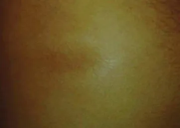
Figure 2. Picture of the skin of a patient affected by localized scleroderma. Typical pigmented skin in the atrophic phase of the disease.
Figure 4. Ultrasonography of the region of atrophic lesion. Decreased dermis thickness, hypoechogenicity and loss of definition between superficial and deep layers can be observed.
Figure 8. Ultrasonography of the region of inflammatory lesion after 30 days of treatment. Despite the decrease as compared to the previous study (Figure 7) still increased thickness of the dermis is observed.
Figure 3. Ultrasonography of the region adjacent to an area of atrophic le-sion. The typical feature of preserved skin can be observed (between crosses).
✛ ✛
Figure 6. Ultrasonography of the region adjacent to the area of inflammatory lesion. A typical feature of preserved skin is observed (between crosses).
✛ ✛
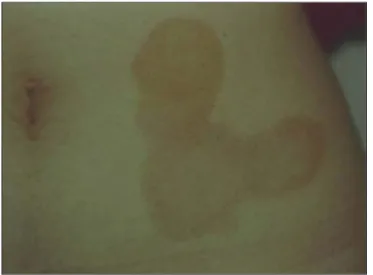
Figure 5. Picture of the skin of a patient affected by localized scleroderma. Typically elevated, slightly hypopigmented lesion in the inflammatory phase of the disease.
Figure 7. Ultrasonography of the region of inflammatory lesion. Note the in-creased thickness of the dermis (hypoechogenic) and loss of definition of the limit between superficial and deep layers (between crosses).
The normal skin is made up of three different layers: a) epidermis, a 0.06–0.6 mm-thick layer with two types of cells – keratinocytes and melanocytes; b) dermis, a 1–4 mm-thick layer constituted of an extensive network of blood and lymphatic vessels, connective tissue, nerves, glands, fibroblasts, histiocytes and mastocytes, di-vided into papillary dermis – more super-ficial and thinner, with loose connective tissue, and reticular dermis, deeper and with a dense connective tissue; c) subcu-taneous tissue, with a variable thickness, consisting predominantly of fat cells.
The image of a normal skin, from the surface to the inner layers, will show:
– The anechogenic gel layer intermingled with hyperechogenic spots typical of its internal component.
– A fine, echogenic line corresponding to the interface between the gel and the skin (epidermis). Because of its typical, very thin thickness, the epidermis ap-pears ill-defines at US scans, except in the foot sole and hypothenar region, where the thickness is higher. Even so, a fine hypoechogenic layer only can be visualized with higher frequency or 20 MHz transducers.
– A hyperechogenic layer corresponding to the dermis, where the superficial and deep layers can be differentiated with transducers of 13 MHz or more — the first layer, slightly hypoechogenic and heterogeneous, and the second one, more hyperechogenic and homoge-neous. Hypoechogenic lines corre-sponding to hair follicles can be ob-liquely seen across this layer. The der-mis echogenicity is variable with age; it appears hypoechogenic in neonates, with a mild increase in echogenicity in older infants, and pronounced increase with the adult age. In the elderly, a more echogenic layer is observed at the level of the superficial dermis, mainly be-cause of the exposure to the sunlight. The dermis is divided into papillary dermis – more superficial and thinner, with loose connective tissue, and reticu-lar dermis, deeper and with a dense connective tissue, the latter being most frequently affected in cases of sclero-derma.
– A hypoechogenic layer (adipous tissue) DISCUSSION
The utilization of ultrasonography with high frequency transducers in the evaluation of dermatologic diseases has in-creased with recent technological develop-ments and the demand for a reliable and non-invasive method to aid the dermatolo-gist in the visualization of abnormalities such as tumors or other less aggressive
dis-eases occurring under the affected skin. In 1979, Alexander & Miller were the first to apply ultrasonography in dermatology to measure the skin thickness(8). From that time on, many other studies about the most different dermatological diseases have been developed, utilizing already available equipment/transducers as well as other devices especially designed for this pur-pose.
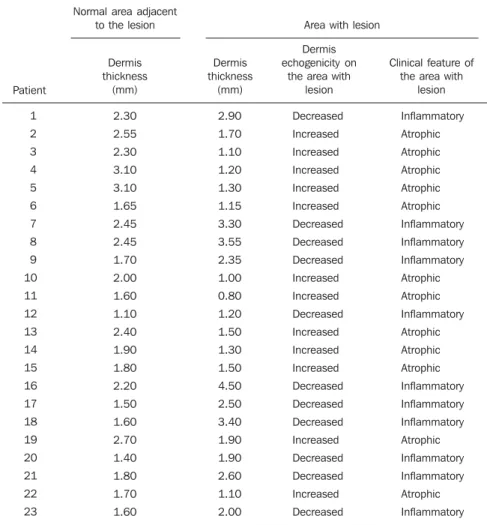
Table 1 Dermis thickness and echogenicity, and corresponding clinical findings for each patient.
Patient 1 2 3 4 5 6 7 8 9 10 11 12 13 14 15 16 17 18 19 20 21 22 23 2.30 2.55 2.30 3.10 3.10 1.65 2.45 2.45 1.70 2.00 1.60 1.10 2.40 1.90 1.80 2.20 1.50 1.60 2.70 1.40 1.80 1.70 1.60 Normal area adjacent
to the lesion Area with lesion
Dermis thickness
(mm)
Dermis echogenicity on
the area with lesion
Clinical feature of the area with
lesion Dermis thickness (mm) 2.90 1.70 1.10 1.20 1.30 1.15 3.30 3.55 2.35 1.00 0.80 1.20 1.50 1.30 1.50 4.50 2.50 3.40 1.90 1.90 2.60 1.10 2.00 Decreased Increased Increased Increased Increased Increased Decreased Decreased Decreased Increased Increased Decreased Increased Increased Increased Decreased Decreased Decreased Increased Decreased Decreased Increased Decreased Inflammatory Atrophic Atrophic Atrophic Atrophic Atrophic Inflammatory Inflammatory Inflammatory Atrophic Atrophic Inflammatory Atrophic Atrophic Atrophic Inflammatory Inflammatory Inflammatory Atrophic Inflammatory Inflammatory Atrophic Inflammatory
Table 2 Pre- and post-treatment measurements of dermis thickness, and corresponding clinical findings.
Patient 1 2 3 4 5 6 7
Clinical feature of the area with lesion
with intermingled hyperechogenic strias (fibrous septae), corresponding to subcutaneous tissue with variable thick-ness. Generally, the limit between this layer and the dermis is well-defined, although nonlinear.
– A hyperechogenic layer, corresponding to the muscular fascia(9).
Scleroderma physiopathology is com-plex and consists in three main factors: vascular damage, mononuclear cell infil-trate, and massive deposition of newly syn-thesized connective tissue, remarkably col-lagen. Apparently, these three factors occur simultaneously, initially with predomi-nance of cellular infiltrate, with an increase in the dermis thickness corresponding to the inflammatory phase of the disease, and latter predominance of deposition of con-nective tissue and fibrosis, with decrease in the dermis thickness, corresponding to the atrophic phase of the disease(1).
The ultrasonographic findings are com-patible with the histological findings: clini-cally atrophic lesions presented a decrease in the dermis thickness, and clinically in-flammatory lesions showed an increase in the dermis thickness. The studies devel-oped by Serup(2), Akesson et al.(3), Myers et al.(4) and Ihn et al.(6) have reported in-crease in the thickness of the dermis in the regions affected by scleroderma, probably because the inflammatory phase predomi-nated in the cases evaluated.
Atrophic lesions presented an increase in the dermis echogenicity, whereas inflam-matory lesions presented a decrease in the echogenicity. Additionally, in the affected
areas, there was a loss of differentiation between the superficial and deep layers of the dermis which has not been described in other studies and that can be explained by the diffuse involvement of the reticular der-mis; in the case of edematous predomi-nance, by the presence of cellular infiltrate and inflammatory process; and in the atro-phic case, by fibrosis and higher collagen concentration.
During the treatment, alterations were observed in the dermis thickness, with in-flammatory lesions presenting a decrease in the thickness of the dermis, and atrophic lesions presenting an increase, however, the differentiation between the superficial and deep layers of the dermis could not be established. Other authors(3,4,7,10,11) have concluded that ultrasonography is useful for a later post-treatment follow-up.
Finally, ultrasonographic findings allow the association between the increase in the dermis thickness/ decrease in the dermis echogenicity and the inflammatory phase of the disease, where edema and inflamma-tory process predominate, and the associa-tion between the decrease in the dermis thickness/increase in the dermis echo-genicity and the atrophic phase of the dis-ease, where loss of the skin elasticity and dermis cells compaction may be observed. Also, one can observe that it is possible to quantify the dermis thickness, utilizing this information in the post-treatment follow-up to indicate, or not, an improvement as-sociated with the clinical evaluation.
The development of transducers with higher frequencies as well as devices with
higher resolution can improve the post-treatment evaluation of the dermis thick-ness, and also de detection of alterations on clinically normal regions.
REFERENCES
1. Sapadin AN, Esser AC, Fleischmajer R. Immuno-pathogenesis of scleroderma – evolving concepts. Mt Sinai J Med. 2001;68:233–42.
2. Serup J. Localized scleroderma (morphoea): thick-ness of sclerotic plaques as measured by 15 MHz pulsed ultrasound. Acta Derm Venereol. 1984;64: 214–9.
3. Akesson A, Forsberg L, Hederstrom E, et al. Ultra-sound examination of skin thickness in patients with progressive systemic sclerosis (scleroderma). Acta Radiol Diagn. 1986;27:91–4.
4. Myers SL, Cohen JS, Sheets PW, et al. B-mode ultrasound evaluation of skin thickness in pro-gressive systemic sclerosis. J Rheumatol. 1986; 13:577–80.
5. Fornage BD, McGavran MH, Duvic M, et al. Imaging of the skin with 20-MHz US. Radiology. 1993;189:69–76.
6. Ihn H, Shimozuma M, Fujimoto M, et al. Ultra-sound measurement of skin thickness in systemic sclerosis. Br J Rheumatol. 1995;34:535–8. 7. Cammarota T, Pinto F, Magliaro A, et al. Current
uses of diagnostic high-frequency US in derma-tology. Eur J Radiol. 1998;27:S215–23.
8. Alexander H, Miller DL. Determining skin thick-ness with pulsed ultrasound. J Invest Dermatol. 1979;72:17–9.
9. Bouer M, Saito OC. Pele, subcutâneo e parede. In: Saito OC, Cerri GG, editores. Ultra-sonogra-fia de pequenas partes. São Paulo: Revinter; 2004. p. 245–68.
10. Hoffmann K, Gerbaulet U, el-Gammal S, et al. 20-MHz B-mode ultrasound in monitoring the course of localized scleroderma (morphea). Acta Derm Venereol Suppl. 1991;164:3–16.