Introduction
A healthy lifestyle has been strongly associated with the practice of regular physical activity, and evidence has shown that physi-cally active subjects have greater longevity with a reduction in morbidity and mortality1. Aerobic exercise prevents and reduces
the deleterious effects of several chronic diseases2.
Resistance exercise (RE) is associated with an increase in muscle strength, power, and rate of force development, conse-quently improving performance in daily activities and quality of life3. More recently, studies have shown that RE can also
prevent diseases, such as obesity, diabetes, dyslipidemia, and hypertension, mainly in elderly and sedentary people3,4. On the
other hand, it has been reported that molecular and cellular abnormalities associated with the development of obesity and insulin resistance are initiated in the youth.5 Indeed, there is an
association between an early onset of physical activity prac-tice and a lower occurrence of endocrine and cardiovascular diseases in adulthood6.
In this context, adipose tissue has been considered as an endocrine organ releasing several substancesincluding leptin, adiponectin, and tumor necrosis factor alpha (TNF-α)7. These
adipocyte-derived cytokines inluence a range of physiological functions, such as energy homeostasis, glucose regulation, and insulin sensitivity8. Some studies have evaluated the effect of
aerobic exercises on adipocyte-derived cytokines in different subjects9. However, there are few studies investigating the
gen-der-speciic responses of blood glucose and lipid proile to RE and their possible association with cytokines in healthy individ-uals. Thus, the aim of this study was to evaluate gender-based differences in healthy, untrained subjects after two bouts of RE in relation to fasting plasma levels of creatine kinase (CK), lipid proile, blood glucose, adiponectin, tumor necrosis factor alpha (TNF-α), and leptin.
Methods
Participants
This study was approved by the Ethics Committee Board of the University of Sao Paulo State (UNESP) and performed in accordance with international ethical standards. All partic-ipants signed an informed consent form. The subjects were Original article (short paper)
Resistance exercise improves metabolic parameters
and changes adipocyte-derived leptin:
a comparison between genders in untrained adults
Gustavo Ribeiro da Mota
Universidade Federal do Triângulo Mineiro, Uberaba, MG, Brasil
Fábio Lera Orsatti
Universidade Federal do Triângulo Mineiro, Uberaba, MG, Brasil
Maria Andréia Delbin
Universidade de Campinas, Campinas, SP, Brasil
Angelina Zanesco
Universidade Estadual Paulista, Rio Claro, SP, Brasil
Abstract––This study examined gender differences after resistance exercise (RE) by measuring fasting plasma levels of creatine kinase, lipid proile, blood glucose, adiponectin, tumor necrosis factor alpha (TNF-α), and leptin. Thirteen women (23.6 ± 7.3 years) and 11 men (29.5 ± 9 years) were enrolled in the study. Two bouts of RE were performed on two different occasions separated by 48 h each. Blood samples were collected and analyzed at baseline and 15 h after the last RE session. Increased creatine kinase levels and improvements in lipid proile and blood glucose were found for both genders. No changes in adiponectin and TNF-α levels were observed for both genders, but leptin levels were reduced (p < 0.05) only for women after RE. Collectively, our indings clearly show that RE was effective in lowering blood glucose and that this effect was not accompanied by changes in adiponectin levels in healthy subjects, indicating that RE is still an important tool for the prevention of metabolic diseases. Furthermore, two sessions of RE promoted a reduction in leptin levels in women, even though no changes in body weight were found, showing that RE is an interesting approach to study obese patients and metabolic regulation.
recruited by advertisement and the inclusion criteria for par-ticipating were as follows: a) No previous engagement in RE programs, b) physically inactive, c) non-smoking, d) nondi-abetic (fasting glucose level < 126 mg/dL), e) normotensive, f) BMI < 29.9 kg/m2, g) no intake of dietary supplements or
potential ergogenic compounds of any kind, h) maintaining a stable weight for at least 6 months before the study, i) having no medical impediment to performance of RE, j) alcohol consumption and k) pregnancy. Twenty-four participants: 13 women (23.6 ± 7.3 years) and 11 men (29.5 ± 9 years) fulilled the inclusion criteria. Subjects were instructed to maintain their usual nutrition habits throughout the study period. Furthermore, they were asked to complete dietary records for all 3 days prior to blood collection. Alcohol and caffeine were prohibited during the week of the experiments.
Protocol design
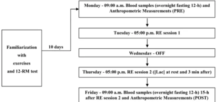
Before starting the RE protocol, all participants were famil-iarized with the types of exercise and 12 repetition maximum (12-RM) test. The familiarization period lasted for six sessions for each participant. The RE protocol was monitored by the researchers and consisted of nine exercises (eight exercises quoted in the 12-RM test, more abdominal - without external load). The two bouts of RE were performed at the same time of the day (05:00 p.m.) to avoid circadian variation. Three sets of 12-RM with 1.5−2 min of rest between sets, and 2−5 min of rest between exercises were performed. Each session lasted nearly 1 h. Total absolute load (sum of load [kg] used in the 12-RM test for the eight exercises) and total relative load (total absolute load [kg] divided per muscle mass [mm] - kg/mm) were determined for of each individual. Two bouts of RE were performed on separate occasions 48 h apart. The protocol design is illustrated in Figure 1.
12-RM strength test
12 RM was deined as the maximum amount of load that could be lifted through a full range motion 12 times, as described previously10. Each subject performed a 12-RM
strength test on eight exercises using resistance machines (Reforce®): (I) 45° leg press, (II) lat pull-down, (III) chest
press, (IV) leg lexion, (V) leg extension, (VI) standing free-weight biceps curl, (VII) triceps pulley and (VIII) dumbbell lateral side shoulder raise. After a warm-up with low weight, the subject performed exercise beginning with 70% of their perceived 12 RM for 12 repetitions. The weight was pro-gressively increased by approximately 5–10 kg after each successful attempt until the 12 RM was reached. A rest inter-val of 2 min was given between attempts and 5 min between exercises. The 12-RM values were then used to determine the corresponding load for the RE sessions. Movement velocity was controlled (approximately 3 s each repetition) through a pacer alarm (D’Accord®). All tests were supervised by the
same investigator.
Anthropometric measurements
Both BMI and percentage of body fat (%BF) were calculated for all participants. Body density was estimated using the three skin fold site method according to a previous study11 and BF%
was predicted by the equation of Brozek and coworkers12.
Blood sample collection
Blood samples were taken before and after the protocol design after a 12-h overnight fast. Venous blood samples were collected using two vacuum tubes (BD vacutainer tubes®): One
for plasma (EDTA K3) and another for serum (clot activator and gel for serum separation). Plasma tubes were immediately centrifuged (8000 g) for 10 min. Serum tubes sat for 30 min at room temperature and then centrifuged. After that, the super-natant (plasma and serum) was stored in aliquots at -80 ºC for future biochemical assays.
Biochemical assays
Total cholesterol, high-density lipoprotein cholesterol (HDL-C), low-density lipoprotein cholesterol (LDL-C), tri-glycerides, and blood glucose were determined using automated standard methods (Cobas Mira Plus).
Adiponectin, TNF-α, and leptin levels were quantiied by enzyme-linked immunosorbent assay (Cay006Dan Chemical, Ann Arbor, MI).
Serum creatine kinase levels were measured enzymatically using commercial kits. Blood lactate concentration ([Lac]) was determined at rest and 3 min after the second RE session using a lactate analyzer (Accutrend–Roche Diagnostics®, Germany).
All biochemical parameters, except [Lac], were assessed in duplicate.
Figure 1. Experimental design. (12-RM = 12 repetition maximum; RE = resistance exercise; [Lac] = blood lactate concentration).
Statistical analysis
Results
Women presented higher body fat (26.9 ± 6%) than men (16.7 ± 3.5%), whereas greater BMI values were found for men (26.2 ± 2.7 kg.m2) than for women (22.4 ± 3.4 kg.m2). No
differences in dietary nutrient composition were found between the two groups (% fat, carbohydrate, protein; P > 0.05, data not shown).
Total load performed and blood lactate
The total absolute load used in RE sessions was signiicantly greater (P < 0.05) for men (352.8 ± 85.5 kg) than for women (185.7 ± 46.8 kg), whereas total relative load was not different (P > 0.05) between genders (men: 5.1 ± 1 kg/mm; women: 4.4 ± 1.1 kg/mm). A signiicant increase in blood lactate levels was found after RE for both men (basal: 1.3 ± 0.2 and after RE: 7.5 ± 1.1 mmol.L-1) and women (basal: 1.2 ± 0.2 and after RE: 5.9
± 0.7 mmol.L-1).
Creatine kinase (CK)
Using RCV analyses, we observed that 9 of the 11 men dis-played signiicantly increased CK levels after RE (152 ± 144 to 728 ± 910 U.L-1), whereas all women presented an increase in
this parameter (51 ± 13 to 1192 ± 1460 U.L-1).
Lipid proile and blood glucose
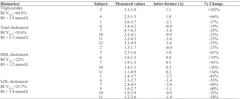
In order to examine the effects of RE on lipid proile by gender, the difference in lipid proile before and after RE sessions was determined. For men, a signiicant decrease in total cholesterol and LDL-C was observed, whereas HDL-C was signiicantly increased after two sessions of RE. No signiicant changes were observed for triglycerides in men. Data are summarized in Table 1. Similarly, RE was effective in lowering total cholesterol and HDL-C in women. HDL-C was also signiicantly increased after the resistance protocol in women. Data are summarized in Table 2.
Regarding blood glucose, two sessions of RE signiicantly re -duced blood glucose (approximately -40%) for both groups (99.9 ± 9 to 60 ± 6 mg/dL in women and 105 ± 9 to 62 ± 4.7 mg/dL in men).
Table 1. Lipid proile evaluated individually before and after two sessions of resistance exercise in men (n = 11).
Biomarker Subject Measured values latter-former (Δ) % Change
Total cholesterol RCV95%=18.6% RI < 5.18 mmol/L
2 3-4.6 -1.6 -35%
5 2.2-2.9 -0.7 -24%
10 3.2-4 -0.8 -21%
11 3.1-4 -0.9 -22%
HDL cholesterol RCV95%=22% RI >1.3 mmol/L
4 0.9-1.3 -0.4 -34%
6 1.1-0.9 0.2 +24%
7 1.4-0.9 0.5 +49%
8 0.9-0.6 0.3 +47%
11 1.0-0.5 0.4 +89%
LDL Cholesterol RCV95%=25.7% RI < 3.37 mmol/L
2 1.6-2.9 -1.3 -45%
3 2.8-1.9 0.9 +46%
5 1.0-1.7 -0.8 -44%
10 2.1-3.3 -1.2 -36%
11 1.9-3.3 -1.3 -41%
RI = Reference interval; Δ = difference between two consecutive analyses.
Table 2. Lipid proile evaluated individually before and after two sessions of resistance exercise in women (n = 13).
Biomarker Subject Measured values latter-former (Δ) % Change
Triglycerides RCV95%=64.8% RI < 3.9 mmol/L
3 2.1-1.0 1.1 +102%
6 2.5-1.5 1.0 +66%
Total cholesterol RCV95%=18.6% RI < 5.1 mmol/L
1 3.6-5.7 -2.1 -37%
6 3.4-4.2 -0.8 -19%
7 4.7-6.3 -1.6 -25%
10 3.2-4.1 -0.9 -23%
11 3.2-4.2 -1.0 -23%
12 3.3-4.9 -1.6 -33%
HDL cholesterol RCV95%=22% RI > 1.2 mmol/L
2 1.3-1.7 -0.4 -23%
5 2.5-1.6 1.0 +61%
6 1.6-1.2 0.4 +35%
7 1.9-1.3 0.5 +41%
10 1.4-1.1 0.3 +26%
11 1.1-0.9 0.2 +24%
LDL cholesterol RCV95%=25.7% RI < 3.4 mmol/L
1 1.4-3.7 -2.3 -62%
6 1.3-2.7 -1.4 -53%
7 2.4-4.5 -2.0 -46%
9 1.6-2.7 -1.1 -40%
10 1.9-2.9 -0.9 -32%
11 1.2-2.6 -1.4 -54%
Pro- and anti-inlammatory mediators
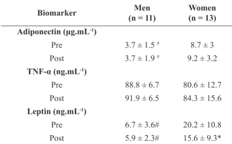
At basal conditions, it was observed that both leptin and adiponectin levels were signiicantly higher, nearly 200% and 135%, respectively, in women as compared with men. Two ses-sions of RE failed to cause any alterations in adiponectin levels in both groups, whereas leptin concentration was signiicantly reduced in women who performed the RE sessions. Regarding the cytokine TNF-α, no gender differences were observed for this pro-inlammatory mediator. In addition, two sessions of RE did not affect this biomarker in either groups. Data are summarized in Table 3.
The correlation analysis showed a positive relationship between leptin levels and BF% for both genders together (r = 0.82) or separately (r = 0.7 for women and r = 0.76 for men).
Table 3. Effects of two bouts of resistance exercise on adiponectin, leptin, and TNF-α levels in healthy women and men.
Biomarker (n = 11)Men Women(n = 13)
Adiponectin (μg.mL-1)
Pre 3.7 ± 1.5 # 8.7 ± 3
Post 3.7 ± 1.9 # 9.2 ± 3.2
TNF-α (ng.mL-1)
Pre 88.8 ± 6.7 80.6 ± 12.7
Post 91.9 ± 6.5 84.3 ± 15.6
Leptin (ng.mL-1)
Pre 6.7 ± 3.6# 20.2 ± 10.8
Post 5.9 ± 2.3# 15.6 ± 9.3*
Data are mean ± SD. # difference between groups. * Difference between pre- and post- exercise resistance sessions.
Discussion
To our knowledge, this is the irst study to verify the adipo -kine responses to a controlled RE protocol in healthy untrained subjects, though other related studies have investigated special populations, such as diabetics13, obese9, elderly14, and athletes15.
Accordingly, the main indings of this report were a) the leptin levels of women are more responsive to RE than those of men b) RE is effective in lowering metabolic parameters in both genders, measured by lipid proile and blood glucose. Taken together these indings suggest that RE could be a tool for preventing chronic diseases in healthy adults.
Anthropometric parameters (BMI and %BF) did not change after RE sessions due the short intervention time and unaltered nutrition habits throughout the study period. Therefore, they cannot explain the signiicant changes that occurred. Despite the similar relative intensity of the RE protocol between gen-ders (i.e., 12-RM for both), men presented higher total absolute load when compared with women. This result was expected because both groups were untrained and it is in accordance with a previous study16.
For both genders [Lac] increased signiicantly after the RE protocol (women: approximately 386% and men: approx-imately 499%) relative to the resting state. The [Lac] found here is commonly seen with RE schemes designed to stimulate muscle hypertrophy because there is a relationship between acute [Lac] response and anabolic hormones17, such as growth
hormone (GH)18. Additionally, the [Lac] observed illustrates a
great contribution from the anaerobic lactic pathway to cover the energetic demand and it was probably due to time of muscle action in our RE protocol (approximately 36 s each set) and brief rest periods between sets (1.5 min).
Serum CK activity is a marker of muscle damage19. Our
results showed that for both genders there were a signiicant increment in CK levels. Because our volunteers were untrained, these results were expected and suggest that RE achieved the “break point of CK release” (i.e., two or three times higher than resting condition)19. Despite the great individual variability
commonly found in CK, our data showed signiicant increases including from RCV analyses, which has been a better guide to interpreting changes in serial results20. Considering that
in-creased CK levels occurred in both genders, the gender-based differences found in other parameters measured cannot be explained by muscle damage.
Plasma lipoproteins play an important role in the genesis of atherosclerosis and evidence has shown that aerobic exercise ameliorates lipid proile and consequently, protects against the atherosclerosis process21. On the other hand, studies evaluating
the effects of RE on the lipid proile are not conclusive. In fact, it was demonstrated that RE promotes an improvement in lipid proile22 or causes no change23. Our indings showed an improve
-ment in lipid proile in response to acute RE in both genders, as evidenced by RCV analyses. This improvement seems to have occurred independently of inlammatory markers (TNF-α and adiponectin). Considering that GH has an important role in lipolysis and that high [Lac] is associated with the release of GH24, we could speculate that the acute improvement in lipid
proile could be related to high [Lac] caused by our RE protocol. Our protocol markedly reduced glucose concentration after two bouts of RE in both groups. These reductions are likely re-lated to glycogen depletion, because our RE protocol used large muscle mass and we had great anaerobic participation (high [Lac])25. In addition, it is well known that physical exercise,
aerobic or resistance, increases insulin sensitivity, increasing glucose uptake in skeletal muscle, either in healthy subjects or diabetic patients26. In fact, it was recently demonstrated that
muscular strength was positively associated with protection against cardiovascular disease in men27. Thus, our study
re-inforces the importance of RE in preventing chronic diseases such as type 2 diabetes.
As expected, adiponectin levels were higher in women than in men at baseline and two sessions of RE failed to change the levels this adipokine. Evidence has shown that physical exercise with high energy expenditure promotes beneicial effects by decreasing leptin and increasing adiponectin concentrations in diabetic28, obese9, and elderly subjects14. Considering that we
contrast, the RE protocol reduced leptin levels in women but not in men, showing that this adipocyte-derived adipokine is more responsive to RE in women than in men. Accordingly, a recent review about RE and adipokines also conirms that women are more responsive than men in relation to reduction after exercise training, but the mechanisms behind are not yet clear29. Regarding TNF-α, no differences were found at baseline
and RE failed to cause any changes in this proinlammatory mediator. TNF-α is considered a key molecule in peripheral insulin resistance and longitudinal as well as cross-sectional studies have shown the inhibitory effects of physical exercise on this proinlammatory biomarker26,30. However, most studies
were conducted in patients with metabolic disorders or elderly patients. In addition, our study is the irst to examine gender comparisons using an RE protocol. The relationship between leptin levels and body fat is well established15 and in agreement
with previous studies, we found a positive relationship in both genders (r = 0.82).
Collectively, our indings clearly show that RE was effective in lowering blood glucose and that this effect was not accom-panied by changes in adiponectin levels in healthy subjects, indicating that RE still an important tool to prevent metabolic diseases. Furthermore, two sessions of RE promoted a reduction in leptin levels in women, even though no changes in body weight were found, showing that RE is an interesting approach to studying obese patients and metabolic regulation.
References
1. Zanesco A, Antunes E. Effects of exercise training on the cardio-vascular system: pharmacological approaches. Pharmacol Ther. 2007; 114(3):307-317.
2. Mota CSA, Mello MAR. Exercise and metabolic syndrome. Motriz J Physical Education. 2006; 12(2):185-193.
3. Phillips SM. Resistance exercise: good for more than just Grandma and Grandpa’s muscles. Appl Physiol Nutr Metab. 2007; 32(6):1198-1205.
4. Xiao T, Fu YF. Resistance training vs. aerobic training and role of other factors on the exercise effects on visceral fat. Eur Rev Med Pharmacol Sci. 2015; 19(10):1779-84.
5. Dwyer T, Magnussen CG, Schmidt MD, Ukoumunne OC, Ponsonby AL, Raitakari OT, et al. Decline in physical itness from childhood to adulthood associated with increased obesity and insulin resistance in adults. Diabetes Care. 2009; 32(4):683-687. 6. Fernandes RA, Zanesco A. Early physical activity promotes lower prevalence of chronic diseases in adulthood. Hypertens Res. 2010; 33(9):926-931.
7. Vazquez-Vela ME, Torres N, Tovar AR. White adipose tissue as endocrine organ and its role in obesity. Arch Med Res. 2008; 39(8):715-728.
8. Kotani K, Sakane N, Saiga K, Sano Y, Kurozawa Y. Lifestyle-related determinants and serum adiponectin concentrations in a general population of Japanese females. Arch Med Res. 2007; 38(8):887-890.
9. Kondo T, Kobayashi I, Murakami M. Effect of exercise on circu-lating adipokine levels in obese young women. Endocr J. 2006; 53(2):189-195.
10. Ortego AR, Dantzler DK, Zaloudek A, Tanner J, Khan T, Panwar R, et al. Effects of gender on physiological responses to strenuous circuit resistance exercise and recovery. J Strength Cond Res. 2009; 23(3):932-938.
11. Jackson AS, Pollock ML. Generalized equations for predicting body density of men. Br J Nutr. 1978; 40(3):497-504.
12. Brozek J, Grande F, Anderson JT, Keys A. Densitometric Analysis of Body Composition: Revision of Some Quantitative Assumptions. Ann N Y Acad Sci. 1963; 110:113-140.
13. Kanaley JA, Fenicchia LM, Miller CS, Ploutz-Synder LL, Weinstock RS, Carhart R, Azevedo JLJr. Resting leptin responses to acute and chronic resistance training in type 2 diabetic men and women. Int J Obes Relat Metab Disord. 2001; 25(10):1474-1480. 14. Fatouros IG, Tournis S, Leontsini D, Jamurtas AZ, Sxina M, Thomakos P, et al. Leptin and adiponectin responses in overweight inactive elderly following resistance training and detraining are in-tensity related. J Clin Endocrinol Metab. 2005; 90(11):5970-5977. 15. Jurimae J, Jurimae T. Leptin responses to short term exercise in college level male rowers. Br J Sports Med. 2005; 39(1):6-9. 16. Miller, AE, MacDougall JD, Tarnopolsky MA, Sale DG. Gender differences in strength and muscle iber characteristics. Eur J Appl Physiol Occup Physiol. 1993; 66(3):254-262.
17. Crewther B, Cronin J, Keogh J. Possible stimuli for strength and power adaptation : acute metabolic responses. Sports Med. 2006; 36(1):65-78.
18. Goto K, Ishii N, Kizuka T, Takamatsu K. The impact of metabolic stress on hormonal responses and muscular adaptations. Med Sci Sports Exerc. 2005 37(6):955-963.
19. Totsuka M, Nakaji S, Suzuki, K, Sugawara K, Sato K. Break point of serum creatine kinase release after endurance exercise. J Appl Physiol. 2002; 93(4): 1280-1286.
20. Ricos C, Cava F, Garcia-Lario JV, Hernandez A, Iglesias N, Jimenez CV, et al. The reference change value: a proposal to interpret laboratory reports in serial testing based on biological variation. Scand J Clin Lab Invest. 2004; 64(3): 175-184. 21. Banz WJ, Maher MA, Thompson WG, Bassett DR, Moore W,
Ashraf M, et al. Effects of resistance versus aerobic training on coronary artery disease risk factors. Exp Biol Med (Maywood). 2003; 228(4):434-440.
22. Hill S, Bermingham MA, Knight PK. Lipid metabolism in young men after acute resistance exercise at two different intensities. J Sci Med Sport. 2005; 8(4):441-445.
23. Nikolaidis MG, Paschalis V, Giakas G, Fatouros IG, Sakellariou GK, Theodorou AA, et al. Favorable and prolonged changes in blood lipid proile after muscle-damaging exercise. Med Sci Sports Exerc. 2008; 40(8):1483-1489.
24. Godfrey RJ, Whyte GP, Buckley J, Quinlivan R. The role of lactate in the exercise-induced human growth hormone re-sponse: evidence from McArdle disease. Br J Sports Med. 2009; 43(7):521-525.
26. Knudsen SH, Pedersen BK. Targeting Inlammation Through a Physical Active Lifestyle and Pharmaceuticals for the Treatment of Type 2 Diabetes. Curr Diab Rep. 2015; 15(10):82.
27. Ruiz JR, Sui X, Lobelo F, Morrow JR, Jr, Jackson AW, Sjostrom M, Blair SN. Association between muscular strength and mortality in men: prospective cohort study. Bmj. 2008: 337:a439. 28. Bluher M, Bullen JW, Jr, Lee JH, Kralisch S, Fasshauer M, Kloting
N, et al. Circulating adiponectin and expression of adiponectin receptors in human skeletal muscle: associations with metabolic parameters and insulin resistance and regulation by physical training. J Clin Endocrinol Metab. 2006. 91(6):2310-2316. 29. de Salles BF, Simao R, Fleck SJ, Dias I, Kraemer-Aguiar LG,
Bouskela E. Effects of resistance training on cytokines. Int J Sports Med. 2010; 31(7): 441-450.
30. Ferguson MA, White LJ, McCoy S, Kim H.W, Petty T, Wilsey J. Plasma adiponectin response to acute exercise in healthy subjects. Eur J Appl Physiol. 2004; 91(2-3):324-329.
Corresponding author
Gustavo Ribeiro da Mota, PhD
Department of Sport Sciences, Federal University of Triângulo Mineiro (UFTM), Uberaba, MG, Brazil
Email: [email protected]
Manuscript received on March 30, 2015 Manuscript accepted on April 10, 2016
Acknowledgments
The authors gratefully acknowledge all the subjects who participated in this study for their cooperation.