DOI: 10.1590/0004-282X20150169
ARTICLE
Surgical technique of retrograde
ventricle-sinus shunt is an option for the
treatment of hydrocephalus in infants after
surgical repair of myelomeningocele
A derivação ventrículo-sinusal retrógrada é uma opção para o tratamento de hidrocefalia
em lactentes após correção de mielomeningocele
Matheus Fernandes de Oliveira1,2, Manoel Jacobsen Teixeira1,3,Karen Andrade Norremose3, Hamilton Matushita4, Marcelo de Lima Oliveira3, Edson Bor Seng Shu3, Fernando Campos Gomes Pinto1,3
Myelomeningocele is a neural tube defect in neurulation embrionary stage by the third week of life. Even in locations with high living standard and supplementation of folic acid, its incidence may reach 1-2 per 1000 live births. Even with all advances in intrauterine and postnatal management, up to
90% of these patients may present hydrocephalus in diferent
degrees, requiring treatment1,2,3.
Treatment consists in cerebrospinal luid (CSF) shunts
with ventricular endoscopy or ventricular catheter and drainage to another body cavity. In the case of hydrocephalus
1Universidade de São Paulo, Hospital das Clínicas, Instituto de Psiquiatria, Divisão de Neurocirurgia Funcional, Sao Paulo SP, Brazil;
2Hospital do Servidor Público Estadual de São Paulo, Instituto de Assistência Médica ao Servidor Público Estadual, Departamento de Neurocirurgia, Sao
Paulo SP, Brazil;
3Universidade de São Paulo, Faculdade de Medicina, Hospital das Clínicas, Divisão de Cirurgia Neurológica, Sao Paulo SP, Brazil; 4Universidade de São Paulo, Hospital das Clínicas, Divisão de Neurocirurgia Pediátrica, Sao Paulo SP, Brazil.
Correspondence: Matheus Fernandes de Oliveira; Rua Loefgren, 700 / apto 103; 04040-000 São Paulo SP, Brasil; E-mail: [email protected]
Conflict of interest: There is no conlict of interest to declare.
Received 18 June 2015; Received in inal form 04 August 2015; Accepted 24 August 2015.
ABSTRACT
Introduction: Treatment of hydrocephalus is accomplished primarily through a ventricular-peritoneal shunt (VPS). This study aims to describe the application of retrograde ventricle-sinus shunt (RVSS) in patients with hydrocephalus after surgical treatment of myelomeningocele.
Method: A prospective, randomized and controlled pilot study. We consecutively enrolled 9 patients with hydrocephalus after surgical repair of myelomeningocele from January 2010 to January 2012. These patients underwent elective RVSS or VPS. Five underwent RVSS and 4 underwent VPS. Patients were followed for one year with quarterly evaluations and application of transcranial Doppler. Results: RVSS group showed outcomes similar to those of VPS group. Doppler revealed signiicant improvement when comparing preoperative to postoperative period. RVSS group had signiicantly higher cephalic perimeter than VPS group. Neuropsychomotor development, complications and subjective outcomes did not differ between groups. Conclusion: RVSS shunt is viable; it is an alternative option for the treatment of hydrocephalus.
Keywords: neurosurgery, hydrocephalus, shunt, myelomeningocele.
RESUMO
O tratamento da hidrocefalia é realizado principalmente através de uma derivação ventrículo-peritoneal (DVP). Nosso objetivo é descrever a aplicação da derivação ventrículo-sinusal retrógrada (DVSR) em pacientes com hidrocefalia após o tratamento cirúrgico de mielomeningocele. Método: Estudo prospectivo, randomizado e controlado. Selecionados consecutivamente 9 pacientes com hidrocefalia após correção cirúrgica de mielomeningocele de janeiro de 2010 a janeiro de 2012. Eles foram submetidos à DVSR ou DVP. Cinco foram submetidos à DVSR e 4 à DVP. Foram seguidos por 1 ano com realização trimestral de avaliações e aplicação do Doppler transcraniano.
Resultados: O grupo DVSR apresentou desfechos semelhantes ao grupo DVP. O Doppler mostrou melhora signiicativa quando comparado o pré-operatório com o pós-operatório. O grupo DVSR apresentou perímetro cefálico signiicativamente maior que o grupo DVP. O desenvolvimento neuropsicomotor e complicações não diferiram entre os grupos. Conclusão: A derivação ventrículo-sinusal retrógrada é viável; ela é uma opção para o tratamento de hidrocefalia.
secondary to myelomeningocele, both methods have consid-erable complications and limited therapeutic success4,5,6,7.
Ventricle-peritoneal shunt (VPS) is the preferred option,
although has a revision rate of more than 70% of patients in
life and until 30% in the irst year. An important complica
-tion is shunt infec-tion, which may occur in up to 5% of the
cases. Another drawback is siphoning phenomenon, leading
to overdrainage and subdural efusions8,9,10,11.
Neuroendoscopy, on the other hand, has increased in experience and indications. Endoscopic third
ventriculos-tomy (ETV), which consists in fenestrating third ventricle
and communicating with interpeduncular and pre-pontine cisterns, is the most applied ventricular endoscopic proce-dure. However, in patients with myelomeningocele, ventricu-lar anatomy is notoriously variable, with patterns that hinder endoscopy and increase operative risks8,9,10,11.
hus, alternatives are needed to treat hydrocephalus in these patients eiciently and safely. Retrograde-ventricular
sinus shunt (RVSS), developed by El-Shafei, shunts later
-al ventricle to superior sagitt-al sinus (SSS) in the opposite direction of venous blood low and is a feasible and rational
alternative for the treatment of hydrocephalus, minimizing
siphon efect12,13,14,15,16,17,18,19,20,21,22,23,24,25,26.
his study aims to describe initial clinical results of RVSS
in hydrocephalus after surgical repair of myelomeningocele.
METHOD
Study design
It is an intervention, prospective, randomized and
con-trolled study. he allocation was parallel with ratio of 2: 2.
Eligibility criteria
We consecutively enrolled patients diagnosed with hy-drocephalus after surgical repair of myelomeningocele from
January 2010 to January 2012, users of Hospital das Clinicas, University of São Paulo. hey were screened after postnatal
surgical treatment for myelomeningocele. hese patients un
-derwent elective RVSS or VPS.
Inclusion criteria
Inclusion criteria were acceptance from parents to par-ticipate in research; myelomeningocele patients in the postoperative period, without infectious complications and without prior ventricular shunts who developed progression
of hydrocephalus after irst month of life, but under 6 months old. Infants performed magnetic resonance imaging (MR) with vascular study showing patency of SSS.
Exclusion criteria
Exclusion criteria: other types of congenital hydrocepha
-lus, previous infectious complications or previous ventricular
shunts, refusal of parents and guardians to participate in
re-search; MR with SSS thrombosis signals.
Ethical adherence
Project and informed consent form were approved without restrictions by the Ethics Committee in Research of HCFMUSP, under number 0178/09.
RVSS technique (Figures 1 and 2)
Surgical technique involves 2 cranial burr holes in the same
arcuate incision in the scalp of parietal region. One burr hole
in posterior parietal bone (Frazier point) and the other in the middle third of the sagittal suture. Small opening is made in
the dura mater of the parietal incision and the lateral ventricle is punctured and then small opening is made in sagittal sinus and the catheter is inserted approximately 2 cm against the
di-rection of blood low. Catheter used to perform shunt was the one in PS Medical valve® (Medtronic) material, which is already in routine use in Hospital das Clínicas12,13,14,15,16,17,18,19,20,21,22,23,24,25,26.
Sample (Table 1)
Nine patients were included in the study for a period of
two years. 3 were allocated to the control group (VPS) and
6 patients in RVSS group. Allocation was made by random
-ization in the operating room. 5 of the 6 patients allocated
to RVSS underwent surgical treatment. Patient 6 had surgery
CT: computed tomography; MR: magnetic resonance; SSS: superior sagital sinus.
Figure1. Image study. (A) Pre operative tomography
revealing ventriculomegaly. (B) CT after RVSS. (C) MR revealing pervious SSS.
A
B
converted to VPS due to intraoperative inaccurate identiica
-tion of SSS and bleeding. 3 patients initially allocated to VPS
group underwent surgery without complications.
VPS group was then composed of four patients, three males (75%) and 1 female (25%). Average age was 3 months. All patients had lumbosacral myelomeningocele. Two of the 3 patients (66%) had club foot. he degree of strength in the lower limbs ranged from 2 to 5. CP ranged from 40 to 45 cm.
Bregmatic fontanelle was hypertense, with 2+/4+ in 3 pa
-tients and 3+/4+ in 1 patient. Neuropsychomotor develop
-ment (NPMD) was adequate for the age group in 3 patients
and delayed in 1. Evans index ranged from 0.47 to 0.50.
RVSS group consisted of 5 patients, 4 male (80%) and 1 female (20%). Average age was 5 months. All patients had lumbosacral myelomeningocele. 3 of 5 patients (60%) had club foot. he degree of strength in the lower limbs ranged from 0 to 5. Cephalic perimeter (CP) ranged from 44 to 47 cm.
Bregmatic fontanelle was hypertensive, with 2+/4+ in 4 pa
-tients and 3+/4+ in 1 patient. NPMD was adequate for the
age group in 3 patients and delayed in 2. Evans index ranged from 0.46 to 0.50.
Follow-up
After surgery, patients were followed with pre-established
consultations at 3 months, 6 months, 9 months and 1 year. In
these consultations, data were evaluated and tabulated for:
CP (3 months, 6 months, 9 months and 1 year); Bregmatic fontanelle(3 months, 6 months, 9 months and 1 year); Transcranial doppler (TD) (3 months and 1 year54,55,56,57):mean velocity of blood low in right and left middle cerebral artery (MCA), basilar artery (BA) and SSS; resistence index in MCA and
BA; pulsatility index in MCA and BA; Computed tomography
(CT) – Evans Index (3 months and 1 year); NPMD - (3 months, 6 months, 9 months and 1 year); Complications and shunt revi-sions; Outcomes focused on patients and caregivers.
Outcome
Primary: transcranial Doppler measurements, including mean velocity in right and left MCA, resistence index and
pulsatility index.
Secondary:evaluation of cephalic perimeter, Evans ratio, fontanelle, neuropsychomotor development, complications and outcomes focused on patients and caregivers.
Table 1. Pre operative patient data.
Group Patient Gender Age (months) MM site Club foot Strength CP (cm) Fontanelle NPMD Evans
RVSS 1 - MFB M 6 Lumbossacral Yes 2 45 Hypertense 2+/4+ Adequate 0.47
RVSS 2 – ENM M 6 Lumbossacral No 5 47 Hypertense 3+/4+ Delayed 0.50
RVSS 3 – JPM M 4 Lumbossacral Yes 0 46 Hypertense 2+/4+ Delayed 0.48
RVSS 4 – RSJ F 3 Lumbossacral Yes 3 44 Hypertense 2+/4+ Adequate 0.46
RVSS 5 – SBS M 6 Lumbossacral No 5 46 Hypertense 2+/4+ Adequate 0.48
VPS 6 – LGS M 1 Lumbossacral No 5 40 Hypertense 2+/4+ Adequate 0.50
VPS 7 - CGG M 3 Lumbossacral Yes 5 44 Hypertense 2+/4+ Adequate 0.47
VPS 8 – AEA M 2 Lumbossacral Yes 3 45 Hypertense 3+/4+ Adequate 0.48
VPS 9 - KAP F 6 Lumbossacral No 2 43 Hypertense 2+/4+ Delayed 0.49
RVSS: retrograde ventricule-sinus shunt; VPS: ventricular-peritoneal shunt; CP: cephalic perimeter; NPMD: neuropsychomotor development. SSS: superior sagital sinus.
Figure 2. Phases of surgical technique. (A) Positioning and arcuate incision. (B) Opening with medial and lateral burr holes. (C) and
(D) Catheterization of lateral ventricle and SSS. (E) Hemostasis. (F) Closure with pericranium and skin closure.
A
B
C
Statistics
In this study, numerical data are presented as mean ±
stan-dard deviation (SD) or as median with range where appropriate.
Categorical data are presented as percentages. When compar
-ing the groups, the signiicance level is considered when p < 0.05.
To determine the distribution of our data, the
Kolmogorov-Smirnov test was used. To evaluate categorical variables,
chi-square tests were used and Mc’Nemar, respectively, as
appro-priate. To evaluate ordinal variables, t-Student, Wilcoxon and
Mann-Whitney tests were applied when applicable.
RESULTS
here were no statistically signiicant diferences be
-tween VPS and RVSS groups (p > 0.05).
Follow-up
Cephalic perimeter (CP)
Figures 3 and 4 show CP before surgery and after sur
-gery. Average perimeter of VPS group patients (42.5 cm) was statistically lower than RVSS group (46.8 cm) in the
late follow-up.
Patients undergoing RVSS showed change in the CP curve,
not abruptly reducing CP, but making it grow at a physiologi
-cal rate (Figure 3).
Patient 3 showed increased head circumference curve
even after RVSS. Transcranial Doppler showed high re
-sistance, pulsatility and maintained speeds, suggesting
non-functioning of the system. A revision surgery found a
non-functioning catheter, without thrombus.
Patients undergoing VPS changed CP percentile, with CP abrupt reduction after surgery and return to growth (Figure 4).
Bregmatic fontanelle
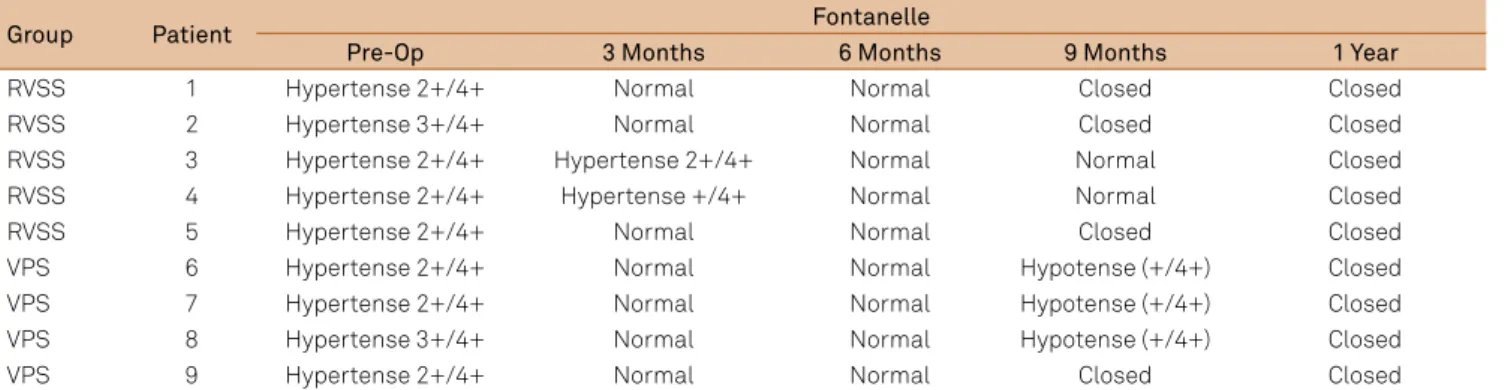
Table 2 shows the evolution of the bregmatic fontanelle characteristics.
Fontanelle characteristics preoperatively did not difer between patients. Postoperatively, there were normotensive patients in RVSS group. In VPS group, fontanelle oscilated between normotensive and mild hypotension. he diference did not reach statistical diference.
49
47 47 47 47 47
46 46 46
45 45
44 44
45
Patient 1 Patient 2 Patient 3 Patient 4 Patient 5
47,5 47,5
46,5
45,5
44,5
47,5
47 46,5 48
45,5 45
43
Pre op 3 months 6 months 9 months 1 year 48
46
44
42
RVSS: retrograde-ventricular sinus shunt; CP: computed tomography.
Figure 3. Cephalic perimeter (cm) curve in RVSS group.There
was no decrease in CP.
46
44
Patient 6 Patient 7 Patient 8 Patient 9 40
36
Pre op 3 months 6 months 9 months 1 year 42
38
34
45 44 43
42 41 40,5
38
43
41,5
40,5 41
42 43,5
40
39,5 41 42 42,5
VPS: ventricular-peritoneal shunt; CP: computed tomography.
Figure 4. Cephalic perimeter curve (cm) in VPS group.There
was immediate decrease in CP, followed by late growth of skull.
Table 2. Fontanelle pattern during follow-up.
Group Patient Fontanelle
Pre-Op 3 Months 6 Months 9 Months 1 Year
RVSS 1 Hypertense 2+/4+ Normal Normal Closed Closed
RVSS 2 Hypertense 3+/4+ Normal Normal Closed Closed
RVSS 3 Hypertense 2+/4+ Hypertense 2+/4+ Normal Normal Closed
RVSS 4 Hypertense 2+/4+ Hypertense +/4+ Normal Normal Closed
RVSS 5 Hypertense 2+/4+ Normal Normal Closed Closed
VPS 6 Hypertense 2+/4+ Normal Normal Hypotense (+/4+) Closed
VPS 7 Hypertense 2+/4+ Normal Normal Hypotense (+/4+) Closed
VPS 8 Hypertense 3+/4+ Normal Normal Hypotense (+/4+) Closed
VPS 9 Hypertense 2+/4+ Normal Normal Closed Closed
Transcranial doppler
Table 3 describes Doppler data of patients before and af
-ter surgery. Preoperative transcranial Doppler did not difer
between VPS and RVSS groups. In both groups, except for pa
-tient 3 (discussed above), there was an increase in mean low
velocity, decreased pulsatility index and decreased resistance index in all vessels analyzed, these data maintained inlate
post operative (1 year). here was no signiicant diference
between groups.
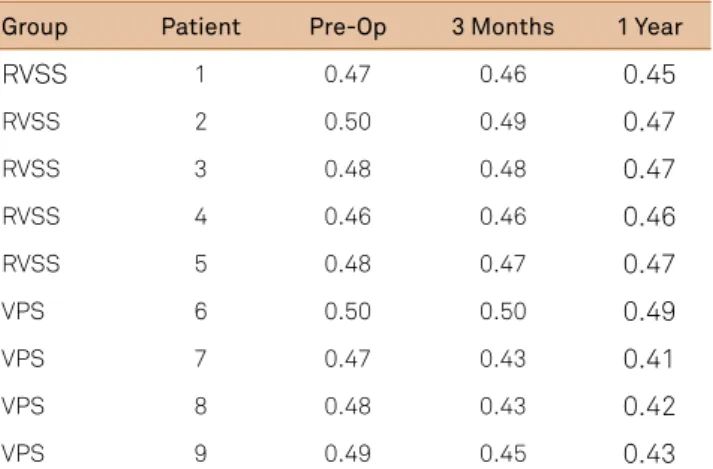
Computed tomography
Table 4 shows the evolution of Evans index in follow-up.
In analogy to what happened with CP, patients undergoing RVSS presented slight reduction in Evans index. In VPS
group, there was a postoperative reduction of ventricular size
greater than in RVSS group, reaching statistical signiicance.
Neuropsychomotor development
In the preoperative period, three patients had delayed psychomotor development for age, patients 2, 3 and 9, two
from RVSS group and 1 from VPS group. After surgical treat
-ment, proile of patients persisted. Patients with adequate
development continued adequacy and patients with devel-opmental delay have evolved, but keeping lagging behind the average age.
During follow-up work, all patients received rehabilita
-tion support in specialized centers with urological, orthope-dic and physiatric monitoring.
Complications and shunt revision
Patient 3 had head circumference curve rising even after RVSS. Transcranial Doppler showed resistance, pulsatility
and maintained speeds, suggesting system dysfunction. A re
-vision surgery found non-functioning catheter, without the presence of thrombus.
Patient 6 had a surgery converted for VPSdue to intraop
-erative inaccurate identiication of SSS and bleeding. In VPS
Table 4. Evans Index during follow-up.
Group Patient Pre-Op 3 Months 1 Year
RVSS 1 0.47 0.46 0.45
RVSS 2 0.50 0.49 0.47
RVSS 3 0.48 0.48 0.47
RVSS 4 0.46 0.46 0.46
RVSS 5 0.48 0.47 0.47
VPS 6 0.50 0.50 0.49
VPS 7 0.47 0.43 0.41
VPS 8 0.48 0.43 0.42
VPS 9 0.49 0.45 0.43
RVSS: retrograde ventricule-sinus shunt; VPS: ventricular-peritoneal shunt.
Tabl e 3. T ranscr anial Dop pl er
. Mean v
el
ocity
, pulsa
tility and r
esis
tence inde
x anal
ysis in s
tudied v essels . P a tient Pr e Op P os t Op 1 Y ear MV
– R MC
A MV – L MC A M V B A MV SSS PI RI MV
– R MC
A MV – L MC A M V B A MV SSS PI RI MV
– R MC
A MV – L MC A M V B A MV SSS PI RI 1 70 61 43 45 1 .1 0 .7 5 80 84 54 40 0 .91 0 .6 80 83 53 40 0 .86 0 .62 2 45 48 32 36 1 .32 0 .7 1 59 58 35 39 0 .9 0 .6 62 64 38 41 0 .89 0 .61 3 53 60 23 30 1 .50 0 .91 54 56 24 28 1. 5 0 .94 70 78 45 36 0 .94 0 .6 4 51 56 36 35 1. 5 0 .7 5 66 62 43 40 1 .1 5 0 .65 65 64 46 40 1 .1 2 0 .64 5 49 54 34 33 1. 4 0 .7 68 64 45 41 1 .1 0 .65 70 68 47 42 1 .1 0 .62 6 47 50 33 37 1 .25 0 .7 5 60 58 35 40 0 .95 0 .6 62 65 39 42 0 .86 0 .61 7 54 58 27 32 1 .45 0 .85 70 72 38 34 1. 0 0 .63 71 73 40 35 0 .96 0 .6 8 46 49 24 32 1. 3 0 .8 62 63 30 33 0 .95 0 .7 64 62 34 36 0 .9 0 .67 9 65 61 38 45 1. 4 0 .7 5 85 87 40 38 1. 0 5 0 .62 84 79 43 41 1 .1 0 .68 MV
– R MC
A:
Mean v
el
ocity (cm/
s) in righ
t mid dl e cer ebr al ar tery; MV – L MC A: Mean v el
ocity in l
e ft mid dl e cer ebr al ar tery; MV B A: Mean v el
ocity in basilar ar
tery;
MV
SSS:
Mean v
el
ocity in superior sa
group, there were no complications during follow-up. No col-lections, infection or dysfunction signals.
Outcomes focused on patients and caregivers
All caregivers reported improvement in the aspects of pa
-tient care, feeling optimistic about the surgery, with no difer
-ence from one group to another. hey reported satisfaction
with procedure, unrepentant and would undergo treatment again if they could choose.
According to caregivers, most of major change and im
-pact aspects were the decrease in head growth, decreased ir-ritability and improvement after surgery.
DISCUSSION
Little has changed over the past decades in terms of shunt revision rate. Numerous series show that over 70% of shunt patients require long-term review and 54% may need four or more revisions, with all additional risks, costs and negative outcomes4,5,6,7,8,9,10. ETV also has known limitations, especially
in patients with myelomeningocele and Chiari type II4,5,6,7,8,9,10,11.
RVSS proposes a safe and more physiological option, us
-ing less prosthetic material. Additionally, only addresses the cranial region. Besides that, procedure uses internal jugular
vein function as a physiological anti-siphon system. In the-ory, it can be used in any hydrocephalus etiology, at any age.
Seven case series with a total of 265 patients undergoing ven
-tricular sinus shunt for hydrocephalus were published since
the introduction of technique. In Egypt, El Shafei reported a
success rate of over 80%12,13,14,15,16,17,18,19,20,21,22,23,24,25,26.
El-Shafei et al pioneered by performing puncture in retro
-grade direction to the direction of blood low, using the impact
pressure of the blood low in the sinuses of the dura to main
-tain the CSF pressure greater than the pressure within the du
-ral regardless of changes posture or intrathoracic pressure.
hey also pioneered use of TD12,13,14,15,16,17,18,19,20,21,22,23,24,25,26,27,28,29,30. Toma et al, in a recent review, showed that, in 265 patients,
there was no sinus thrombosis, air embolism, uncontrollable operative bleeding or nephritis associated with shunt25.
In our sample, transcranial Doppler revealed improve
-ment in patterns similarly in both treat-ment groups. Head circumference, tomography and fontanelle were
characteristic in each group. In RVSS group, there was no abrupt reduction in head circumference. CP assumed a physiologic contour but remained high. In VPS group, CP
reduced abruptly after surgery, with subsequent regrowth.
Similarly, fontanelle in RVSS group remained normotensive, meaning cranial normotension. In VPS group, they became
normotensive or hypotensive, especially in older children and may infer a siphoning component in older children with a more upright posture.
In RVSS group, there was need for one revision and one conversion to VPS. In VPS group there was no revision or complications. Psychomotor development after surgery was
similar in both groups. However, further evaluation is
need-ed with speciic neuropsychological tests which can detect
subtle changes.
Caregivers opinions were unanimous in accepting the
surgery and conirm its beneits. However, it is diicult to es
-tablish a clear distinction between the clinical improvement due to aging and consequent development of children and clinical improvement as a result of treatment.
Some limitations of the study should be mentioned.
Initially, our number of patients, although enough to draw up a pilot study, is small for us to make more robust inferences. We believe that our number was lower than planned due to strict inclusion criteria. In addition, our assessment of
psy-chomotor development was brief and lacks objective data
and neuropsychological tests. We must also remark that our follow up was of 1 year and should be larger.
We applied in our protocol computed tomography (CT) instead of transfontanellar ultrasound (US) to evaluate Evans index. Although US is cheaper and avoids using radiation, we preferred to apply CT in accordance to protocol proposed by El Shafei et al.
Additionally, RVSS is still relatively unknown in neu
-rosurgical community, often feared due to manipulation of a potentially hazardous structure, superior sagittal sinus. Because of this, few centers have performed this technique even experimentally and thus the learning curve in our coun-try is still early despite the simple technical concept.
In summary, clinical evaluation and Doppler data showed that both treatment modalities were efective, with diferent CP standards postoperatively. We believe that RVSS can be a
viable and safe alternative for the treatment of
hydrocepha-lus in selected cases. Future increase in number of patients
treated might disclose further results.
In conclusion, surgical technique of RVSS is feasible.
Clinical results are comparable with VPS, being a viable alter
References
1. Heuer GG, Adzick NS, Sutton LN. Fetal myelomeningocele closure: technical considerations. Fetal DiagnTher. 2014;37(3):166-71. doi:10.1159/000363182
2. Saadai P, Farmer DL. Fetal surgery for myelomeningocele. Clin Perinatol. 2012;39(2):279-88. doi:10.1016/j.clp.2012.04.003 3. Adzick NS, Thom EA, Spong CY, Brock JW 3rd, Burrows PK, Johnson
MP et al. MOMS Investigators. A randomized trial of prenatal versus postnatal repair of myelomeningocele. N Engl J Med. 2011;364(11):993-1004. doi:10.1056/NEJMoa1014379
4. Browd SR, Ragel BT, Gottfried ON, Kestle JR. Failure of cerebrospinal luid shunts: part I: obstruction and mechanical failure. Pediatr Neurol. 2004;34(2):83-92. doi:10.1016/j.pediatrneurol.2005.05.020 5. Lo P, Drake JM. Shunt malfunctions. Neurosurg Clin N Am.
2001;2(4):695-701.
6. Browd SR, Gottfried ON, Ragel BT, Kestle JR. Failure of cerebrospinal luid shunts: part II: overdrainage, loculation, and abdominal complications. Pediatr Neurol. 2006;34(3):171-6. doi:10.1016/j.pediatrneurol.2005.05.021
7. Choux M, Camboulives J, Rigaut F. Prevention of infections in ventriculoperitoneal shunts in children. Ann Fr Anesth Reanim. 1992;11(6):699-704. doi:10.1016/S0750-7658(05)80793-2 8. Oliveira MF, Saad F, Reis RC, Rotta JM, Pinto FC. Programmable valve
represents an eficient and safe tool in the treatment of idiopathic normal-pressure hydrocephalus patients. Arq Neuropsiquiatr. 2013;71(4):229-36. doi:10.1590/0004-282X20130007
9. Pinto FC, Pereira RM, Saad F, Teixeira MJ. Performance of ixed-pressure valve with antisiphon device SPHERA(®) in hydrocephalus treatment and overdrainage prevention. Arq Neuropsiquiatr. 2012;70(9):704-9. doi:10.1590/S0004-282X2012000900011
10. Pavez A, Salazar C, Rivera R, Contreras J, Orellana A, Guzman C et al. Description of endoscopic ventricular anatomy in myelomeningocele. Minim Invasive Neurosurg. 2006;49(3):161-7. doi:10.1055/s-2006-932193
11. Kadri H, Mawla AA. Variations of endoscopic ventricular anatomy in children suffering from hydrocephalus associated with myelomeningocele. Minim Invasive Neurosurg. 2004;47(6):339-41. doi:10.1055/s-2004-830127
12. Fox JL, McCullough DC, Green RC. Effect of CSF shunt on ICP and CSF dynamics: 2. a new technique of pressure measurement, results and concepts. J Neurol Neurosurg Psychiatry. 1973; 36(2):302-12. doi:10.1136/jnnp.36.2.302
13. Portnoy HD, Schulte RR, Fox JL. Antisiphon and reversible occlusion valves for shunting in hydrocephalus and preventing post shunt subdural hematomas. J Neurosurg. 1973;38(6):729-38. 14. El Shafei IL, El Shafei HI. The retrograde ventriculo-sinus shunt
(El Shafei RVS shunt). Rationale, evolution, surgical technique and long-term results. Pediatr Neurosurg. 2005;41(6):305-17. doi:10.1159/000088733
15. El-Shafei IL, El-Shafei HI. The retrograde ventriculovenous shunts: the El-Shafei retrograde ventriculojugular and ventriculosinus shunts. Pediatr Neurosurg. 2010;46(3):160-71. doi:10.1159/000316639
16. El-Shafei IL, El-Shafei HI. The retrograde ventriculosinus shunt: concept and technique for treatment of hydrocephalus by shunting the cerebrospinal luid to the superior sagittal sinus against the direction of blood low: preliminary report. Childs Nerv Syst. 2001;17(8):457-65. doi:10.1007/s003810100456
17. El-Shafei IL, El-Rifaii MA. Ventriculojugular shunt against the direction of blood low. II. Theoretical and experimental basis for shunting the cerebrospinal luid against the direction of blood low. Childs Nerv Syst. 1987;3(5):285-91. doi:10.1007/BF00271825 18. El-Shafei IL, El-Rifaii MA. Ventriculojugular shunt against the direction
of blood low. I. Role of the internal jugular vein as an antisiphonage device. Childs Nerv Syst. 1987;3(5):282-4. doi:10.1007/BF00271824 19. El-Shafei IL. Ventriculojugular shunt against the direction of
blood low. III. Operative technique and results. Childs Nerv Syst. 1987;3(6):342-9. doi:10.1007/BF00270704
20. El-Shafei IL. Ventriculovenous shunt against the direction of blood low: a new approach for shunting the cerebrospinal luid to the venous circulation. Childs Nerv Syst. 1985;1(4):200-7. doi:10.1007/BF00270762
21. Souza RB, Pinto FC. Could craniometric measurements explain the growth of the superior sagittal sinus? Pediatr Neurosurg. 2012;48(4):225-8. doi:10.1159/000348555
22. Børgesen SE, Pieri A, Cappelen J, Agerlin N, Gjerris F. Shunting to the cranial venous sinus using the Sinu Shunt. Childs Nerv Syst. 2004;20(6):397-404. doi:10.1007/s00381-004-0914-6
23. Børgesen SE, Gjerris F, Agerlin N. Shunting to the sagittal sinus. Acta Neurochir Suppl. 2002;81:11-4.
24. Eklund A, Koskinen LO, Malm J. Features of the Sinushunt and its inluence on the cerebrospinal luid system. J Neurol Neurosurg Psychiatry. 2004;75(8):1156-9. doi:10.1136/jnnp.2003.023168 25. Toma AK, Tarnaris A, Kitchen ND, Watkins LD. Ventriculosinus shunt.
Neurosurg Rev. 2010;;33(2):147-52. doi:10.1007/s10143-010-0242-0 26. Van Canneyt K, Kips J, Mareels G, Baert E, Van Roost D, Verdonck
P. Experimental and numerical modelling of the ventriculosinus shunt (El-Shafei shunt). Proc Inst Mech Eng H. 2008;222(4):455-64. doi:10.1243/09544119JEIM299
27. Alexandrov AV, Sloan MA, Tegeler CH, Newell DN, Lumsden A, Garami Z et al. Practice standards for transcranial Doppler (TCD) ultrasound. Part II. Clinical indications and expected outcomes. J Neuroimaging. 2012;22(3):215-24. doi:10.1111/j.1552-6569.2010.00523.x 28. Rainov NG, Weise JB, Burkert W. Transcranial Doppler sonography
in adult hydrocephalic patients. Neurosurg Rev. 2000;23(1):34-8. doi:10.1007/s101430050029
29. Jindal A, Mahapatra AK. Correlation of ventricular size and transcranial Doppler indings before and after ventricular peritoneal shunt in patients with hydrocephalus: prospective study of 35 patients. J Neurol Neurosurg Psychiatry. 1998;65(2):269-71. doi:10.1136/jnnp.65.2.269