PRIMARY FILUM TERMINALE EPENDYMOMA
A series of 16 cases
Murilo S. Meneses
1, André Giacomelli Leal
2, Larissa B. Periotto
6, Jerônimo Buzetti Milano
4,
Maurício Coelho-Neto
4, Ana Cristina Sobral
5, Ricardo Ramina
3Abstract – Filum terminale ependymomas are slow growing tumors of the cauda equina with a high incidence in young adults. Although a complete microsurgical resection can lead to a cure, recurrence is not uncommon. Sixteen cases of filum terminale ependymomas treated at the Instituto de Neurologia de Curitiba were analyzed. Eleven patients were females and 5 males, their age ranging from 7 to 84 years. Symptoms and signs included lumbar pain (31.25%), radicular pain (56.25%) and neurological deficits (12.5%). In three cases, patients had previously undergone surgery in other hospitals. All were tested through MRI and were operated on. Two underwent a laminoplasty and 14 a laminectomy. The last 8 patients of this series had neuro-physiological monitoring during surgery. In all patients a total microsurgical resection was achieved. Histologically, 2 cases were cellular ependymomas and 14 cases myxopapillary ependymomas. There was no recurrence during a 2 to 84 month follow-up period.
KEy woRdS: ependymoma, filum terminale, spinal tumor.
Ependimoma primário de filum terminale: análise de uma série de 16 casos
Resumo – os ependimomas do filum teminale são tumores da cauda eqüina de crescimento lento com maior incidência em adultos jovens. A ressecção microcirúrgica total possibilita a cura da doença, recidivas, entretanto, apresentam sérias dificuldades no tratamento. Com o objetivo de estudar os aspectos clínicos, anatomopatológicos e do tratamento, analisaram-se 16 casos de ependimomas do filum teminale tratados no Instituto de Neurologia de Curitiba, 11 do sexo feminino e 5 do sexo masculino, com idade entre 7 e 84 anos, que apresentavam dor lombar (31,25%), radiculopatia (56,25%) e déficits neurológicos (12,5%). Em 3 casos, os pacientes tinham sido operados em outro serviço anteriormente. Em todos os casos o diagnóstico foi confirmado pela ressonância magnética. Em 2 pacientes realizou-se laminoplastia e em 14 laminectomia. Nos últimos 8 pacientes empregou-se monitorização neurofisiológica. Em todos os casos a ressecção microcirúrgica foi total. do ponto de vista histológico, demonstraram-se 2 casos de ependimoma celular e 14 casos mixopapilares. Não houve recidiva do tumor em um seguimento entre 2 e 84 meses.
PAlAvRAS-CHAvE: ependimoma, filum terminale, tumor raquimedular.
Instituto de Neurologia de Curitiba, Curitiba PR, Brazil: 1Neurosurgeon, Chief of the department of Stereotactic and Functional Neurosurgery; 2Resident in Neurosurgery, Post-graduation in Surgery, PUC-PR; 3Neurosurgeon, Chief of the department of Neurosurgery; 4Neurosurgeon; 5Pathologist, Pathology laboratory; 6Medical Student (Universidade Federal do Paraná, UFPR).
Received 18 September 2007, received in inal form 25 April 2008. Accepted 20 May 2008.
Dr. Murilo S. Meneses – Department of Neurosurgery / Instituto de Neurologia de Curitiba - Rua Jeremias Maciel Perreto 300 - 81210-310 Curitiba PR - Brasil. E-mail: [email protected]
Tumors of the ilum terminale are rare, constituting less than 6% of all spinal tumors1. Myxopapillary ependymomas are a distinct variant of ependymomas and were described by Kernohan in 19322. They are slow-growing gliomas that occur in young adults, located in the conus medullaris and the ilum terminale. Myxopapillary ependymomas are clas-siied as Grade I, according to the world Health organiza-tion classiicaorganiza-tion3 of central nervous system (CNS) tumors. The clinical manifestations frequently observed are lumbago and radicular pain, which can delay diagnosis
be-cause of its low speciicity. The results of surgery on cau-da equina tumors depend not only on histologic indings but also on the size of the tumor, as large lesions are gen-erally more dificult to remove without causing further neurological damage4. Early recognition is, therefore, fun-damental for optimal management. An MRI is essential in the diagnosis and the indication of treatment.
In-stituto de Neurologia de Curitiba (INC) between 1999 and 2006 were retrospectively analyzed.
METhOD
Between 1999 and 2006, 16 patients with ilum terminale ep-endymomas were surgically treated at the Instituto de Neurolo-gia de Curitiba. These cases were studied after having received approval from the Ethics Committee at the INC.
of the 16 cases, 11 patients were female and 5 were male, with the age at presentation ranging between 7 and 84 years (average 41.4 years). The clinical picture that leads to the diagnosis was lumbar pain in 5 cases (31.25%), radicular pain in the lower limbs in 9 cases (56.25%), and neurological deicits in 2 cases (12.5%). All patients were submitted to an MRI for evaluation. The lesions were found in the ilum terminale in all cases, from l1 to S2 (Table).
RESULTS
All patients underwent microsurgical treatment (Figs 1, 2, 3 and 4). The surgical approach was done through a lam-inectomy and laminoplasty in two cases and a laminecto-my in 14 cases. Starting in May 2005, all 8 patients under-went neuro-physiological monitoring. The microsurgical resection was considered radical in all patients (Fig 5). In three cases, patients had previously had operations in oth-er hospitals and presented with recurrent or poth-ersistent tu-mors. There were no post-operative neurological deicits;
however, two cases were re-operated for cerebrospinal luid (CSF) istula with success. There was no recurrence of tumors in this series in the pursuing 2 to 84 months. In 14 patients, the histological diagnosis was myxopap-illary ependymoma, while two cases were deined as cel-lular ependymomas.
DIScUSSION
Ependymal tumors are lesions with a moderate cell density, originating from the neuro-epithelium that covers the cerebral ventricles and the central spinal cord canal. The new classiication by the world Health organization5 for tumors of the nervous system characterizes ependy-mal tumors as follows: 1 – ependymomas, with four sub-types – cellular, papillary, clear cells and tanycytic; 2 – an-aplastic ependymoma; 3 – myxopapillary ependymoma; 4 – subependymoma.
The myxopapillary ependymoma2 arises from a well-deined site within the neuro-axis and has a characteristic histological appearance. It occurs most often in the region of the cauda equina, originating from the ilum terminale, although there is one report of a myxopapillary ependy-moma located in the brain6. As for its microscopic pattern, it is described as a proliferation of columnar or cubical cells, organized in a papillary arrangement around a vascu-lar core against a mucoid matrix. Scattered mitoses can be
Table. Data from 16 patients with ilum terminale ependymoma.
Case Sex Age (years)
level date of surgery
Pathology indings
Complications Clinical presentation
Intra-operative monitoring 1 M 39 l2–l3 09 Nov 99 Myxopapillary – lumbago No 2 F 54 l2–l3 22 Feb 99 Myxopapillary – Radicular pain No
3 F 53 l2–l3 08 Jan 01 Cellular – lumbago No
4 F 46 l3–l4 05 Jun 02 Myxopapillary – Radicular pain No 5 M 40 l2–l3 16 Jul 03 Myxopapillary CSF istula Radicular pain No 6 F 45 l1–l2 05 Apr 03 Myxopapillary – Radicular pain No 7 F 24 l2–l3 29 Jan 04 Myxopapillary – lumbago No 8 F 37 l4–S1 18 Jul 04 Myxopapillary – lumbago No 9 F 84 l1–l2 31 May 05 Myxopapillary – Radicular pain yes 10 F 51 l2–l3 15 Aug 05 Myxopapillary – Radicular pain yes 11 M 40 l3–l4 06 oct 05 Cellular – lumbago yes 12 F 60 l2–l3 13 dec 05 Myxopapillary – Radicular pain yes 13 F 54 l1– l2 20 May 06 Myxopapillary CSF istula Radicular pain yes 14 F 07 l1–l2
S1–S2
28 Jan 07 Myxopapillary – Motor deicit yes
15 M 08 l1–l2 l5–S1
01 Jun 07 Myxopapillary – Radicular pain yes
observed, with the proliferative index being generally low (there is no apparent relationship with recurrence rate). The glial origin of the neoplastic cells can be conirmed by glial ibrillary acidic protein (GFAP) staining.
The differential diagnoses include papillary tumors, cordomas, mixoid condrossarcomas and mesotheliomas.
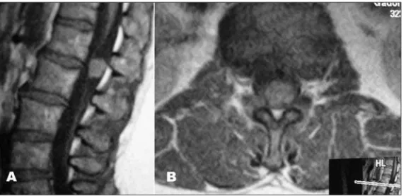
From a clinical point of view, lumbago and radicular pain in the lower limbs, were the main symptoms that lead to further investigation in our series. MRI, without a doubt, is the method of choice for the evaluation of these lesions7 (Figs 6A and 6B). In more than 50% of the cases of the present series, the most common symptom was radicular pain. lumbar tumors cannot be adequately diag-nosed with computerized mielo-tomography or mielog-raphy. In the MRI, these tumors almost always enhance after contrast media administration. Certain cases present with associated lipomas of the ilum terminale, where T1-weighted images demonstrate the hyper-intensive char-acteristic of fat8,9.
Microsurgery is the treatment of choice for ilum ter-minale ependymomas, however, in some cases adjuvant treatments are employed.
The best adjuvant treatment of ependymomas has been the cause of intense discussion in literature. Until the
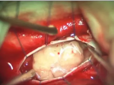
Fig 1. Surgical operating microscope view of the ilum terminale ep-endymoma after opening of the dura mater.
Fig 2. Magnified view depicting the filum terminale next to the tumor.
Fig 3. Image of the ilum terminale being sectioned. Fig 4. Image of the removal of the dissected tumor.
mid of 1990s, there was no consensus on the indication of radiotherapy and even less of chemotherapy10. only now, after the completion of long follow-up studies, could cer-tain conclusions be drawn. Radiotherapy should be re-served only for those cases in which the tumor has not been totally resected. In small and medium sized tumors when a tumor capsule is kept intact, the lesion is less like-ly to disseminate through the neuro-axis11. In large tumors, whether through manipulation or a partial resection, ra-diotherapy has been indicated as the treatment of choice12.
Recent articles show that radiotherapy reduces the progress of the illness, improving the overall survival rates10,11,13. Initially, a dose of 50 to 60 Gy is used on the tumor bed. Radiotherapy in the neuro-axis was reserved for cases in which a diagnosis of dissemination already existed prior to the surgery or as rescue therapy after con-ventional radiotherapy in cases of late spreading11.
Chemotherapy is also a matter of discussion. How-ever, with the selection of speciic patients within deined parameters, the earlier diagnosis of CNS spreading and higher local control rates of the disease, the optimiza-tion of the treatment has been made possible. The drug of choice is etoposide at 50 mg/m2, which has a good level of tolerance and is easy to administer. Nonetheless, several schemes have been tested without any one dem-onstrating superiority14,15. The current indications for che-motherapy are the neuro-axis dissemination and as rescue therapy for recurrence. An increase in life expectancy and the quality of life has been reported14-16.
Although initially developed for the prevention of cer-vical deformities in children17, the use of laminotomy with replacement of the posterior elements of the vertebra removed during surgery can be useful in many cases. The theoretical advantages described by some authors18,19 are: restoration of bone protection in the spinal canal, better esthetic results, a lesser risk of the formation of epidural ibroses20, which can be related to pain, and support for
the function of the intra-spinal musculature. Adequate fu-sion of the spine seems to occur with a greater probability in the thoracolumbar region18. Although there is no Class I evidence that a laminotomy prevents the occurrence of a deformity, we believe that the thoracolumbar region may theoretically beneit from this technique due to its transi-tional characteristics and higher risk for late onset defor-mities. The situations in which we prefer laminotomy to laminectomy are: children and young adults; when more than two levels of laminectomy are necessary and when there is an existing degeneration of the affected segments.
The objective of the microsurgery in ilum terminale ependymomas is a total resection. The intra-operative neuro-physiological monitoring is a valuable tool that optimizes the procedure. depending on their size, the ependymomas can involve both the nerves of the cau-da equina and the conus medullaris. Further, the tumor causes local inlammation and arachnoiditis of the nerves and medullar tissue, making it imperative to dissect tumor from these structures with maximum care. As such, two types of monitoring can be employed: the direct stimula-tion of the roots and the monitoring of somato-sensory evoked potentials (SSEP’s) of the lower limbs. The proto-col of nerve stimulation used in our unit involves bipolar stimulation, varying from 0.2 to 2mA in intensity, a fre-quency of 60 Hz, the duration of each pulse being 1.5ms, delivered through the MSC-02B stimulator (Micromar, Brazil). This protocol avoids the spreading of electrical activity through the CSF, reducing the unwanted stimula-tion of remote roots and nerves.
The involvement of the conus medullaris is a good indication for the SSEP’s. The anterior tibial nerve area is stimulated and the evoked potential is registered from the somato-sensitive cortex area (Cz’ according to the in-ternational 10–20 assembly system) or preferably at the epidural space of the thoracic (d8-d10) region. The moni-tor used is the MK-15 (Amplaid, Italy), with latency
ence of 20 ms and 38-40 ms for the lower thoracic region and contra-lateral cerebral cortex respectively.
The radical excision of the tumor through microsur-gery techniques is what makes the cure possible for this disease and this should be the goal of the initial surgery, since a second approach can be hazardous and less likely to result in a total resection.
REFERENcES
1. Fearnside MR, Adams CBT. Tumors of the cauda equina. J Neurol Neu-rosurg Psychiatry 1987;41:24-31.
2. Kernohan JW. Primary tumors of the spinal cord and intradural ilum terminale. In: Penield W (Ed). Cytology and cellular pathology of the
nervous system, Vol 3, New York: Paul B.Hoeber, 1932:993-1035. 3. Wiestler OD, Schiffer D, Coon SW, Prayson RA, Rosenblum MK.
Myxo-papillary ependymoma. In: WHO pathology and genetics tumors of the nervous system. IARC Press, 2000;78-79.
4. Shimada Y, Miyakoshi N, Kasukawa Y, Hongo M, Ando S, Itoi E. Clin -ical features of cauda eqüina tumors requiring surg-ical treatment.
To-hoku J Exp Med 2006;209:1-6.
5. WHO Classiication of Tumors of the Nervous System. Geneva, WHO, 1999. 6. Maruyama R, Koga K, Nakahara T, Kishida K, Nabeshima K. Cerebral
myxopapillary ependymoma. Hum Pathol 1992;23:960-962.
7. Hallacq P, Labrousse F, Streichenberger N, Lisii D, Fisher G. Bifocal myxopapillary ependymoma of the ilum terminale: the end of a spec
-trum? Case report. J Neurosurg (Spine 3) 2003;98:288-289.
8. Gallia GL, Burger PC, Suk I, et al. Concomitant comus medullaris epen
-dymoma and ilum terminale lipoma: case report. Neurosurgery 2006;
58:1214-1215.
9. Adamson C, Cummings TJ, Friedman AH. Myxopapillary
ependymo-ma and fatty ilum in an adult with tethered cord syndrome: a shared embryological lesion? Case report. Neurosurgery 2005;57:373-376.
10. Sgouros S, Malluci CL, Jackowski A. Spinal ependymomas - the value of postoperative radiotherapy for residual disease control. Br J
Neuro-surg 1996;10:559-566.
11. Lin YH, Huang CI, Wong TT, et al. Treatment of spinal cord ependy-momas by surgery with or without postoperative radiotherapy. J Neu-rooncol 2005;71:205-210.
12. Akyurek S, Chang EL, Yu TK, et al. Spinal myxopapillary ependymo -ma outcomes in patients treated with surgery and radiotherapy at M.D.
Anderson Cancer Center. J Neurooncol 2006;80:177-183.
13. Schild SE, Nisi K, Scheithauer BW, et al. The results of radiotherapy
for ependymomas: the Mayo Clinic experience. Int J Radiat Oncol Biol Phys 1998;42:953-958.
14. Chamberlain MC. Etoposide for recurrent spinal cord ependymoma.
Neurology 2002;58:1310-1311.
15. Chamberlain MC. Salvage chemotherapy for recurrent spinal cord ep-endymona. Cancer 2002;95:997-1002.
16. Chamberlain MC. Ependymomas. Curr Neurol Neurosci Rep 2003;3:
193-199.
17. Raimondi AJ, Gutierrez FA, Di Rocco C. Laminotomy and total recon -struction of the posterior arch for spinal canal surgery in childhood. J
Neurosurg 1976;45:555-560.
18. Wiedemayer H, Sandalcioglu IE, Aalders M, Wiedemayer H, Floerke M, Stolke D. Reconstruction of the laminar roof with miniplates for a posterior approach in intraspinal surgery: technical considerations and
critical evaluation of follow-up results. Spine 2004;29:E333-E342.
19. Kawahara N, Tomita K, Shinya Y, et al. Recapping T-saw laminoplasty
for spinal cord tumors. Spine 1999;24:1363-1370.