ARTICLE
Intraoperative ultrasonography for presumed
brain metastases: a case series study
Uso de ultrassonografia intraoperatória para metástases cerebrais presumidas: estudo
de uma série de casos
Helder Picarelli1, Marcelo de Lima Oliveira2, Edson Bor-Seng-Shu2, Eduardo Santamaria Carvalhal Ribas2,
Alexandre Maria Santos1, Manoel Jacobsen Teixeira2
Brain metastases (BM) represent more than 50% of intra-cranial tumors in the adult population and have shown an increasing incidence over recent decades1,2. Around 170,000
patients per year are diagnosed with BM in the United States of America. In addition, 10% to 40% of oncologic patients de-velop BM in the course of the disease1-8. he factors
associ-ated with this increasing incidence include advances in neu-roimaging techniques, easier access to health care systems, and increased survival of cancer patients. he best strategy for treating BM remains controversial, but the management includes radiotherapy, stereotactic radiosurgery and surgi-cal resection. Patients are typisurgi-cally treated according to age, functional performance, neurological status, type of cancer,
number and localization of BM, radiosensitivity and chemo-sensitivity of the tumors, and systemic control of the primary cancer5-8. Local control of BM can restore neurological
func-tional status and increase survival of patients that may die due to extracranial progression of the disease9-12. Some trials
suggest that neurosurgical resection is a good option, pre-senting low recurrence rates especially when followed by ra-diotherapy6-8,11,12. BM resection can rapidly decrease the mass
efect and surrounding edema, improve symptoms and qual-ity of life, control epileptic seizures and provide tissue for histological analysis and diagnosis of suspected lesions6-8.
In addition, resection of multiple BM can also bring positive outcomes if systemic cancer is controlled13-15. he objective
1Division of Neurological Surgery, Instituto do Câncer do Estado de São Paulo “Octavio Frias de Oliveira”, São Paulo SP, Brazil;
2Division of Neurological Surgery, Hospital das Clínicas da Faculdade de Medicina da Universidade de São Paulo, São Paulo SP, Brazil.
Correspondence: Helder Picarelli; Rua Itacolomi 222 / conj. 5; 01239-020 São Paulo SP - Brasil, e-mail: [email protected] / [email protected] Conflict of interest: There is no conflict of interest to declare.
Received 21 December 2012; Received in final form 08 June 2012; Accepted 15 June 2012
ABSTRACT
Brain metastases (BM) are one of the most common intracranial tumors and surgical treatment can improve both the functional outcomes and patient survival, particularly when systemic disease is controlled. Image-guided BM resection using intraoperative exams, such as in-traoperative ultrasound (IOUS), can lead to better surgical results. Methods: To evaluate the use of IOUS for BM resection, 20 consecutives patients were operated using IOUS to locate tumors, identify their anatomical relationships and surgical cavity after resection. Technical difficulties, complications, recurrence and survival rates were noted. Results: IOUS proved effective for locating, determining borders and defining the anatomical relationships of BM, as well as to identify incomplete tumor resection. No complications related to IOUS were seen. Conclusion: IOUS is a practical supporting method for the resection of BM, but further studies comparing this method with other intraopera-tive exams are needed to evaluate its actual contribution and reliability.
Key words: intraoperative ultrasound, brain metastases, neurosurgery.
RESUMO
As metástases cerebrais (MC) são os tumores intracranianos mais frequentes e seu tratamento cirúrgico pode melhorar a sobrevida e a funcionalidade do paciente, especialmente quando a doença sistêmica está controlada. A ressecção das MC guiada por imagens de exames intraoperatórios, como ultrassom intraoperatório (USIO), pode levar a melhores resultados cirúrgicos. Métodos: Avaliar o uso do USIO nas ressecções de MC de 20 pacientes para localizar os tumores, avaliar suas relações anatômicas e a cavidade cirúrgica após o procedimento. As dificuldades técnicas, complicações, recorrência e taxa de sobrevivência foram anotadas em cada caso. Resultados: USIO foi eficaz para localizar, delinear e definir as relações anatômicas das MC, assim como a ocorrência de ressecção incompleta. Não foram encontradas com-plicações relacionadas ao uso do USIO. Conclusão: USIO é um método auxiliar prático para as ressecções de MC, porém outros estudos ainda se fazem necessários para avaliar sua real contribuição nesses procedimentos.
of surgical treatment is to achieve complete tumor resec-tion with minimal damage to adjacent structures. he use of intraoperative image guidance, such as neuronavigation, magnetic resonance imaging (MRI) or ultrasonography (US), allows a more accurate detection of BM and reveals their anatomical relationships, possibly leading to better surgical results16-19. he present study reports a series of twenty
pa-tients with BM in whom intraoperative ultrasound, hence-forth referred to as IOUS, was used to locate and guide tumor resection, as well as to prevent vascular injuries. After the mor resection, IOUS was performed to identify residual tu-mor cells, blood clots and foreign bodies in the surgical bed.
he aims of this study were to evaluate the efectiveness of IOUS in locating and accurately deining tumors and to disclose relationships with adjacent tissues. Additionally, the use of IOUS to identify residual tumors, clots and foreign bodies in the surgical cavity immediately after tumor resec-tion was also assessed.
METHODS
Twenty consecutive patients with medical history and/ or lesions in MRI consistent with BM were treated by sur-gical resection aided by IOUS and followed up prospectively for one year. he decision to perform surgical resection was taken by a neuro-oncology multidisciplinary group from our institution, according to a protocol approved by the lo-cal Ethics Committee. All patients were given phenytoin (300 mg/d), dexamethasone (16 mg/d) and a prophylactic antibiotic (cefuroxime) before and during the procedure. he patient position during the operation and craniotomy was decided according to the tumor location shown by radiologi-cal exams, MRI (Fig 1), and the surgery was performed using classical neurosurgical and stereotactic techniques. Doppler ultrasonography was performed immediately after the open-ing of the dura mater and once again at the end of the surgery using a high-resolution system (MicroMax® model, Sonosite®,
Bothel, WA) with broadband phased array transducers. he transducers (6-13 and 4-8 MHz) were protected by sterile plastic covers (Fig 1) and saline irrigation was used to im-prove images. IOUS was irst used to determine the tumor location and to evaluate its volume, as well as to deine its borders and relationships with adjacent tissues, and the best surgical route to reach the BM (Figs 1–5). After surgery, IOUS was used to identify any residual tumor cells, hematomas or foreign bodies (Fig 4A). Technical diiculties, duration of the surgery, time required to locate the lesions by IOUS, and sur-gical complications during or after the surgery were all noted. he accuracy of IOUS for locating tumors was compared to that of stereotactic localization by Aimsystem®
Stereotactic System (Micromar®
). he lesions’ volume estimated by IOUS were compared to the estimates based on the preopera-tive MRI using the formula V = π/6xAxBxC (where A, C and
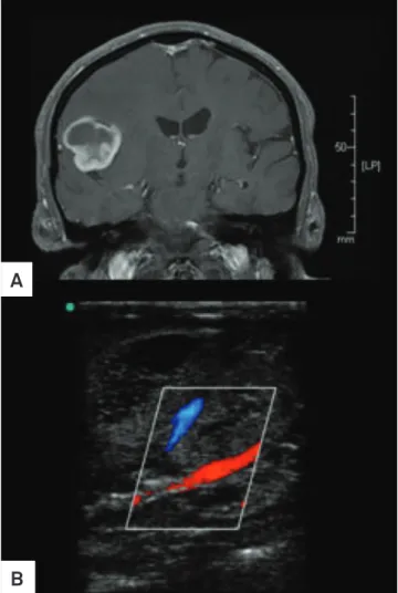
Fig 1. Case 2: Cavernoma. (A) Magnetic Resonance Images, (B) Intraoperative ultrasound, (C) IOUS Images of subcortical and hypoechoic tumor, (D) Tumor was resected em bloc.
A
C
B
D
D correspond to the largest tumor diameter on sagittal, cor-onal and axial views). he presence of residual tumor cells in the surgical cavity was assessed by IOUS after the resec-tion; the imaging was later on compared to the brain CT-scan
Fig 2. Case 10: Glioblastoma Multiforme. (A) Contrast enhanced T1 MRI image; (B) IOUS showing middle cerebral artery branch within tumor.
B
A
Fig 4. Brain metastasis of breast cancer. (A) IOUS image after the lesion resection discloses another small tumor below the surgical cavity, not seen under the microscope.
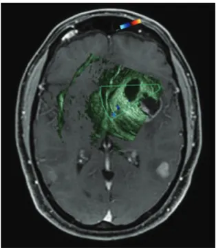
Fig 5. Case 4: Brain metastasis of melanoma. Merged IOUS and MRI images: the IOUS oriented the corticectomy and the best route to approach the tumor through the insula. Images clearly show ventricles, midline shift, tumor and relationships with middle artery branches. This patient had a good neurological recovery immediately after the surgery with no motor deficit and he’s submitted to radiotherapy. Two hundread and thirty days later he died of gastrointestinal bleeding secondary to bowel metastases and meningeal carcinomatosis. There was not local recurrence.
images, performed in all cases within 24 hours postopera-tively. Diferences between tumor volumes calculated using IOUS compared to preoperative MRI greater than or equal to 5% were considered discordant predicted volumes (p-value). Contrast-enhancing brain lesions on the postoperative CT-scan were regarded as probable residual tumor cells and con-sidered discordant if IOUS did not detect the same inding. he suspected areas identiied by IOUS were biopsied and sent to the laboratory for anatomopathological examination. All pa-tients received the same radiotherapy irradiation dose to the surgical site given within the irst ive weeks of surgery.
RESULTS
Patients’ mean age was 53.4 years (26–76 years), twelve of them were males (60%), and three (15%) had no previous on-cological history. Twenty-three BM were operated in the study (Table 1). Histological analysis conirmed BM in 85% of the pa-tients. Two of them were diagnosed with glioblastoma multi-forme and one with cavernoma (Fig 1 and Table 2). In all cas-es, IOUS indicated the tumor localization in the irst minute of the exam, and the results were in agreement with stereotactic localization. In all cases, ultrasonographic characterization of
the lesions was in agreement with the preoperative MRI ind-ings in terms of tumor borders and anatomical relationships to ventricular and vascular structures. he volume of the lesion was calculated based on intraoperative US images in ive pa-tients, and a signiicant correspondence to the volume calcu-lated from the preoperative MRI was observed in all cases. Six lesions (26.1%) were supericial, at the cortex, being identiied by direct visualization; IOUS was then used to determine the echographic characteristics and anatomical relationships, and to search for residual tumor cells after resection. Seventeen lesions were subcortical, and intraoperative US was used in these cases to guide the surgeon in electing the best route to reach the lesion (Figs 4 and 5).
In eleven cases (55%), IOUS identiied suspected areas of residual tumor cells at the surgical bed, eight of which (72.7%) were later conirmed by anatomopathological examination (Fig 4A). he three remaining suspected lesions were diag-nosed as gliosis, necrosis and thermal lesion. No contrast-en-hancing lesions supposedly missed by IOUS were identiied in the postoperative CT-scan.
Doppler color low imaging mode revealed important re-lations between the vascular structures and the tumor in ive cases (25%), but accidental vascular injuries were not found. In four cases (20%), the surgical strategy was modiied based on
Fig 3. Case 5: Thalamic Brain Metastasis of colon adenocarcinoma. (A) Contrast enhanced T1 MRI image, (B) IOUS showing corticectomy and optimal route to reach tumor, (C) Intraoperative view.
C
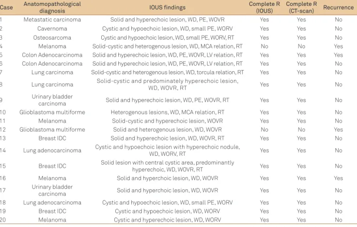
Table 2. Surgical findings, IOUS image characteristics, and postoperative evolution.
PE: perilesional edema, WD: well-delimited, WOVR: without important vascular relations, MCA: middle cerebral artery, LV: lateral ventricle, R: resection, IOUS: intraoperative ultrasound, RT: residual tumor, IDC: breast invasive ductal carcinoma.
Case Anatomopathological
diagnosis IOUS findings
Complete R (IOUS)
Complete R
(CT-scan) Recurrence
1 Metastatic carcinoma Solid and hyperechoic lesion, WD, PE, WOVR Yes Yes No
2 Cavernoma Cystic and hypoechoic lesion, WD, small PE, WORV Yes Yes No
3 Osteosarcoma Cystic and hypoechoic lesion, WD, small PE, WORV, RT Yes Yes No
4 Melanoma Solid-cystic and heterogenous lesion, WD, MCA relation, RT No No Yes
5 Colon Adenocarcinoma Solid and hyperechoic lesion, WD, PE, WOVR, LV relation, RT Yes Yes Yes 6 Colon Adenocarcinoma Solid and hyperechoic lesion, WD, PE, WOVR, LV relation, RT Yes Yes No 7 Lung carcinoma Solid-cystic and heterogenous lesion, WD, torcula relation, RT Yes Yes No
8 Lung carcinoma Solid-cystic and predominately hyperechoic lesion,
WD, WOVR, RT Yes Yes No
9 Urinary bladder
carcinoma Solid and hyperechoic lesion, WD, PE, WOVR, RT Yes Yes No
10 Glioblastoma multiforme Heterogenous lesions, WD, MCA relation, RT Yes Yes No
11 Melanoma Solid-cystic and hyperechoic lesion, WOVR Yes Yes No
12 Glioblastoma multiforme Solid and heterogenous lesion, WD, WOVR No No Yes
13 Breast IDC Solid and hyperechoic lesion, WD, WOVR, RT Yes Yes No
14 Lung adenocarcinoma Cystic and hypoechoic lesion with hyperechoic nodule,
WD, WORV, RT Yes Yes No
15 Breast IDC Solid lesion with central cystic area, predominantly
hyperechoic, WD, WOVR, RT Yes Yes No
16 Melanoma Solid and hyperchoic lesion, WD, WOVR Yes Yes Yes
17 Urinary bladder
carcinoma Solid and hyperchoic lesion, WD, WOVR Yes Yes No
18 Lung adenocarcinoma Cystic and hypoechoic lesion, WD, small PE, WORV Yes Yes No
19 Breast IDC Cystic and hypoechoic lesion, WD, WORV Yes Yes No
20 Melanoma Cystic and hyperechoic lesion, WD, WORV Yes Yes No
Table 1. Patients and data.
C: cortical, SC: subcortical, D: deep, ASA: American Society of Anesthesiologists classification, ECOG: Eastern Cooperative Oncology Group performance status, SIR: Score Index for Radiosurgery of brain metastases, KPS: Karnofsky Performance Scale.
*Volume: π/6xAxBxC, where A, B and C correspond to the lesion’s largest diameter on axial, coronal and sagittal MRI images.
Case Age
(years) Oncological history
N° of Lesions/
volume (cc3)* Localization ASA ECOG SIR KPS
1 42 Breast Invasive ductal
carcinoma 2/ 6.56 C-SC, occipital II 1 6 80
2 57 Lymphoma 1/ 3.81 precentral gyrus III 4 7 40
3 26 Osteosarcoma 1/ 77.71 C-SC-P, frontal lobe III 1 6 80
4 45 Melanoma 1/ 17.08 D, insula III 1 4 90
5 61 Colon adenocarcinoma 1/ 15.5 D, thalamus II 2 3 80
6 41 Colon adenocarcinoma 1/ 13.13 D, parieto-occipital II 1 6 70
7 76 X 1/ 14.97 C-SC, parieto-occipital III 2 2 40
8 70 X 1/ 7.24 C-SC, parietal III 3 3 50
9 52 Clear cell renal carcinoma 3/ 1.98 C-SC, occipital III 3 3 50
10 70 Prostate adenocarcinoma 1/ 22.08 D, frontal III 1 6 90
11 34 Melanoma 1/ 3.04 C-SC, frontal I 1 7 90
12 49 X 1/31.34 C-SC-D, parieto-occipito-temporal II 1 7 80
13 67 Breast Invasive ductal
carcinoma 1/ 8.05 D, cerebellum II 1 5 90
14 65 Lung adenocarcinoma 1/2.84 C, frontal II 3 3 40
15 64 Breast Invasive ductal
carcinoma 1/9.97 C-SC, frontal III 2 6 80
16 26 Melanoma 1/12.23 D, temporal II 1 7 90
17 72 Urinary bladder carcinoma 1/56.16 C-SC, frontal III 4 2 30
18 63 Lung adenocarcinoma 1/23.47 D, parieto-occipital III 3 2 50
19 42 Breast Invasive ductal
carcinoma 1/ 3.78 SC, frontal III 1 6 90
IOUS indings, which revealed an alternative route for the neu-rosurgeon to reach the lesion and perform the resection (Fig 4). Mean total operation time for a single lesion was 289 minutes and total usage of IOUS never exceeded thirty minutes.
here were no complications related to the surgical proce-dure per se, such as neurological worsening, wound infections, meningitis, abscess, contusions or hematomas. Patient survival time ranged from ifteen days (death related to sepsis and central venous access infection) to 365 days (maximum follow-up time). Functional performance improved after surgery in thirteen pa-tients (65%), and was unchanged in six of them. he most rel-evant surgical and follow-up indings are shown in Table 2.
Survival rates in the irst, second and third months were 95%, 95% and 80%, respectively. Mean follow-up time was 180 days (15 to 365 days), and three patients presented with local tumor recurrence after BM resection. All recurrences were related to meningeal carcinomatosis (2 patients with mela-noma and 1 patient with colon adenocarcimela-noma) (Fig 3), and these patients died during follow-up.
DISCUSSION
Brain metastases are more common than primary brain cancer and there is debate over the best treatment protocol. Well-designed studies conducted with patients with single BM, good functional performance, and controlled system-ic cancer suggest that neurosurgery is an efective option for local disease control14,15. BM happens by expansion and can
reach a signiicant volume, increasing intracranial pressure and promoting inlammation in adjacent tissues and brain vasogenic edema. he complete resection of BM can resolve some of these processes, restoring neurological functions and solving symptoms. Additionally, surgery allows conirmation of the histological diagnosis of the tumor. In our series, BM was conirmed in 85% of the patients, which enabled their referral, as well as the 15% non-conirmed cases, to appropriate treat-ment. Although surgery are very beneicial for patients, the de-cision should always be made by weighing up the beneits and risks. he surgical morbidity and mortality associated to the procedure are estimated to be less than 3 and 5%, respective-ly. he most common complications are neurological worsen-ing, wound infections, intracranial hematomas, deep venous thrombosis, pulmonary embolism and pneumonia.
he extension of tumor resection is an important fac-tor when it comes to decreasing the local tumor progression rate and increasing patient survival. Complete tumor resec-tion is possible provided that the lesion borders are well de-ined, the tumor is removed en bloc, and potential residual cells are recognized during surgery. Intraoperative radiolog-ical exams such as MRI or IOUS may promptly identify le-sion borders, anatomical relationships and residual tumor cells with high sensitivity, leading to more complete and safe
tumor removal16-19. he choice of the method also depends
on their availability and on the neurosurgeon’s personal expe-rience. Intraoperative MRI provides high-quality images, but is still an expensive option that is rarely available in Brazil. he technique also requires complex infrastructure, and im-age acquisition time is longer. Lower cost, more practical us-age, ability to repeat the exam several times and faster image acquisition are some of the characteristics that make IOUS a more attractive method of choice. All lesions in our series were located in less than one minute using this method, and their size, shape, borders, anatomical relations, as well as identiication of necrosis, edema and calciications were de-termined in most cases and proved concordant with preop-erative exam indings. IOUS also indicated where it was best to open the dura-mater, the direction and relationships of each sulcus with the tumor, the optimal point to perform the corticectomy, and the best route to approach the tumor. he use of Doppler color low helped identify important vascular structures, preventing possible injuries.
1. Landis SH, Murray T, Bolden S, et al. Cancer statistics, 1999. CA Cancer J Clin 1999;49:8-31.
2. Patchell RA, Posner JB. Neurologic complications of systemic cancer. Neurol Clin 1985;3:729-750.
3. Klos KJ, O’Neill BP. Brain metastases. Neurologist 2004;10:31-46.
4. Norden AD, Wen P, Kesari S. Brain Metastases. Curr Opin Neurol 2005;18:654-661.
5. Nguyen T, Abrey LE. Brain metastases: old problem, new strategies. Hematol Oncol ClinNorth Am 2007;21:369-388.
6. Richards GM, Khuntia D, Mehta MP. Therapeutic management of metastatic brain tumors. Crit Rev Oncol Hematol 2007;61:70-78.
7. Posner JB, Chernik NL. Intracranial metastases from systemic cancer. Adv Neurol 1978;19:579-592.
8. Posner JB. Management of brain metastases. Rev Neurol 1992;148:477-482.
9. Delattre JY, Krol G, Thaler HT, et al. Distribution of brain metastases. Arch Neurol 1998;45:741-744.
10. Chao ST, Barnett GH, Liu SW, et al. Five-year survivors of brain metastases: A single-institution report of 32 patients. Int J Radiat Oncol Biol Phys 2006;66:801-809.
11. Patchell RA, Tibbs PA, Walsh JW, et al. A randomized trial of surgery in the treatment of single metastases to the brain. [abstract] N Engl J Med 1990;322(8):494.
12. Patchell RA, Tibbs PA, Walsh JW, et al. A randomized trial of surgery in the treatment of single metastases to the brain. N Engl J Med 1990;322:494-500.
13. Bindall RK, Sawaya R, Leavens ME, et al. Surgical treatment of multiple brain metastases. J Neurosurg 1993;79:210-216.
14. Ranasinghe MG, Sheehan JM. Surgical management of brain metastases. Neurosurg Focus 2007;22:E2.
15. Ammirati M, Cobbs CS, Linskey ME, et al. The role of retreatment in the management of recurrent/progressive brain metastases: a systematic review and evidence-based clinical practice guideline. J Neurooncol 2010;96:85-96.
16. Hammoud MA, Ligon BL, Elsouki R, et al. Use of intraoperative ultrasound for localizing tumors and determining the extent of resection: a comparative study with magnetic resonance imaging. J Neurosurg 1996;84:737-741.
17. Solheim O, Selbekk T, Lindseth F. Navigated resection of giant intracranial meningiomas based on intraoperative 3D ultrasound. Acta Neurochir 2009;151:1143-1151.
18. Gerganov VM, Samii A, Akbarian A. Reliability of intraoperative high-resolution 2D ultrasound as an alternative to high–field strength MR imaging for tumor resection control: a prospective comparative study. J Neurosurg 2009;111:512-519.
19. Tronnier VM, Bonsanto MM, Staubert A, et al. Comparison of intraoperative MR imaging and 3D-navigated ultrasonography in the detection and resection control of lesions. Neurosurg Focus 2001;10:1-5. References
Incorrect patient positioning or craniotomy procedures, a poor quality IOUS device and little experience of the surgeon with the method can decrease the efectiveness of the exam. However, the skill needed to use this harmless and efective intraoperative guidance tool can be easily acquired with a lit-tle dedication. Recent studies have demonstrated that three-dimensional IOUS images can be used in conjunction with other intraoperative guidance methods, but further studies are needed to evaluate their advantages.