829
Arq Neuropsiquiatr 2012;70(10):826-830 attempt, two for two attempts, one for three attempts, and
none if the problem cannot be solved with three attempts. Descriptive analysis of the participants was performed and then linear regression analysis was used for assessing the inluence of age, education, and gender on test performance. Group comparisons (performed by one-way ANOVA) were used for determining the normative data divisions. Statistical signiicance was established at 0.05.
he participant’s description and test performance are shown in Table. he linear regression model was signiicant (F=7.00, p<0.001, R2=0.07), showing inluence of age (β=-0.19,
p=0.001) and education (β=0.18, p<0.001) on test perfor-mance, but not of gender (p>0.05). he ANOVA results sug-gested a normative data division based on age and education,
since the proposed age-education groups presented signii-cant diferences (F=8.59, p<0.01, η2=0.07).
he TOL is a well-validated classical neuropsychologi-cal test for the assessment of planning skills. As other stud-ies suggested, age and education were related to task per-formance4. he development of adequate normative data is
essential for cognitive assessment in clinical setting. When a precise characterization of executive/planning performance is necessary, as in the assessment of diferent conditions like dementia, neuropsychiatry disorders, and mild cognitive im-pairment, stratiied data for sociodemographic factors as age and education allow a more accurate interpretation of test performance and neuropsychological hypothesis testing in the clinical setting5.
1. Lezak MD, Howieson DB, Loring DW. Neuropsychological Assessment. 4th ed. New York: Oxford University Press; 2004.
2. Shallice T. Specific impairment of planning. Phil Trans R Soc Lond B. 1982;298:199-209.
3. Krikorian R, Bartok J, Gay N. Tower of London procedure: a standard method and developmental data. J Clin Exp Neuropsychol 1994;16:840-850.
References
4. Pena-Casanova J, Quiñones-Úbeda S, Quintana-Aparicio M, et al. Spanish Multicenter Normative Studies (NEURONORMA Project): norms for the Stroop Color-Word Interference Test and the Tower of London-Drexel. Arch Clin Neuropsychol 2009;24:413-429.
5. de Paula JJ, Moreira L, Nicolato R, et al. The Tower of London Task: different scoring criteria for diagnosing Alzheimer’s disease and mild cognitive impairment. Psychol Reports 2012;110:477-488.
Ibuprofen-induced unilateral optic neuritis
Neurite óptica unilateral induzida por ibuprofeno
Josef Finsterer1, Simon Brunner2
1MD, PhD; Krankenanstalt Rudolfstiftung, Vienna, Austria;
2 MD; Department of Ophthalmology, Krankenanstalt Rudolfstiftung, Vienna, Austria.
Correspondence: Josef Finsterer; Postfach 20; 1180 Vienna; Austria - Europe; E-mail: [email protected]
Conflict of interest: There is no conflict of interest to declare.
Received 26 April 2012; Received in final form 10 May 2012; Accepted 17 May 2012
Visual disturbances have been reported to occur as side efects from ibuprofen (2-4’-isobutylphenyl-propionic acid) in therapeutic dosages, in <1% of the cases1-3. he most
com-mon visual disturbances include amblyopia, scotomata, or changes in color vision3. Also, the contrast sensitivity may
be depressed at low spatial frequencies during treatment with ibuprofen, 800 mg/d3. Visual side efects occur more
frequently in adults as compared to children and they are dose-dependent4.
CASE REPORT
A 61-year-old Caucasian female, referred by the depart-ment of ophthalmology for acute and constant visual deicit
(Fig 1), and papillary edema on the right eye for one week. She took ibuprofen 1,600 mg/d during seven days for lum-balgia. At the last day of ibuprofen administration, the pa-tient experienced visual deicits on the right eye, which she described as blurring and dimming. Initially, she went to her ophthalmologist who found a visual acuity of -0.5 (left) and -0.5 (right) and fuzzy papilla, and referred her to a second-ary ophthalmology center, which found a prominent right papilla due to edema and swelling (Fig 2), but did not reveal an ophthalmologic cause of the abnormality either. To ex-clude a central nervous system lesion, she was referred to the neurologist.
830 Arq Neuropsiquiatr 2012;70(10):826-830
Clinical exam and blood tests were noninformative. he magnetic resonance imaging (MRI) of the cerebrum did not show any abnormalities. Visually-evoked potentials, howev-er, gave a prolonged latency of the P100 component on the right side. She was advised to refrain from misusing ibupro-fen in the future and to treat lumbalgia by other means than nonsteroidal anti-rheumatic drugs. Two days after the irst neurological exam and nine days after ibuprofen discontinu-ation, visual ield defect had markedly improved (Fig 2), and further six days later, vision was normal again. he probabil-ity that the adverse reaction was attributable to ibuprofen was 4 on the Naranjo ADR scale.
DISCUSSION
Visual deicits following intake of ibuprofen have been occasionally reported1-4. However, the patient described
here difers in several aspects from the previous cases. Contrary to previous reports2,3, this patient was taking a
higher dosage of ibuprofen. Previously reported patients took dosages of 800 mg3 or 1,200 mg1. Contrary to previous
reports, our patient developed visual deicits one week after
starting ibuprofen. In Hamburger et al. case, visual acuity and color vision decreased not earlier than two months af-ter starting ibuprofen2. In Ridder’s3 case, contrast
sensitiv-ity depressed after two days taking 800 mg/d3. Duration
of impaired vision was also diferent between the studies. Again, our patient did not exhibit decreased color vision or depressed contrast sensitivity. Compared to Hamburger et al. case, our patient did not exhibit a reduced N75/P100 am-plitude. Gamulescu et al.1 reported a case with optic
neu-ritis, which lasted until two days after discontinuation of 1,200 mg ibuprofen/daily. his case was a 41-year-old male who complained about right-sided blurred vision and ocu-lar pain during voluntary movements of the eyes or head. Vision was markedly reduced, there was a quadrant visual ield defect, and absent response of visually-evoked poten-tials1. Visual disturbances associated with ibuprofen have
also been reported in patients who received the drug over the counter5. Although the described cases indicate that
some patients may develop ophthalmologic side efects, there are also studies on larger series of patients under ibu-profen for osteoarthritis, which could not ind any ocular complications from the drug6.
This case shows that high daily dosage of ibuprofen during one week may result in unilateral, transient le-sion of the visual pathway. Withdrawal of ibuprofen may be followed by immediate recovery of the disturbed visual functions.
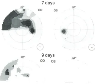
Fig 1. Visual field defects in seven (upper panels) and nine days (lower panels) after discontinuation of 1,600 mg of ibuprofen. Within nine days after ibuprofen administration, the deficits had almost completely disappeared.
Fig 2. Optical coherence tomography of the right eye seven days after discontinuation of 1,600 mg of ibuprofen showing a prominent papilla.
1. Gamulescu MA, Schalke B, Schuierer G, Gabel VP. Optic neuritis with visual field defect - possible Ibuprofen-related toxicity. Ann Pharmacother 2006;40:571-573.
2. Hamburger HA, Beckman H, Thompson R. Visual evoked potentials and ibuprofen (Motrin) toxicity. Ann Ophthalmol 1984;16:328-329. 3. Ridder WH 3rd, Tomlinson A. Effect of ibuprofen on contrast sensitivity.
Optom Vis Sci 1992;69:652-655.
4. Hall AH, Smolinske SC, Conrad FL, et al. Ibuprofen overdose: 126 cases. Ann Emerg Med 1986;15:1308-1213.
5. Nicastro NJ. Visual disturbances associated with over-the-counter ibuprofen in three patients. Ann Ophthalmol 1989;21:447-450. 6. Melluish JW, Brooks CD, Ruoff G, Cross CJ, Sanborn EC. Ibuprofen
and visual function. Prospective evaluation. Arch Ophthalmol 1975;93:781-782.