INTRODUCTION
Severe Acute Liver Failure (SALF) is a syndrome with high morbidity and mortality rates and low prevalence. It is charac-terized by sudden onset in patients with previously normal liver with rapid progress, leading to hepatocellular insuficiency, which translates into extensive metabolic disturbances, particular sus-ceptibility to bacterial or fungal infections, collapse of multiple organs, coagulopathy, and central nervous system disorders, with mortality reaching 80%(6,24,27). The severe acute attack on the hepatic parenchyma can have different etiologies, such as drugs, xenobiotics and viruses. At present, the treatment of excellence in most cases is
Action of vitamin E on experimental severe
acute liver failure
Fabiano Moraes
MIGUEL
1,5, Elizângela Gonçalves
SCHEMITT
2,5,6, Josieli Raskopf
COLARES
2,5,6,
Renata Minuzzo
HARTMANN
2,5,6, Maria Isabel
MORGAN-MARTINS
4and Norma Possa
MARRONI
1,2,3,5,6Received 15/8/2016 Accepted 28/11/2016
ABSTRACT – Background – Severe Acute Liver Failure (ALF) is a life-threatening clinical syndrome characterized by hepatocyte necrosis, loss of hepatic architecture, and impairment of liver functions. One of the main causes of ALF is hepatotoxicity from chemical agents, which damage hepatocytes and result in increase of reactive oxygen species. The vitamin E isoform is the one with the strongest biological antioxidant activity. Objective – To evaluate the antioxidant effect of vitamin E in this ALF model. Methods – We used 56 rats (mean weight of 300 g) divided into eight groups, four groups assessed at 24 hours and 4 assessed at 48 hours after induction: control group (CO); Vitamin E (Vit. E); Thioacetamide (TAA) and Thioacet-amide + Vitamina E (TAA+Vit.E). Rats were submitted to injections of thioacetThioacet-amide (400 mg/kg i.p.) at baseline and 8 hours later. Vitamin E (100 mg/kg ip) was administered 30 minutes after the second dose of thioacetamide. The 48-hour group rats received two additional doses of vitamin E (24h and 36h). At 24h or 48 hours after the administration of the irst dose of TAA, rats were weighed and anesthetized and their blood sampled for evaluation of liver integrity through enzymes aspartate aminotransferase (AST) and alanine aminotransferase (ALT). Liver tissue was sampled for assessment of lipid peroxidation (LPO) by the technique TBARS, antioxidant enzymes SOD, CAT, GPx and GST activity, levels of the NO2/NO3 and histology by H&E in two times. The results were expressed as mean ± standard deviation and statistically analyzed by ANOVA followed by Stu-dent-Newman-Keuls, with P<0.05 considered as signiicant. Results – After treatment with vitamin E, we observed a reduction in liver enzymes AST (U/L) (101.32±19.45 in 24 hours and 97.85±29.65 in 48 hours) related to the TAA group (469.56± 0.69 in 24 hours and 598.23±55.45 in 48 hours) and ALT (U/L) (76.59±8.56 in 24 hours and 68.47±6.49 in 48 hours) compared to the TAA group (312.21±10.23 in 24 hours and 359.15±17.58 in 48 hours). There was a reduction of LPO (nmol/mg Prot) in the TAA+Vit.E group (0.77±0.07 in 24 hours and 0.95±0.08 in 48 hours) compared to the TAA group (1.50±0.07 in 24 hours e 1.65±0.16 in 48 hours). SOD decreased in the TAA+Vit.E group (49.48±9.47 in 24 hours and 62.45±18, 47 in 48 hours), related to the TAA group (98.46±15.48 in 24 hours and 154.13±21.46 in 48 hours), as well as GST (nmol/min/mg Prot) in the TAA+Vit.E group (350.57±36.93 in 24 hours and 453.29±13.84 in 48 hours) compared to the TAA group (561.57±64.56 in 24 hours and 673.43±38.13 in 48 hours). There was an increase in CAT (pmol/min/mg Prot) in the TAA+Vit.E group (3.40±0.44 in 24 hours and 3.0±0.35 in 48 hours) compared to the TAA group (1.65±0.21 in 24 hours and 1.86±0.42 in 48 hours). The GPx (nmol/min/mg Prot) increased in 24 hours in the TAA+Vit.E group (1.01±0.16) compared to the TAA group (0.41±0.04) and decreased in 48 hours (1.19±0.17) compared to the TAA group (1.76±0.21). There was a reduction in NO2/NO3 (mmol/L) levels in the TAA+Vit.E group (31.47±4.26 in 24 hours and 38.93±5.20 in 48 hours) compared to the TAA group (49.37±5.12 in 24 hours and 53.53±5.97 in 48 hours). The histopathological evaluation showed a decrease in liver injury (necrosis and inlammation) in both studied times. Conclusion – These results suggest that vitamin E was able to protect the liver from lesions caused by thioacetamide.
HEADINGS – Acute liver failure. Thioacetamide. Oxidative stress. Antioxidants.
Declared conflict of interest of all authors: none Disclosure of funding: no funding received
1 Programa de Pós-Graduação BioSaúde, Universidade Luterana do Brasil, Canoas, RS, Brasil; 2 Programa de Pós-Graduação em Medicina: Ciências Médicas, Universidade Federal do Rio
Grande do Sul, Porto Alegre, RS, Brasil; 3 Programa de Pós-Graduação em Ciências Biológicas: Fisiologia - Universidade Federal do Rio Grande do Sul, Porto Alegre, RS, Brasil; 4 Programa
de Pós-Graduação Pró-Saúde, Universidade Luterana do Brasil, Canoas, RS, Brasil; 5 Laboratório de Estresse Oxidativo e Antioxidantes, Universidade Luterana do Brasil, Canoas, RS, Brasil; 6 Laboratório de Hepatologia e Gastroenterologia Experimental, Hospital de Clínicas de Porto Alegre, RS, Brasil.
Correspondence: Norma Possa Marroni. Rua José Kanan Aranha, 102. Bairro Ipanema – CEP: 91760-170 – Porto Alegre, RS, Brasil. E-mail: [email protected]
of its course. Thioacetamide (TAA) is a xenobiotic known as a potent hepatotoxic, carcinogenic and cirrhosis-inducing agent in rats(5,12). Its administration causes the death of hepatic cells by both centrilobular necrosis and apoptosis(10,14,20). This process involves reactive oxygen species (ROS), which leads to oxidative stress (OS), with increased damage to DNA, proteins and lipids from the exces-sive generation of free radicals (FR)(15,29).
Both ROS and reactive nitrogen species (RNS) as well as other free radicals are critical intermediaries in the physiopathogenesis and physiopathology of hepatocyte lesion(9). Bioactive products resulting from lipoperoxidation are highly implicated as being key abnormalities responsible for the hepatic injury(11).
The organism relies on an antioxidant defense system against ROS and RNS, which is divided in two main types: enzymatic, such as enzymes superoxide dismutase (SOD), glutathione peroxidase (GPx), catalase (CAT) and glutathione S-Transferase (GST); and non-enzymatic, such as glutathione (GSH), ascorbic acid (vitamin C), lavonoids, vitamin E, among others(26).
Vitamin E is a component of vegetable oils that is found in nature in four different forms: α, β, γ, and δ-tocopherol. Vitamin E is the main antioxidant vitamin transported in the blood low by the lipid phase of plasma lipoprotein particles(16).
The α-tocopherol isoform is the one with the strongest bio-logical antioxidant activity and is widely distributed in tissues and plasma. In the non-hydrophobic portion of a-tocopherol there is the hydroxyl radical (HO), whose atom of hydrogen is easily removable. So, when peroxyl and/or alcoxyl radicals are generated during lipid peroxidation, they are likely to combine with fatty acids of the tail of vitamin E, thus stopping to withdraw electrons from membrane fatty acids. Therefore, vitamin E, owing to its structural character-istics, acts as chain breaker, i.e. a scavenger of free radicals, thus precluding lipoperoxidation (LPO)(30).
Given the physiopathogeny of severe SALF involving the formation of ROS and RNS, the hepatotoxic ability of TAA and the antioxidant effects of Vitamin E, this work was designed to investigate the action of this vitamin on SALF in rats.
METHODS
Ethical considerations
Animal handling complied with the ethical principles estab-lished by Federal Law No. 11.794, which regulates the scientiic use of experimental animals in Brazil. This project was approved by the Ethical Research Committee of Universidade Luterana do Brasil (ULBRA) for Animal Use (CEUA- Protocol 2012 – 43P).
Animals and research design
Fifty-six male Wistar rats with mean weight of 300 g were used, divided in two experiments according to time of interest, 24h and 48h (28 animals per experiment). Each experiment comprised four groups: control (CO) group, Vitamin E (Vit. E) group, Tioacetamide (TAA) group and Tioacetamide + Vitamin E (TAA+Vit. E). Each experimental group was composed of 7 animals (n=7 based on sampling calculation) obtained from the animal facility of ULBRA. Along the study period the animals were kept in plastic boxes lined with wood shavings on a 12h light/dark cycle and room tempera-ture between 20 and 25ºC. They had free access to food and water. Thioacetamide (Sigma Chemical Co., St. Luis, MO, USA) was diluted in 1 mL of 0.9% NaCl vehicle and administered with intraperitoneal injection (i.p.). Vitamin E (α–tocopherol),
sup-plied in gelatinous capsules with oil by Importadora Química DELAWERE®, was administered at a dose of 125 mg/kg (i.p.)
Experimental protocol
The CO-24h group received three doses of 0.9% NaCl vehicle, with the second dose given 8 hours after the irst and the third and last dose 30 minutes after the second. Thioacetamide was admin-istered at two doses of 400mg/Kg (i.p.) each with an interval of 8 hours, while vitamin E was given at a dose of 125 mg/kg (i.p.) 30 minutes after the second dose of TAA. In the 48-hour experiment, two additional doses of vitamin E were administered, with the sec-ond dose given 24 hours after the start of the experiment and the third, 36 hours after it. Doses of vehicle at the same dilution (0.9% NaCl, 1 mL) were administered to the groups in both experiments in order to expose the animals to the same number of administra-tions. At the end of each experiment, animals were weighed and anesthetized with ketamine 95 mg/kg and xylazine hydrochloride 8 mg/kg (i.p.). Blood samples were collected from the retro-orbital plexus for hepatic integrity assays and livers were dissected out for posterior analyses. At the end of each experiment (24h and 48h), animals were killed by exsanguination under deep anesthesia.
Plasma analyses
Liver integrity was determined by evaluation of enzymes aspartate aminotransferase (AST) and alanine aminotransferase (ALT) in plasma using the commercial kit Boehriner Mannheim (Germany). AST (340 nm) and ALT (340 nm) activities were ob-tained by kinetic assay using the commercial liquiform kit Labtest®.
Liver homogenates
Nine mL of phosphate buffer solution (1.15% KCL) per gram of tissue (liver) and phenylmethylsulfonyl luoride (PMSF) at a concentration of 100 mM in isopropanol (10 µL/mL of KCl) were used. The tissue was homogenized in ULTRA-TURRAX for 40 seconds at 0-2ºC and subsequently centrifuged for 10 min at 3000 rpm in refrigerated centrifuge. The precipitate was discarded and the supernatant removed and frozen at -80ºC for posterior bio-chemical analyses(19).
Protein
The Bradford method (1976) was used to quantify proteins, with bovine albumin as standard (SIGMA). The samples were spectrophotometrically measured at 595 nm, and the concentrations expressed in mg/mL and used to calculate thiobarbituric acid reac-tive substances (TBARS) levels and antioxidant enzyme activity.
Lipoperoxidation
Analyses of antioxidant enzymes
Glutathione S-transferase (GST) is based on an enzyme that catalyzes the formation of 1 mmol of DNP-SG per minute at 30°C using 1mM of the concentration of (reduced) GSH and CDNB, detected spectrophotometrically at 340 nm, values expressed in mmol/min/mgprot(21). The analysis of superoxide dismutase (SOD) activity is deined as its ability to inhibit a detection system that reacts with O
-2. The technique of measuring SOD is based on the inhibition of this reaction with adrenalin, detected spectrophoto-metrically at 480 nm. The data were expressed as units of SOD per milligram of protein (USOD/ mg prot.)(25). The analysis of catalase activity (CAT) is deined by the breakdown of hydrogen peroxide in water and oxygen, being directly proportional to its enzymatic activity, detected spectrophotometrically at 240 nm. The results were expressed in µmoles per milligram of protein (mmoles of H2O2)(3). Glutathione peroxidase (GPX) can be studied by measuring the rate of consumption of nicotinamide adenine dinucleotide (NADPH) in the reduction of glutathione oxidase, detected spectrophotometrically at 340 nm and its activity expressed in nmoles per minute per milligram of protein (nmol/ min/mg prot)(13).
Evaluation of nitric oxide metabolites – nitrites e nitrates
Nitric oxide production was measured indirectly through a colorimetric quantitative test by the Griess reaction. It is based on the enzymatic reduction of nitrates to nitrites in the presence of nitrate reductase and NADPH, and posterior reaction of the formed nitrites (or initially present in the samples) with Griess reagent (mixture of sulfanilamide and naphthyl ethylenediamine, speciic for NO2-). However, as the excess of NADPH used inhibits the Griess reaction, it is necessary to oxidize all of the NADPH not used in the reduction of nitrates. This is achieved by adding nitrate reductase. The reading was performed in a microplate reader at 540nm and the results expressed in mmol of NO2/NO3.
Histological analysis
Histological analyses were performed on liver samples preserved in 10% formaldehyde solution for 24h, which were then embed-ded in parafin and cut in 3 mm slices using a rotating microtome. Histological examinations were performed using hematoxylin-eosin staining. A single pathologist, blinded to experimental protocol, analyzed all livers under a binocular Labophot NIKON microscope, at 100X magniication.
Statistical analysis
The results were expressed as mean ± standard error for each experimental group. The software GrapPad Instat, version 3.0 was used for the statistical analysis. For symmetrical data, simple ANOVA was used to compare the differences found in each studied parameter. The complementary Student-Newman-Keuls test was used as well for multiple comparisons. The level of signiicance for each comparison was at least 5% (P<0.05).
RESULTS
Liver integrity
Table 1 shows the results of AST and ALT evaluation at 24h and 48h. The TAA groups showed a signiicant increase (P<0.001) as compared to the CO group at both times, while the TAA groups receiving Vit. E (125 mg/kg) reduced these enzymes signiicantly
(P<0.001) at both these times, decreasing and protecting from the damage triggered by TAA. It was thus demonstrated that Vit. E doses contributed to a protective effect on the liver tissue.
TABLE 1. Evaluation of enzymes AST and ALT (U/L) in the different
experimental groups at the two studied times (24h and 48h)
Grupos CO VIT. E TAA TAA + Vit. E
24 h
AST 39.05±6.55 39.99±5.23 469.56±0.69a 101.32±19.45b
ALT 22.36±3.45 21.56±2.64 312.21±10.23a 76.59±8.56b
48 h
AST 43.12±5.63 41.56±3.45 598.23±55.45a 97.85±29.65b
ALT 29.48±3.12 32.45±3.05 359.15±17.58a 68.47±6.49b
CO: control group, Vit. E: Vitamin E group; TAA: Thiocetamide group; TAA+Vit.E: Thio-cetamide with Vit. E group). Data are expressed as mean ± standard error of mean. n=7 per group, where a= signiicant increase (P<0.01) of TAA vs Controls, and b= indicates signiicant decrease (P<0.001) of TAA+Vit.E vs TAA.
Lipoperoxidation measurement
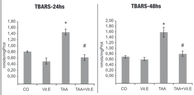
LPO results for the 24h and 48h groups can be seen in Figure 1, where a signiicant increase (P<0.001) of LPO was found in TAA groups as compared to the others. As Vit. E is administered at 24 h, LPO is reduced as compared to the TAA group (P<0.001), and the same was found at 48h. This is clear evidence that Vit. E ad-ministration in the TAA group reduces LPO at both studied times.
Antioxidant enzymes
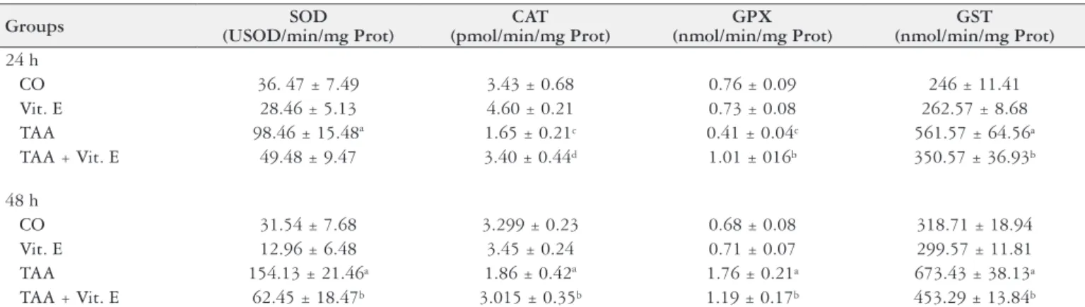
SOD, CAT, GPx and GST activities were assessed at 24h and 48h (Table 2). GST and SOD activities were signiicantly increased in the TAA groups as compared to control groups at 24h and 48h, and decreased in the TAA+Vit.E groups as compared to the TAA groups at both times. Cat activity was signiicantly reduced in the TAA groups as compared to controls at 24h and 48 h, and increased in the TAA+Vit.E groups as compared to TAA at both studied times. GPx showed a different pattern across the studied times. At 24h GPx activity was reduced in the TAA as compared to controls and the TAA+Vit.E group had increased GPx activity as compared to the TAA group. At 48h, GPx activity was increased in the TAA group as compared to controls and was reduced in the TAA+Vit.E group as compared to TAA.
FIGURE 1. Evaluation of lipoperoxidation through TBARS (nmol/
mg prot) in the different experimental groups assessed at 24h and 48h. CO: control group, Vit. E: Vitamin E group; TAA: Thiocetamide group; TAA+Vit.E: Thiocetamide with Vit. E group. Data are expressed as mean ± standard error of mean. n=7 per group. *Signiicant increase (P<0.05) of TAA vs Controls and #signiicant decrease (P<0.05) of TAA+Vit.E vs TAA.
1,80 1,60 1,40 1,20 1,00 0,80 0,60 0,40 0,20 0,00 -TBARS-24hs
CO Vit.E TAA TAA+Vit.E
n mo ls/ mg Pro t * # TBARS-48hs 2,00 1,80 1,60 1,40 1,20 1,00 0,80 0,60 0,40 0,20 0,00
-CO Vit.E TAA TAA+Vit.E
Nitrites e Nitrates
Figure 2 shows that nitric oxide metabolites increased signii-cantly (P<0.001) in the TAA groups at both times and were signii-cantly reduced in the Vitamin E-supplemented groups (P<0.001) at both studied times.
TABLE 2. Evaluation of antioxidant enzymes SOD, CAT, GPX and GST activities in the different studied groups and two studied times: 24h and 48h
Groups SOD
(USOD/min/mg Prot)
CAT (pmol/min/mg Prot)
GPX (nmol/min/mg Prot)
GST (nmol/min/mg Prot)
24 h
CO 36. 47 ± 7.49 3.43 ± 0.68 0.76 ± 0.09 246 ± 11.41
Vit. E 28.46 ± 5.13 4.60 ± 0.21 0.73 ± 0.08 262.57 ± 8.68
TAA 98.46 ± 15.48ª 1.65 ± 0.21c 0.41 ± 0.04c 561.57 ± 64.56a
TAA + Vit. E 49.48 ± 9.47 3.40 ± 0.44d 1.01 ± 016b 350.57 ± 36.93b
48 h
CO 31.54 ± 7.68 3.299 ± 0.23 0.68 ± 0.08 318.71 ± 18.94
Vit. E 12.96 ± 6.48 3.45 ± 0.24 0.71 ± 0.07 299.57 ± 11.81
TAA 154.13 ± 21.46a 1.86 ± 0.42ª 1.76 ± 0.21a 673.43 ± 38.13a
TAA + Vit. E 62.45 ± 18.47b 3.015 ± 0.35b 1.19 ± 0.17b 453.29 ± 13.84b
CO: control group, Vit. E: Vitamin E group; TAA: Thiocetamide group; TAA+Vit.E: Thiocetamide with Vit. E group. Data are expressed as mean ± standard error of mean. n=7 per group.
a= signiicant increase (P<0.001) TAA vs Controls; b= signiicant decrease (P<0.001) of TAA+Vit.E vs TAA; c= signiicant increase (P<0.05) of TAA vs Control groups, and d= signiicant decrease (P<0.01) TAA+Vit.E vs TAA.
Histology
Histological evaluation of liver tissue was performed by he-matoxylin & eosin (HE) staining at 200x magniication. As can be seen in Figures 3A and B (24h) and 4-A and B (48h), the CO and Vit.E groups showed normal hepatic parenchyma, with clear-cut hepatocyte cordons with well-preserved cytoplasms and nuclei. In the histology of the TAA group, shown in Figures 3C (24h) and 4C (48h), there is evidence of hepatocyte cordon disorganization, inlammatory iniltrate, and necrosis. In the TAA+Vit.E group, seen in Figures 3D (24h) and 4D (48h), note the preservation of hepatocyte cordons and decreased incidence of necrosis and inlam-matory iniltrate in response to Vitamin E.
FIGURE 2. Evaluation of nitric oxide metabolites - nitrites and
ni-trates (mmol/L) in the different experimental groups at 24h and 48h. CO: control group, Vit. E: Vitamin E group; TAA: Thiocetamide group; TAA+Vit.E: Thiocetamide with Vit. E group. Data are ex pressed as mean ± standard error of mean. n=7 per group. a = signiicant incre-ase (P<0.001) TAA vs Controls; b = signiicant decrease (P<0.001) of TAA+Vit.E vs TAA.
FIGURE 3. Photomicrograph of hepatic tissue by hematoxylin & eosin
(HE) stain in the different experimental groups at 24h. Magniication: 200X. A = CO group, B = Vit. E group, C = TAA group and D = TAA+Vit.E group. Arrow 1: Necrosis. Arrow 2: Inlammatory iniltrate.
FIGURE 4. Photomicrograph of hepatic tissue by hematoxylin & eosin
(HE) stain in the different experimental groups at 48h. Magniication: 200X. A = CO group, B = Vit. E group, C = TAA group and D = TAA+Vit.E group. Arrow 1: Necrosis. Arrow 2: Inlammatory iniltrate.
CO Vit.E TAA TAA+Vit.E 60
50
40
30
20
10
0
-mmo
l/
L
NO2/ NO3 - 24h
*
#
NO2/ NO3 - 48h
CO Vit.E TAA TAA+Vit.E 80
60
40
20
0
-mmo
l/
L #
DISCUSSION
Severe SALF is an acute hepatic disorder with varied, poorly known physiopathogeny and established physiopathology with variants concerning etiology. Knowledge of these mechanisms is important to determine the pathways of the lesion and any potential effects of external intervention. The utilization of ex-perimental models and antioxidant drugs comes to contribute to its understanding. Drugs that can delay the progress of the disease or reorganize the hepatic parenchyma are potential therapeutic agents for the disease.
In the present study, vitamin E was found to attenuate oxida-tive stress and inlammation in TAA-induced SALF. Enzymes AST and ALT were increased by about 1200% in the groups receiving TAA as compared to controls at 24h and 48h. Other authors also obtained similar results by inducing toxicity in rat livers through administration of AA doses (350 mg/kg). In Vitamin E-treated groups, there was a signiicant decrease of about 75% in AST and ALT as compared to TAA groups at 24h and 48h(7,28). In another study also observed a reduction of these parameters in an experimental model of toxicity, treating with lavonoid quercitin(8).
In LPO evaluation, TBARS presented an increase of 59.5% and 94.1% at 24h and 48h, respectively, in the TAA group. In the Vit. E group, however, values were similar to those of the control group and significantly reduced as compared to TAA groups, with reductions of 51.3% and 42.4% at 24h and 48h, respectively. Such reduction demonstrates decreased LPO in the hepatic tissue due to the antioxidant action of vitamin E administered to these animals, which can be evidenced by histology, where one notes reorganization of the hepatic tissue and reduction of the inlam-matory iniltrate and necrosis(2,17,28).
Administration of Vitamin E signiicantly reduced SOD ac-tivity by 49.7% at 24h and 59.48% at 48h, suggesting decreased production of O2•, and consequent decreased LPO, as demon-strated by histology and TBARS levels. In a study investigating the preventive effect of quercetin in animals with TAA-induced SALF, de Oliveira et al., showed signiicantly increased SOD activity from the administration of the lavonoid. Other works have reported decreased SOD activity from the administration of other antioxidants(8,22,23).
CAT activity was significantly reduced in TAA groups as compared to CO groups by 51.9% and 43.6% at 24h and 48 h, respectively. TAA acts directly on the formation of O2•-, which act on membrane lipids and form hydroperoxide lipids, thus accounting for reduced CAT activity. The use of Vit. E signiicantly restored CAT activity at 24h and 48h, with increases of 106% and 61.8%, respectively, making it similar to CO groups and reinforcing the antioxidant effect of Vitamin E. Another study found signiicant reduction of CAT activity in rats, evaluating the damage caused by oxidative stress induced by high-carbohydrate diet in a model of hepatic encephalopathy(1).
The behavior of GPx activity varied across the studied times. While in the TAA group there was a reduction of 46% in GPx activity in relation to the CO group at 24h, there was an increase of it (158.8%) at 48h, possibly explained by the reestablishment of the hepatic tissue. As compared to the TAA group, GPx activity in the TAA+Vit.E group presented an increase of 146.3% at 24h and a decrease of 32.3% at 48h, which can be explained by the decreased tissue aggression due to the protective effect of Vit. E,
evidenced by lower TBARS levels and histology. The increase in GPx appears to accompany the accumulation of organic peroxides formed by LPO caused by TAA. Similar results were reported in a work using antioxidant quercetin and glutamine(7,31).
The high toxicity of TAA in hepatocytes triggered an increase in enzyme GST, through its detoxifying protective action, with increases of 128.2% and 111% at 24h and 48h, respectively. In the Vitamin E-treated group, however, the reduction was of 37.5% and 32.6% at 24h e 48h, respectively, owing to the preservation of hepatocytes by the exogenous antioxidant, as evidenced again by decreased TBARS levels and liver histology. Our indings cor-roborate those of other authors who evaluated GST activity in experimental model, using TAA as hepatotoxic inducer(31).
On histological evaluation of hepatic tissue using hematoxylin & eosin (HE) staining at a magniication of 200x, CO and Vit. E groups showed hepatic cells arranged in hepatocyte cordons around capillaries with aspects of normal distribution, and visible nuclei without inlammatory iniltrates or necrosis. On the other hand, the hepatic tissue in the TAA group displayed disorganization of hepatocytes, inlammatory iniltrates, and necrosis, which accounts for increased TBARS levels at both times. Histological evaluation of the Vit.E-treated group showed a reorganization of hepatocyte cordons and smaller incidence of inlammatory iniltrate or necrosis. These evidences coincide with signiicant reduction of TBARS and lower lipoperoxidation in the hepatic parenchyma at both studied times, promoted by Vitamin E antioxidant action. Another author also demonstrated Vitamin E antioxidant role when administered at 200 mg/Kg/ day for 3 days(28).
The rise of metabolites NO2 and NO3 indicates an increase in NO production, which participates closely in the inlammatory and destructive process of the hepatic tissue(9). Animals in TAA groups presented increase of NO metabolites that associate with the su-peroxide anion to form peroxinitrite, which is extremely damaging to hepatocytes. This increase was of 476% (24h) and 646% (48h) in relation to control groups, while Vitamin E-supplemented groups showed a reduction of 36.2% (24h) and 29.8% (48h) as compared to groups receiving TAA, which in a way can be explained by the improvement of the hepatic parenchyma. Similar indings were reported by other authors who assessed nitric oxide levels(7, 22).
From the indings it is possible to see that the Vitamin E group at 48h, despite having received two additional 100 mg/Kg doses of Vitamin E, presents values that are close to those obtained at 24h, suggesting that the dose of 100 mg/Kg administered intra-peritoneally thirty minutes after TAA administration was already suficient to protect the liver against the oxidative stress triggered by the drug.
CONCLUSION
consideration apoptosis markers, inlammatory route, and DNA damage caused by oxidative stress will be helpful in elucidating these mechanisms.
ACKNOWLEDGMENT
Dr. Cláudio Augusto Marroni and Mariana do Couto Soares, by aid during the conduct of the study. Conselho Nacional de Desenvolvimento Cientíico e Tecnológico (CNPq), Coordenação
de Aperfeiçoamento de Pessoal de Nível Superior (CAPES) and Fundação de Amparo à Pesquisa do Estado do Rio Grande do Sul (FAPERGS) for inancial support.
Authors’ contributions
Miguel FM and Schemitt EG performed all of the research work; Colares JR and Hartmann RM were in charge of the experi-mental model; Morgan-Martins MI and Marroni NP designed the research work and the article review.
Miguel FM, Schemitt EG, Colares JR, Hartmann RM, Morgan-Martins MI, Marroni NP. Ação da vitamina E sobre a insuiciência hepática aguda grave experimental. Arq Gastroenterol. 2017;54(2):123-9.
RESUMO – Contexto – A Insuiciência Hepática Aguda Grave (IHAG) é uma síndrome clínica potencialmente fatal, na qual ocorre necrose dos
hepatócitos, perda da arquitetura hepática e deterioração de suas funções. Dentre as principais causas da IHAG está a hepatotoxicidade decorrente de agentes químicos, que lesam os hepatócitos e acarretam aumento das espécies reativas de oxigênio. A vitamina E tem alta atividade antioxidante biológica e é amplamente distribuída nos tecidos. Objetivo – Avaliar o efeito antioxidante da Vitamina E no modelo de IHAG. Métodos – Foram utilizados 56 ratos, com peso médio de 300 g, divididos em oito grupos, quatro grupos avaliados em 24 horas e quatro em 48 horas após a indução: grupo controle (CO); Vitamina E (Vit.E); Tioacetamida (TAA) e Tioacetamida + Vitamina E (TAA+Vit.E). Os ratos foram submetidos a injeções de tioacetamida, na dose de 400 mg/Kg de peso i.p., no início do experimento e, posteriormente, após 8 horas. A vit E (100 mg//Kg i.p.) foi administrada 30 minutos após a segunda dose de tioacetamida. Os animais do tempo 48 horas receberam mais duas doses de vit. E (24h e 36h). Transcorridas 24 ou 48 horas após a administração da primeira dose de TAA, os animais foram pesados, anestesiados e o sangue retirado para a avaliação da integridade hepática através das enzimas Aspartatoaminotransferase (AST) e Alanina aminotransferase (ALT). O tecido hepático foi retirado para avaliação da lipoperoxidação através da técnica de TBARS, atividade das enzimas antioxidantes SOD, CAT, GPx, e GST, avaliação de NO2/NO3 e avaliação histológica pela coloração de hematoxilina e eosina nos dois tempos. Os resultados foram expressos como média ± erro padrão e a análise estatística utilizada foi ANOVA, seguido de teste de Student-Newman-Keuls, considerado signiicativo P<0,05. Resultados – Após o tratamento com a vit. E, observamos uma redução nas enzimas de integridade hepática AST (U/L) (101,32±19,45 em 24h e 97,85±29,65 em 48h) relacionado ao grupo TAA (469,56±20,69 em 24h e 598,23±55,45 em 48h) e ALT (U/L) (76,59±8,56 em 24h e 68,47±6,49 em 48h) comparado ao grupo TAA (312,21±10,23 em 24h e 359,15±17,58 em 48h). Houve uma redução da LPO (nmol/mg Prot), no grupo TAA+Vit.E (0,77±0,07 em 24h e 0,95±0,08 em 48h) com-parado ao grupo TAA (1,50±0,07 em 24h e 1,65±0,16 em 48h). A SOD (USOD/min/mg Prot) diminuiu no grupo TAA+Vit.E (49,48±9,47 em 24h e 62,45±18,47 em 48h) relacionado ao grupo TAA (98,46±15,48 em 24h e 154,13±21,46 em 48h), assim como a GST (nmol/min/mg Prot) no grupo TAA+Vit.E (350,57±36,93 em 24h e 453,29±13,84 em 48h) comparado ao grupo TAA (561,57±64,56 em 24h e 673,43±38,13 em 48h). Houve aumento da CAT (pmol/min/mg Prot) no grupo TAA+Vit.E (3,40±0,44 em 24h e 3,01±0,35 em 48h) em relação ao grupo TAA (1,65±0,21 em 24h e 1,86±0,42 em 48h). A GPx (nmol/min/mg Prot) aumentou em 24h no grupo TAA+Vit.E (1,01±0,16) comparado ao grupo TAA (0,41±0,04) e diminuiu em 48h (1,19±0,17) em relação ao grupo TAA (1,76±0,21). Veriicou-se redução nos níveis de NO2/NO3 (mmol/L) no grupo TAA+Vit.E (31,47±4,26 em 24h e 38,93±5,20 em 48h) em relação ao grupo TAA (49,37±5,12 em 24h e 53,53±5,97 em 48h). A avaliação histopatológica mostrou diminuição da lesão hepática (necrose e inlamação) em ambas os tempos estudados. Conclusão – Estes resultados sugerem que a vitamina E foi capaz de proteger o fígado de lesões causadas por tioacetamida.
DESCRITORES – Falência hepática aguda. Tioacetamida. Estresse oxidativo. Antioxidantes.
REFERENCES
1. Alzoubi KH, Khabour O F, Salah H A, Hasan Z, et al. Vitamin E prevents high-fat high –carbohidrates diet-induced memory impairment: The role of oxidative stress. Physiol Behav. 2013;119:72-8.
2. Bhatti FR, Mehmood A, Wajid N, Rauf M, Khan S, Riazuddin S, et al. Vitamin E protects chondrocytes against hydrogen peroxide-induced oxidative stress in vitro. Inlamm Res. 2013;62:781-9.
3. Boveris A, Chance B. The mitochondrial generation of hydrogen peroxide. General properties and effect of hyperbaric oxygen. Biochem J. 1973;134:707-16. 4. Buege JA, Aust SD. Microsomal lipid peroxidation. Methods Enzymol.
1978;52:302-9.
5. Chieli E, Malvadi G. Role of Cyt P-450 dependent and FAA containing mono oxygenases in the bioactivation of thioacetamide, thiobezamide and their sulo-phoxides. Biochem Pharmacol. 1985; 34:395-1.
6. Chu CJ, Hsiao CC, Wang TF, Chan CY, Lee FY, Chang FY, et al. Prostacyclin inhibition by indomethacin aggravates hepatic damage and encephalopathy in rats with thioacetamide-induced fulminant hepatic failure. World J Gastroenterol. 2005;11:232-6.
7. David C, Rodrigues G, Bona S, Meurer L, González-Gallego J, Tuñón M, et al. Role of quercetin in preventing thioacetamide-induced liver injury in rats. Toxicol Pathol. 2011;39:949-57.
8. De Oliveira CR, Ceolin J, De Oliveira RR, Schemitt EG, Colares JR, Bauermann LF, et al. Effects of quercetin on polychlorinated biphenyls-induced liver injury in rats. Nutr Hosp. 2014; 1141-8.
9. Diesen DL, Kuo PC. Nitric oxide and redox regulation in the liver: Part I. Gen-eral considerations and redox biology in hepatitis. J Surg Res. 2010;162:95-109. 10. Diez-Fernandez C, Sanz N, Cascales M. Changes in glucose-6-phosphate dehy-drogenase and malic enzyme gene expression in acute hepatic injury induced by thioacetamide. Biochem Pharmacol. 1996;51:1159-63.
11. Fedstein AE. Novel insights into the pathophysiology of nonalcoholic fatty liver disease. Semin Liver Dis. 2010;30:391-401.
12. Fitzhugh OG, Nelson AA. Liver tumours in rats fed thiourea or thioacetamide. Science, 1948; 108: 626–28.
14. Fontana L, Moreira E, Torres MI, Fernández MI, Ríos A, De Medina FS, et al. Serum amino acid changes in rats with thioacetamide-induced liver cirrhosis. Toxicology.1996;106:197-206.
15. Halliwell B, Gutteridge J. Free Radicals in Biology and Medicine. 4th edn. New York: Oxford University Press Inc; 2007.
16. Herrera E, Barbas C. Vitamin E: action, metabolism and perspectives. J Physiol Biochem. 2001; 57:43-56.
17. Jeon YH. Vitamin E, an antioxidant, as a possible therapeutic agent for treating pain. Korean J Pain. 2013;3:314-5.
18. Lee, WM. Acute liver failure in the United States. Semin. Liver Dis. 2003;23:217-26. 19. Llesuy SF, Milei J, Molina H, Boveris A, Milei S. Comparison of lipid peroxi-dation and myocardial damage induced by adriamycin and 4’- epiadriamycin in mice. Tumori. 1985,71:241-9.
20. Mangipudy RS, Chanda S, Mehendale HM. Tissue repair response as a function of dose in thioacetamide hepatotoxicity. Environ Health Perspect. 1995;103:260-7. 21. Mannervick BGC, Guthenberg C. Glutationa Transferase (human placenta).
Methods Enzymol. 1981;77:231-5.
22. Marques C, Licks F, Zattoni I, Borges B, De Souza LER, Marroni, CA, et al. Antioxidant properties of glutamine and its role in VEGF-Akt pathways in portal hypertension gastropathy. World J Gastroenterol. 2013;19:4464-74.
23. Marques C, Mauriz JL, Simonetto D, Marroni CA, Tuñon MJ, González-Gallego J, Marrón NP. Glutamine Prevents gastric oxidative stress in an animal model of portal hypertension gastropathy. Ann Hepatol. 2011;4:531-39.
24. Mattos AA, Dantas-Corrêa EB. Tratado de Hepatologia. 4 Ed. Rubio: São Paulo, 2010. 1.024 p.
25. Misra HP, Fridovich I. The role of superoxide anion in the autoxidation of epinephrine and a simple assay for superoxide dismutase. J Biol Chem. 1972;247:3170-5.
26. Morgan-Martins MI. Estresse Oxidativo e Antioxidantes. In Marroni, NP. Radicais Livres no processo saúde-doença: da bancada à clínica/ Norma Possa Marroni, Maria Isabel Morgan-Martins, Marilene Porawski (organizadoras) – Curitiba, PR: CRV, 2012.
27. Muñoz RS, Maroja JL, Vasconcelos FR, Melo J. Avaliação cognitiva breve para detecção de encefalopatia em pacientes com doença hepática crônica. GE Port J Gastroenterol. 2013;20:255-60.
28. Mustafa HN, Awdan AS, Hegazy GA. Protective role of antioxidants on thioac-etamide-induced acute hepatic encephalopathy: Biochemical and Ultrastructural study. Tissue and Cell. 2013;45:350-62.
29. Neal RA. Halpert, J. Toxicology of thionosulfer compounds. Annu Rev Phar-macol Toxicol. 1982;22:321-9.
30. Niki E. Role of vitamin E as a lipid-soluble peroxil radical scavenger: in vitro and in vivo edidence. Free Radical Biol Med. 2014;66:3-12.