1
Arquivos Brasileiros de Cardiologia - Volume 84, Nº 2, Fevereiro 2005
Original Article
Maximal Functional Capacity, Ejection Fraction,
and Functional Class in Chagas Cardiomyopathy.
Are these Indices Related?
Charles Mady, Vera Maria Cury Salemi, Barbara Maria Ianni, Felix José Alvarez Ramires,
Edmundo Arteaga
São Paulo, SP - Brazil
Chagas’ disease is an infectious disease caused by the proto-zoan Trypanosoma cruzi, which may result in a dilated form of cardiomyopathy, usually complicated by congestive heart failure1.
The extent of myocardial involvement and its functional conse-quences are the fundamental determinants of the natural history2,3.
Similarly to that in other forms of heart failure 4-7, mortality
in-creases as myocardial function deteriorates 3. In addition, left
ventricular ejection fraction (LVEF) and maximal functional capacity (VO2max) have both been shown to be related to a poor long-term survival in Chagas’ disease patients 3.
Surprisingly, however, some studies on other forms of dilated cardiomyopathy have demonstrated that, despite having a severe left ventricular impairment, a rather normal functional capacity may be found 8,9. In addition, some reports show a lack of
connec-tion between LVEF and VO2max in these patients 10-13. These
findings indicate that the traditional indices widely used for the evaluation of cardiac function and functional capacity are not entirely appropriate for all patients with heart failure. Contrasting with these reports, it has been previously shown in patients with Cha-gas’ cardiomyopathy that VO2max progressively worsens as left ventricular chamber dimension increases 14,15.
Therefore, in view of these conflicting data, the aim of the present study was to investigate the potential association of VO2max and LVEF with functional class in Chagas’ disease.
Methods
All procedures were carried out in accordance with institutional guidelines, and the protocol was approved by our institutional review committee. Before entering the study, patients underwent complete clinical and biochemical evaluations. Those with a sug-gestive history of ischemic heart disease, valvular heart disease, diabetes, alcohol abuse, renal failure (serum creatinine >1.4 mg/ dL), anemia (serum hemoglobin <12 g/L), and systemic hyper-tension were excluded from the study. The diagnosis of Chagas’ disease was suggested by positive epidemiological evidence, clinical history, and physical and laboratory findings indicating a dilated form of cardiomyopathy concurrently with typical ECG changes 16
and ultimately confirmed by specific serological tests (Machado-Guerrero and immunofluorescence).
One hundred four male patients aged 40.3±9.0 years (range, 18 to 65) with congestive heart failure due to Chagas’ cardio-myopathy were selected and gave informed consent to enter the study. All patients were admitted to the hospital and received
Instituto do Coração (InCor) do Hospital das Clínicas - FMUSP Mailing address: Charles Mady - InCor - Av. Dr. Enéas de Carvalho Aguiar, 44 - Cep 05403-000 - São Paulo, SP - Brazil
E-mail: [email protected] / [email protected] Received for publication: 01/08/04
Accepted for publication: 08/13/04
Objective
Left ventricular ejection fraction (LVEF) and maximal func-tional capacity (VO2max) have both been shown to be related to a poor long-term survival in Chagas’ disease patients. The aim of this study was to estimate the potential association of VO2max, LVEF, and NYHA functional class in patients with Chagas’ disease cardiomyopathy.
Methods
One hundred four male patients, aged 40.3±9.0 years (range, 18 to 65), with a definite diagnosis of Chagas disease cardiomyopathy were studied. LVEF and VO2max were both clas-sified into 3 degrees: LVEF ≤0.30, 0.30< LVEF ≤0.50, and LVEF> 0.50 and VO2max ≤10, 10<VO2max ≤20, and VO2max >20 ml.kg-1.min-1, respectively.
Results
Thirty-one patients (29.8%) were in NYHA functional class II, 41 (39.4%) in functional class III, and 32 (30.8%) in functional class IV. The corresponding values of VO2max and LVEF for func-tional classes II, III, and IV were 21.5±4.0 ml.kg-1.min-1, 18.3±5.8 ml.kg-1.min-1, and 14.7±4.9 ml.kg-1.min-1 and 0.50±0.6, 0.35±0.9, and 0.29±0.7, respectively. LVEF =
≤0.30 and VO2max = ≤10 ml.kg
-1.min-1 were found in the
ma-jority of patients in NYHA functional class IV. Conversely, pa-tients in functional class II were mostly those with LVEF >0.50 as well as VO2max >20 ml.kg-1.min-1.
Conclusions
A convincingly good association exists between NYHA func-tional class, funcfunc-tional capacity and LVEF in patients with Chagas’ disease cardiomyopathy. These data may be helpful in the ma-nagement of heart failure in Chagas’ disease patients.
Key words
2
Arquivos Brasileiros de Cardiologia - Volume 84, Nº 2, Fevereiro 2005
Maximal Functional Capacity, Ejection Fraction, and Functional Class in Chagas Cardiomyopathy. Are these Indices Related? traditional measures aimed at heart failure treatment, which
in-cluded bed rest, digitalis, diuretics, and vasodilator drugs, including angiotensin-converting enzyme inhibitors. Heart failure status was classified at the time of entering the study according to the criteria established by the New York Heart Association (NYHA) 17.
All patients after having been stabilized for heart failure symp-toms underwent ergospirometry for VO2max (ml.kg-1.min-1)
deter-mination using a Metabolic Cart (MGC CAD/NET 1988, Minnea-polis, MN, USA). Respiratory variables were obtained under con-ditions of standard temperature, pressure, and humidity (StPD) with appropriate correction factors being used. All patients under-went a maximal exercise test based on the modified Naughton protocol 18 using monitoring treadmill equipment (Quinton, model
Q65, Seattle, Washington) with variable speed and inclination. We used conventional echocardiographic equipment. The echo-cardiographic parameters were determined, based on the recom-mendations of the American Society of Echocardiography 19.
Maximal functional capacity and cardiac function were cate-gorized as follows: VO2max ≤10, 10< VO2max ≤20, VO2max >20 ml.kg-1.min-1, and LVEF≤0.30, 30< LVEF ≤0.50, LVEF >0.50,
respectively.
Patients stratified according to functional class were compared regarding VO2max and LVEF by using the analysis of variance and the Duncan’s multiple comparison method 20.
The potential associations of NYHA functional class with VO2max and LVEF were tested using the analysis of corresponden-ce21, which is a multivariate exploratory method of categorical
data for converting a contingency table into a 2-dimensional chart. In this chart, a distinctive vector for each category represents each one of the 3 variables studied. The potential association of a pair of categories studied is expressed by a small angle between their corresponding vectors.
All statistical calculations were performed using SAS software (Statistical Analysis System). The significance level was established at 0.05. Continuous variables are expressed as mean value ± SD.
Results
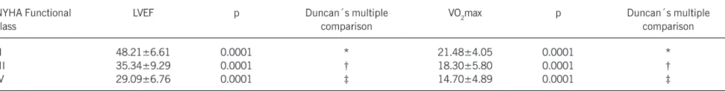
Thirty-one patients aged 41.0±7.9 years were in NYHA func-tional class II, 41 patients aged 41.2±8.6 years were in funcfunc-tional class III, and 32 patients aged 39.8±8.1 years were in functional class IV. LVEF and VO2max values according to patient’s functional class are depicted in table I. As can been observed, each one of the 3 categories of heart failure studied showed statistically different LVEF and VO2max.
Figure 1 shows the chart representing the results of the analysis of correspondence. We can observe that the variables LVEF and VO2max were both associated with NYHA functional class. As a matter of fact, the vectors corresponding to LVEF >0.50, VO2max >20 ml.kg-1.min-1, and functional class II are within the same
qua-drant, and the angles between each 2 of them are all small. A similar association is evident for the following 2 groups of categories analyzed: 0.30< LVEF ≤0.50, 10< VO2max ≤20 ml.kg-1.min-1 and
functional class III; and LVEF ≤0.30, VO2max <10 ml.kg-1.min-1,
and functional class IV.
In short, the results of this analysis are in agreement with that of the analysis of variance showing an evident association of
functional class, LVEF, and VO2max, indicating that the more ad-vanced the functional class, the more impaired both functional capacity and cardiac function are.
Discussion
In general, it is agreed that functional exercise capacity is significantly related to the severity of heart failure. This represents the basis of the universally adopted subjective clinical classification of heart failure recommended by the New York Heart Association17.
This observation is based on the well-documented relationship found between functional capacity and cardiac performance, ma-nifested by a positive correlation between VO2max and cardiac output 22,23. However, some investigators were unable to
demons-trate such a relationship between LVEF and VO2max 10-13. Moreover,
exercise capacity was found to be quite normal in a significant number of patients with severe left ventricular dysfunction 8,9.
Therefore, the premise that functional exercise capacity represents a good index for evaluating and predicting cardiac performance has been questioned. We speculate that these contradictory findings are the result of improperly classifying cardiac function merely as normal or abnormal in some of these studies. To avoid such a shortcoming, in the present study patients were classified ac-cording to 3 different categories of LVEF and maximal exercise capacity. Accordingly, the 37 patients with VO2max values over 20 ml.kg-1.min-1 were actually those who were the least
sympto-matic and with the highest values of LVEF.
Currently, data concerning functional exercise capacity and cardiac function provide the essential information for making con-fident clinical and surgical therapeutic decisions in patients with heart failure. Recently, it has been shown that clinical history alone is far from being a reliable method for determining functional capacity in patients with ischemic heart disease and other forms of cardiomyopathy 24. The heterogeneity of the population studied
might have represented an important drawback in that investiga-tion. It has long been recognized, for instance, that survival curves may differ according to the underlying cause of heart disease 25. It
is also well known that men compared with women with equiva-lent heart failure status show a higher exercise capacity 26. In the
present study, only male patients, all with Chagas’ cardiomyopathy, were included. However, a previous study showed that in male patients individuals with Chagas’ disease were at higher risk of progression of the disease, defined as death or the presence of a new ECG or left ventricular echocardiographic abnormality on follow-up, making these patients a subgroup with increasing risk of adverse events 27.
The association between the impairment of VO2max and poor long-term prognosis has been debated 28,29. A strikingly limited
short-term survival has been demonstrated in patients with a marked exercise limitation, corresponding to a VO2max below 10 ml.kg-1.min-1 28. When patients with such a degree of exercise
capacity impairment are excluded from the analysis, exercise capacity provides little prognostic information 29. In contrast, it
has been previously demonstrated that VO2max is in fact a good predictor of survival in patients with heart failure due to Chagas’ disease 3. The prognostic significance of VO
2max remained strong
3
Arquivos Brasileiros de Cardiologia - Volume 84, Nº 2, Fevereiro 2005
Maximal Functional Capacity, Ejection Fraction, and Functional Class in Chagas Cardiomyopathy. Are these Indices Related?
previous study showed that the functional capacity of patients in the initial phase of chronic Chagas’ disease is higher than that of patients in an advanced phase, and that this functional capacity decreases following the myocardial functional impairment 30.
LVEF is also recognized as a valuable index for estimating outcome in patients with heart failure 31. Yet again, as previously
pointed out for both functional class and exercise capacity, con-flicting data exist regarding the ability of LVEF in predicting survival.
Dimension 2
Dimension 1
EF>0.50 0.30<EF≤≤≤≤≤0.50 EF≤≤≤≤≤0.30 VO2max>20 10<VO2max≤≤≤≤≤20 VO2max≤≤≤≤≤10 Functional class II Functional class III Functional class IV
Fig. 1 - Two-dimensional graphic representation of the association found among NYHA functional class, left ventricular ejection fraction (LVEF), and maximal exercise functional capacity (VO2max.).
Table I - Comparison of left ventricular ejection fraction (LVEF) and maximal functional capacity (VO2max) among the three different groups of NYHA functional class
NYHA Functional LVEF p Duncan´s multiple VO2max p Duncan´s multiple
class comparison comparison
II 48.21±6.61 0.0001 * 21.48±4.05 0.0001 *
III 35.34±9.29 0.0001 † 18.30±5.80 0.0001 †
IV 29.09±6.76 0.0001 ‡ 14.70±4.89 0.0001 ‡
Groups with different symbols (*, †, ‡) are statistically different.
Although LVEF has been reported not to differ between survivors and nonsurvivors 32, a significant and positive relation between
LVEF and long-term survival has been documented 33,34. Once
again, it became apparent from previous reported data that LVEF might have in fact a marked influence on survival of patients with heart failure due to Chagas’ cardiomyopathy 3. The present results,
demonstrating a significant association of LVEF with both functional class and exercise capacity, underscores one of the major mecha-nisms involved in the predictive value of LVEF on survival, namely impairment in functional capacity.
In the present study, we were able to demonstrate that NYHA functional class is related to functional exercise capacity and cardiac function in patients with Chagas’ cardiomyopathy. Accordingly, the more advanced the functional class, the more severely both exercise capacity and cardiac performance are affected. Based on these results, we may, thus, infer that the traditional indices currently used for clinical evaluation of patients with heart failure are very consistent for patients with Chagas’ disease.
We are convinced that these data will be helpful in the mana-gement of heart failure in the setting of Chagas’ disease. The consistency of the traditional indices used for cardiac evaluation herein demonstrated certainly will help making more appropriate therapeutic decisions for each individual patient, taking into ac-count, firstly, the severity of the disease and, ultimately, bearing in mind the new modalities of treatment.
1. Laranja FS, Dias E, Nóbrega G, Miranda A. Chagas’ disease: a clinical, epidemio-logical and pathologic study. Circulation 1956;14:1035-60.
2. Mady C, Ianni BM, Arteaga E, et al. Relation between interstitial myocardial col-lagen and the degree of clinical impairment in Chagas’ disease. Am J Cardiol 1999;84:354-6.
3. Mady C, Cardoso RHA, Pereira-Barretto AC, da Luz PL, Bellotti G, Pileggi F. Sur-vival and predictors of surSur-vival in patients with congestive heart failure due to Chagas’ cardiomyopathy. Circulation 1994;90:3098-102.
4. Franciosa JA, Wilen M, Ziesche S, Cohn JN. Survival in men with severe chronic left ventricular failure due to either coronary heart disease or idiopathic dilated car-diomyopathy. Am J Cardiol 1983;51:831-6.
5. Wilson JR, Schwartz JS, St John Sutton M, et al. Prognosis in severe heart failure: relation to hemodynamic measurements and ventricular ectopic activity. J Am Coll Cardiol 1983;2:403-10.
6. Fuster V, Gersh BJ, Giuliani ER, Tajik AJ, Brandenburg RO, Frye RL. The natural history of idiopathic dilated cardiomyopathy. Am J Cardiol 1981;47:525-31. 7. Massie BM, Conway M. Survival of patients with congestive heart failure: past,
present and future prospects. Circulation 1987;75(suppl. IV):IV-11 (abstr). 8. Benge W, Litchfield RL, Marcus ML. Exercise capacity in patients with severe left
ventricular dysfunction. Circulation 1980;61:955-9.
9. Litchfield RL, Kerber RE, Benge J, et al. Normal exercise capacity in patients with severe left ventricular dysfunction: compensatory mechanisms. Circulation 1982;66:129-34.
10. Franciosa JA, Park M, Levine TB. Lack of correlation between exercise capacity and indexes of resting left ventricular performance in heart failure. Am J Cardiol 1981;47:33-9.
References
11. Marantz PR, Tobin JN, Wassertheil-Smoller S, et al. The relationship between left ventricular systolic function and congestive heart failure diagnosed by clinical cri-teria. Circulation 1988;77:607-12.
12. Cohen-Solal A, Caviezell B. Cardiopulmonary exercise testing in chronic heart fai-lure. Heart Failure 1994;10:46-57.
13. Francis GS, Goldsmith SR, Cohn JN. Relationship of exercise capacity to resting left ventricular performance and basal plasma norepinephrine levels in patients with congestive heart failure. Am Heart J 1982;104:725-31.
14. Mady C, Yazbek Jr. P, Pereira-Barretto AC, et al. Estudo da capacidade funcional máxima pela ergoespirometria em pacientes portadores da doença de Chagas. Arq Bras Cardiol 1986;47:201-5.
15. Mady C, Ianni BM, Arteaga E, Salemi VMC, Frimm C de C. Maximal functional ca-pacity in patients with Chagas’ cardiomyopathy without congestive heart failure. J Cardiac Failure 2.000;6:220-4.
16. Macedo V. Inquérito eletrocardiográfico nacional para Doença de Chagas. Rev Soc Bras Med Trop 1993;26:12-13.
17. Criteria Committee of the New York Heart Association. Nomenclature and criteria for diagnosis of the heart and great vessels. 6.ed. Boston, Little Brown 1964:1-23. 18. Naughton J, Balke B, Nagle F. Refinement in methods of evaluation and physical
conditionning before and after myocardial infarction. Am J Cardiol 1964;14:837-43. 19. Sahn DJ, De Maria A, Kisslo J, Weymann A. The Committee on M-Mode Standar-dization of The American Society of Echocardiography. Recommendations regar-ding quantitation in M-mode echocardiography: results of a survey of echocardio-graphic measurements. Circulation1978;58:1072-83.
4
Arquivos Brasileiros de Cardiologia - Volume 84, Nº 2, Fevereiro 2005
Maximal Functional Capacity, Ejection Fraction, and Functional Class in Chagas Cardiomyopathy. Are these Indices Related?
21. Greenacre MJ, Hastie T. The geometric interpretatiom of correspondence analysis. JASA 1987;82:437-47.
22. Weber KT, Kinasewitz GT, Janicki JS, Fishman AP. Oxygen utilization and ventila-tion during exercise in patients with chronic cardiac failure. Circulaventila-tion 1982;65: 1213-23.
23. Higginbotham MB, Morris KG, Conn EH, Coleman RE, Cobb FR. Determinants of variable exercise performance among patients with severe left ventricular dysfunc-tion. Am J Cardiol 1983;51:52-60.
24. Wilson JR, Hanamanthu S, Chomsky DB, Davis SF. Relationship between exertio-nal symptoms and functioexertio-nal capacity in patients with heart failure. J Am Coll Cardiol 1999;33:1943-7.
25. Massie BM, Ports T, Chatterjee K, et al. Long-term vasodilator therapy for heart fai-lure: clinical response and its relationship to hemodynamic measurements. Circu-lation 1981;63:269-78.
26. Higginbotham MB, Morris KG, Coleman RE, Cobb FR. Sex-related differences in the normal cardiac response to upright exercise. Circulation 1984;70:357-66. 27. Basquiera AL, Sembaj A, Aguerri AM, et al. Risk progression to chronic Chagas
car-diomyopathy: influence of male sex and of parasitaemia detected by polymerase chain reaction. Heart 2003;89:1186-90.
28. Szlachcic J, Massie BM, Kramer BL, Topic N, Tubau J. Correlates and prognostic implication of exercise capacity in chronic congestive heart failure. Am J Cardiol 1985;55:1037-42.
29. Franciosa JA. Exercise testing in chronic congestive heart failure: Am J Cardiol 1984;53:1447-50.
30. Oliveira FP, Pedrosa RC, Giannella-Neto A. Gas exchange during exercise in different evo-lutional stages of chronic Chagas’ heart disease. Arq Bras Cardiol 2000;75: 481-98.
31. Nelson GR, Cohn PF, Gorlin R. Prognosis in medically-treated coronary artery di-sease. Influence of ejection fraction compared to other parameters. Circulation 1975;52:408-12.
32. Unverferth DV, Magorien RD, Moeschberger ML, Baker PB, Fetters JK, Leier CV. Factors influencing the one-year mortality of dilated cardiomyopathy. Am J Cardiol 1984;54:147-52.
33. Burggraf GW, Parker JO. Prognosis in coronary artery disease: Angiographic, he-modynamic and clinical factors. Circulation 1975;51:146-56.
34. Schwarz F, Mall G, Zebe H, et al. Determinants of survival in patients with conges-tive cardiomyopathy: quantitaconges-tive morphologic findings and left ventricular hemo-dynamics. Circulation 1984;70:923-8.
35. Bestetti RB. Predictors of unfavourable prognosis in chronic Chagas’ disease. Trop Med Int Health 2001;6:476-83.
36. Bestetti RB, Dalbo CRM, Freitas OC, Teno LAC, Castilho OT, Oliveira JSM. Nonin-vasive predictors of mortality for patients with Chagas’ heart disease: a multiva-riate stepwise logistic regression study. Cardiology 1994;84:261-7.
37. Bestetti RB, Dalbo CRM, Arruda CA, Correia-Filho D, Freitas OC. Predictors of sudden cardiac death for patients with Chagas’ disease: a hospital-derived cohort study. Cardiology 1996;87:481-7.
38. Bestetti RB, Rossi MA. A rationale approach for mortality risk stratification in Chagas’ heart disease. Int J Cardiol 1997;58:199-209.
39. Carrasco HA, Parada H, Guerrero L, Duque M, Duran D, Molina C. Prognostic im-plications of clinical, electrocardiographic and hemodynamic findings in chronic Chagas’disease. Int J Cardiol 1994;43:27-38.