BACTEREMIA IN CIRRHOTIC
PATIENTS SUBMITTED TO
ENDOSCOPIC BAND LIGATION OF
ESOPHAGEAL VARICES
Eduardo Balzano
MAULAZ
1, Angelo Alves de
MATTOS
1, Júlio
PEREIRA-LIMA
1and Judite
DIETZ
2ABSTRACT – Background – Endoscopic procedures can develop bacteremia. Patients with chronic liver disease are more predisposed to undergo bacteremia and infections because they are immunocompromised. Aims – The purpose of this study was to determine the incidence of bacteremia in cirrhotics submitted to endoscopic variceal ligation. Methods – Three groups of 40 patients each were studied. One group was made up of patients with cirrhosis who were submitted to ligation, a second group was composed of cirrhotics who underwent esophagogastroduodenoscopy only, and a third group was composed of patients without liver disease who underwent esophagogastroduodenoscopy. Blood was sampled from all patients for culture, both in aerobic and in anaerobic mediums, immediately before endoscopy and at 5 and 30 minutes after its completion. Results – Blood culture was positive in 6 samples. In 4 of these, the bacteria (Staphylococcus hominis hominis, Staphylococcus auricularis, Acinetobacter lwoffii, and coagulase-negative staphylococcus) were isolated before the endoscopic procedure and thus were considered as contamination. In the ligation group, a streptococcus of the viridans group was isolated 5 minutes after the procedure, and in the cirrhosis without ligation group, a Staphylococcus epidermidis was isolated at 30 minutes. None of the patients showed clinical evidence of infection. Conclusions – The bacteremia incidence in cirrhotic patients submitted to variceal ligation was 2.5%, showing no difference from the control groups.
HEADINGS – Bacteremia. Esophageal and gastric varices. Esophagoscopy. Ligation. Liver cirrhosis.
AR
TI
GO
O
RI
GI
NA
L
/ O
RI
GI
NA
L
AR
TI
CL
1
Department of Gastroenterology and Hepatology of the Porto Alegre School of Medical Sciences (FFFCMPA), Santa Casa Hospital and 2Nossa Senhora da Conceição Hospital, Porto Alegre, RS, Brazil.
Address for correspondence: Dr. Angelo A. de Mattos – Rua Cel. Aurélio Bittencourt, 35/ 201 – 90430-080 – Porto Alegre, RS, Brazil. E-mail: [email protected]
INTRODUCTION
The incidence of bacterial infection in hospitalized cirrhotic patients is higher than in other patient populations(19), ranging from 33% to 61%(41). The most frequent infections are urinary tract infections (12%-29%), spontaneous bacterial peritonitis (SBP) (7%-23%), respiratory infections (6%-10%), skin infections (2%-11%), and bacteremia (4%-9%)(28, 29, 37, 41). Bacterial infections account for up to 38% of deaths in patients with chronic liver disease, both directly or indirectly(10, 42).
The frequent occurrence of bacterial infections in cirrhotic patients(37, 42) probably results from def icient defense mechanisms. Accordingly, systemic alterations have been described such as complement deficiency(3, 17, 33, 57), alterations in immunoglobulins levels(1, 3, 48, 57), defects in opsonization activity as well as in serum bactericide activity(1, 48, 57), a
decrease in the phagocytic activity of the reticuloendothelial system(37), and neutrophilic dysfunction(14, 15, 35).
Transitory bacteremia is a frequent event and may even occur following a minor trauma; it seems to accrue from the formation of solution of continuity in the epithelial surfaces which shelter a great number of bacteria(43). Most bacteremias do not cause symptoms and have no clinical significance. However, they become important in immunodef icient patients(37) and in those at risk for the development of infectious endocarditis(13, 26).
Endoscopic variceal ligation (EVL) is a first line therapy for variceal bleeding and bleeding prophylaxis(52). Six prospective studies(7, 21, 24, 25, 40, 55) have evaluated blood cultures after EVL and showed a post-EVL bacteremia incidence ranging from 0% to 16%, yet one of them found a discrepant incidence of 25%(21).
Due to the paucity of studies on this topic, the variable incidence reported, and the growing utilization of elastic band ligation of esophageal varices in patients with cirrhosis, we deemed it important to evaluate this population of patients regarding the risk of bacteremia, since its presence could mean a change in the management of these patients, specifically regarding antibiotic prophylaxis.
Therefore, the present study aimed to determine the incidence of bacteremia in patients with cirrhosis submitted to EVL.
PATIENTS AND METHODS
From January 1999 to January 2001, we prospectively enrolled three groups of patients: a) cirrhotics submitted to esophago-gastroduodenoscopy followed by EVL, b) cirrhotics submitted to esophagogastroduodenoscopy only (EGD), and c) patients with no liver disease submitted to EGD without biopsy (controls).
Cirrhosis was confirmed by liver biopsy and/or by clinical, laboratory, and ultrasonographic data in all cases. For the control group, the criterion for inclusion was absence of alterations in “liver function tests” (aminotransferases, bilirubins, alkaline phosphatase, prothrombin time and albumin) and markers of hepatitis B and C.
Patients in the EGD group underwent EGD for varices detection and those in the EVL group underwent the procedure electively, i.e., not in the bleeding episode. All patients in the control group presented dyspeptic symptoms as the indication of endoscopy.
Exclusion criteria for all groups were as follows: a) evidence of infection, detected by skin examination, urinalysis and urine culture, blood count (above 10,000 leukocytes/mm3), and chest X-rays. Also excluded were those patients with ascites who presented polymor-phonuclear leukocytes counts over 250 cells/mm3 in their peritoneal fluid and/or bacterial growth in culture examination; b) fever (axillary temperature higher than or equal to 37.1oC) within 48 hours before endoscopy; c) use of antimicrobials within 14 days before endoscopy; d) previous report of cardiopathy that formally indicated the use of antibiotic prophylaxis; e) insertion of probes, tubes, or catheters within 6 hours before endoscopy; f) previous knowledge that the patient had some neoplasia or was HIV positive; and use of immunosuppressors and/or corticosteroids.
Among the chronic liver disease patients excluded, 21 were from the ligation group and 32 were from the EGD group. Of these, 52 were on antibiotic therapy (32 as prophylaxis for SBP, 12 for treatment of urinary tract infection, and 8 for SBP treatment) and one was using an immunosuppressor for autoimmune hepatitis.
After screening patients according to the exclusion criteria, 40 patients were included in each group. Cirrhotics were classified by Child’s criteria modif ied by Pugh(37) and the technique used for esophageal variceal ligation was the same as described by STIEGMANN et al.(56), yet no overtube was used.
Venous blood was collected immediately before endoscopy and at 5 and 30 minutes after its completion. Skin antisepsis was performed with 70o GL ethanol and samples were obtained from separated punctures of the vena mediana. All blood samples were collected by one of the authors (EBM) using throwaway gloves. Twenty mL of blood was collected in each sample: 10 mL for the aerobic and 10 mL for the anaerobic analysis. Blood was transferred to the culture flasks immediately after sampling, with particular attention given to replacing the original needle with a new one and disinfecting the flask top with 70o GL ethanol.
For blood culture mediums, we used the BACTEC® system: aerobic medium (BACTEC Plus Aerobic/F®) and the anaerobic medium (BACTEC Plus Anaerobic/F®) manufactured by Becton Dickinson and Company, USA. Cultures were immediately incubated at 35oC in the BACTEC 9240® equipment (Becton Dickinson and Company, USA), where they remained under continuous shaking for up to 4 days.
When the reading of the BACTEC 9240® system indicated the presence of bacteria, the bacterioscopic examination was performed using Gram’s stain and the blood was introduced in a Petri dish with chocolate-agar, which was kept in an incubator. Upon colonies formation, microorganisms identification was done by automation through MicroScan WalkAway-96® (Dade MicroScan Inc., USA).
Hospitalized patients were checked for the presence of fever or any symptom or sign of infection within 7 days after endoscopy, and in case they were discharged before completion of one week, they were booked to be evaluated on an outpatient basis. For the outpatients included in the study, an appointment was made for the 7th day after the examination to check for any infection manifestation.
Informed consent was obtained from the patients or their relatives before they were included in the study, which was approved by the Ethics Committee of our Institution.
The statistical package for the social sciences (SPSS®) was used for statistical analysis of the data. ANOVA with Post Hoc (Tukey) test was used for comparing means, and the Chi square test – with Yates correction as necessary – or Fisher’s exact test was used for comparing proportions. The significance level assumed was 5%.
RESULTS
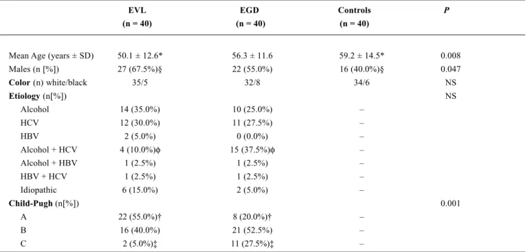
When comparing the two groups of cirrhotics (EVL and EGD) regarding etiology of liver disease, no significant difference was found between them, except for the category of alcohol associated with HCV. On the other hand, when the role of alcohol as an etiologic agent was assessed, it was present in 19 cases (47.5%) in the EVL group and in 26 (65%) in the EGD group (NS).
Regarding the Child-Pugh classification, overall comparison of the groups showed a significant difference, which also occurred between classes A and C, as shown in Table 1. As Child-Pugh A patients were separately analyzed from Child B or C cases, the following results were observed 22 (55.0%) and 18 (45.0%) in the EVL group for Child A and B/C patients, respectively, and 8 (20.0%) and 32 (80.0%) in the EGD group, respectively (P = 0.001).
The mean number of elastic bands used was 3.6 per session, varying from one to eight.
As each of the 120 patients was punctured three times, 360 samples resulting in 720 flasks for blood cultures were obtained (360 aerobic and 360 anaerobic cultures).
Only six blood cultures became positive (Table 2), all of them from different patients. Four of these were detected before the procedure: three in the EVL group (Staphylococcus hominis hominis, Acinetobacter lwoffii and coagulase-negative Staphylococcus) and one in the control group (Staphylococcus auricularis). At 5 minutes post-procedure, only one culture became positive; the germ, a Streptococcus of the viridans group, was detected in a patient of the EVL group, Child-Pugh A, who received three rubber bands. Staphylococcus epidermidis was isolated at 30 minutes after endoscopy in a Child-Pugh B patient of the EGD group. No endoscopic biopsy was performed in the cases which presented a positive blood culture.
If we consider only blood cultures that became positive after the endoscopic procedure, a bacteremia incidence of 2.5% is obtained for the EVL group, with no statistical difference in relation to control groups (P > 0.05). No evidence of infection or hyperthermia was detected in the patients evaluated 7 days after the procedure.
TABLE 1 – Characteristics of the groups
EVL EGD Controls P
(n = 40) (n = 40) (n = 40)
Mean Age (years ± SD) 50.1 ± 12.6* 56.3 ± 11.6 59.2 ± 14.5* 0.008 Males (n [%]) 27 (67.5%)§ 22 (55.0%) 16 (40.0%)§ 0.047
Color (n) white/black 35/5 32/8 34/6 NS
Etiology (n[%]) NS
Alcohol 14 (35.0%) 10 (25.0%) – HCV 12 (30.0%) 11 (27.5%) – HBV 2 (5.0%) 0 (0.0%) – Alcohol + HCV 4 (10.0%)φ 15 (37.5%)φ – Alcohol + HBV 1 (2.5%) 1 (2.5%) – HBV + HCV 1 (2.5%) 1 (2.5%) – Idiopathic 6 (15.0%) 2 (5.0%) –
Child-Pugh (n[%]) 0.001
A 22 (55.0%)† 8 (20.0%)† – B 16 (40.0%) 21 (52.5%) – C 2 (5.0%)‡ 11 (27.5%)‡ –
SD = standard deviation; HCV = hepatitis C virus; HBV = hepatitis B virus; NS = not significant * The P value between these groups is 0.007
§ The P value between these groups is 0.013
DISCUSSION
A search in literature reveals no studies about the incidence of bacteremia following upper digestive endoscopy in cirrhotics. However, when endoscopy is followed by more invasive procedures such as esophageal variceal sclerotherapy (EVS), a variable incidence of bacteremia is reported(7, 9, 11, 12, 20, 21, 25, 40, 45, 50). Considering that in recent years EVL has replaced EVS in the treatment of esophageal varices, and that only a few studies have analyzed the presence of bacteremia after EVL(21, 24, 25, 40, 55), this study aimed at investigating the incidence of bacteremia in cirrhotics submitted to such therapeutic procedure.
Even though the mean age and sex of our patients were statistically different between the groups, there is no evidence that males or females are more prone to suffer from bacteremia(36). However, bacteremia is indeed more common in the elderly(18). Here, no difference in mean age was found between the EVL and its control, the EGD group. Thus, immunodeficient populations, who could be more susceptible to the effect of age, were similar.
In view to the fact that bacterial infections are more common in cirrhotics of alcoholic origin(42, 54), the groups of patients with chronic liver disease were stratified according to the presence of alcohol in its etiology. The results obtained revealed no difference in the frequency of alcoholic etiology in the chronic liver disease groups, thus avoiding a possible bias in patient selection.
Four types of bacteria were detected before the endoscopic procedure, all of them in separate patients. One of them was isolated in the control group (Staphylococcus auricularis) and the other three in the EVL group (Staphylococcus hominis hominis, Acinetobacter lwoffii and an negative Staphylococcus). This
coagulase-negative Staphylococcus was not thoroughly identif ied in the MicroScan WalkAway-96®. Both Staphylococcus auricularis and Staphylococcus hominis hominis are coagulase-negative staphylococci. Bacteria of this group, when isolated, are generally interpreted as contamination, since only a minority of these bacteria represent true bacteremia(56) and both are naturally found in the human skin and oropharynx(47). Acinetobacter lwoffii is a bacterium belonging to the acinetobacter genus, which colonize up to 25% of the skin of healthy adults(2) and is the gram-negative organism more commonly isolated from the skin of health professionals in hospital settings(22). The isolation of this type of bacteria has already been reported after diagnostic endoscopic procedures(30, 47), and is also considered as contamination.
We infere that these bacteria that were detected pre-procedure resulted from contamination, since they are normally present in the human skin; they were not isolated after the endoscopic procedure, and their carriers showed no clinical evidence of bacteremia or infection immediately after or one week after endoscopy.
It should be stressed that, despite all the aseptic measures taken to collect blood, a 1%-5% rate of blood cultures are observed to result false-positive, i.e., contamination(4). One possible explanation is that nowadays culture mediums are very sensitive. In the present study specifically, culture mediums were used that have resins in their composition, which diminish the action of certain antibiotics and affect phagocytosis by inhibiting lysozyme action and complement formation, thus increasing the retrieval of many microorganisms(4). SMITH et al.(49) found that the mediums containing such resins are significantly superior to standard mediums (without resin) regarding sensitivity, since a higher number of microorganisms were isolated from resin-containing mediums than from the standard ones, when
TABLE 2 – Positive blood cultures
EVL EGD Controls
BC pre-endoscopy 3 0 1
(Staphylococccus hominis h.) (Staphylococcus auricularis) (Acinetobacter lwoffii)
(Staphylococcus coagul.-neg.)
BC at 5 min 1 0 0 (Streptococcus viridans)
BC at 30 min 0 1 0 (Staphylococcus epidermidis)
the same blood was simultaneously inoculated into both culture mediums.
After EVL, there was bacterial growth in one case, at 5 minutes after endoscopy completion. The isolated bacterium, a Streptococcus of the viridans group, was not detected at the culture of the 30th minute. The viridans group includes several strains of streptococci, most of which are part of the normal microbiota of the upper respiratory tract and of the oropharynx(16). They are observed in 30%-40% of the cases of infectious endocarditis(39), but are also responsible for transient bacteremias, i.e., those that do not result in infection. These bacteria are observed after endoscopic procedures such as EGD(5, 31, 46, 51), esophageal dilation(51), endoscopic retrograde cholangiopancreatography (ERCP), endoscopic mucosal resection(23), EVS(12, 40, 46, 50, 52) and EVL(24). The explanation for bacteremia would be that, as the endoscope moves along the oropharynx it can injure the mucosa, thus favoring bacteremia(12, 46). In the case of EVL, in view of the device attached to the endoscope tip, and particularly due to the pressure exerted by the elastic band on the mucosa, it is understood that a greater trauma is likely to occur, thus increasing the likelihood of bacteremia.
In the present study, the bacteremia observed was transient, as a microorganism could not be isolated from the culture performed later. Moreover, the bacteremia had no clinical impact, as the patient remained asymptomatic. The incidence of 2.5% of bacteremia following EVL found in this study is in line with published reports(7, 24, 25, 40, 55).
We also found that among 40 cirrhotic patients submitted to diagnostic EGD, just one (2,5%) positive blood culture was obtained. The microorganism detected was Staphylococcus epidermidis, which is a coagulase-negative staphylococcus similar to those isolated prior to the endoscopic procedure as described above. Thus, this germ may have arisen from contamination of the sample. In addition, another reinforcing finding for this view is the favorable clinical outcome shown by the patient. Other authors, however, have previously described this group of bacteria determining transient bacteremia following EGD(27), esophageal dilation(34), ERCP(32), EVS(7, 11, 20, 21, 25), and EVL(21, 24, 55), yet without causing infectious sequelae. It is therefore hard to infer the probable entryway for S. epidermidis in this patient, as it may have been introduced by the endoscope or may be contamination of the blood sample during its collection. A definite stand cannot be taken in this case.
Some authors have demonstrated that transient bacteremia and infections secondary to it are more often found in patients with severe liver disease(8, 21, 25). It has even been suggested that liver dysfunction
is an independent risk factor for bacteremia after EVS or EVL(6, 40), yet this claim is not supported by all(46). The present study cannot support this issue because of the limited number of cases for comparison, as only one patient showed positive blood culture after EVL, and nevertheless he was of the Child-Pugh A class. In the other positive blood culture obtained here, although the possibility of contamination was not ruled out, it occurred in a Child-Pugh B patient after the performance of EGD.
Even if we had evaluated more cases, however, the analysis would be impaired because there was a significant difference between the Child-Pugh categories of the groups. This difference is explained by the fact that we assessed a consecutive population of patients with cirrhosis and also because indication of EVL is disputable in Child-Pugh C patients(44). Thus, it would be hard to avoid this selection bias. Moreover, an attempt to pair the cases according to the Child-Pugh classification would extend case collection for too long.
A review of the studies relating EVL to bacteremia(7, 21, 24, 25, 40, 55) revealed that the mean number of bands per session was around three, except for one study reporting a mean of eight(25). ROHR et al.(40) reported that their two patients with positive blood cultures following EVL received three bands each, an information that is not provided by other authors. All these studies used a single-shot band ligator. We report herein a mean of 3.6 bands per session and the only patient showing post-EVL bacteremia also received three bands from a single-shot band ligator.
In this study, none of the 80 cirrhotics submitted to diagnostic or therapeutic EGD, including those who presented a positive blood culture after the endoscopic procedures developed clinical features of bacterial infection in the follow-up. LIN et al.(24) diagnosed bacterial peritonitis due to Escherichia coli in two patients (3%) following EVL, though the blood cultures were negative. One case of pneumonia (2%) without isolated germ was reported by STIEGMANN et al.(53). LO et al.(25) observed one event of sustained bacteremia associated with fever and one case of SBP (3.3%) following EVL, yet they do not clarify whether the germ detected in the ascitic fluid was the same as the one isolated from blood. We therefore find that infectious sequelae following EVL, if any, are very rare.
Maulaz EB, Mattos AA, Pereira-Lima J, Dietz J. Bacteremia em pacientes cirróticos submetidos a ligadura elástica endoscópica de varizes esofágicas. Arq Gastroenterol 2003;40(3):166-172.
RESUMO – Racional – Os procedimentos endoscópicos são passíveis de favorecerem o desenvolvimento de bacteremia. Por serem imunodeprimidos, os hepatopatas crônicos estão mais predispostos a essa complicação e, conseqüentemente, a infecções. Objetivo – Determinar a incidência de bacteremia em pacientes cirróticos submetidos a ligadura elástica endoscópica de varizes esofágicas. Pacientes e métodos – Foram estudados prospectivamente 120 pacientes divididos em três grupos: um grupo composto por pacientes cirróticos submetidos a ligadura elástica endoscópica, o segundo grupo composto por pacientes cirróticos submetidos a esofagogastroduodenoscopia exclusivamente, e o terceiro grupo de pacientes sem doença hepática submetidos a esofagogastroduodenoscopia. Amostra sangüínea para cultura em meios aeróbico e anaeróbico foi coletada desses pacientes imediatamente antes do procedimento endoscópico e após 5 e 30 minutos de seu término. Resultados – Hemocultura positiva foi obtida em seis amostras. Em quatro destas, a bactéria (Staphylococcus hominis hominis, Staphylococcus auricularis, Acinetobacter lwoffii, e Staphylococcus coagulase-negativo) foi isolada antes do procedimento endoscópico. No grupo submetido a ligadura elástica, foi isolado Streptococcus do grupo viridans 5 minutos após o procedimento endoscópico, e no grupo de pacientes cirróticos não submetidos a ligadura elástica Staphylococcus epidermidis foi isolado após 30 minutos. Nenhum dos pacientes estudados apresentou sinais de infecção. Conclusões – A incidência de bacteremia em pacientes cirróticos submetidos a ligadura elástica endoscópica de varizes esofágicas foi de 2,5%, não diferindo da incidência encontrada nos grupos-controle.
DESCRITORES – Bacteremia. Varizes esofágicas e gástricas. Esofagoscopia. Ligadura. Cirrose hepática.
REFERENCES
1. Akalin HE, Lateli Y, Telatar H. Bactericidal and opsonic activity of ascitic fluid from cirrhotic and noncirrhotic patients. J Infect Dis 1983;147:1011-7. 2. Al-Khoja MS, Darell JH. The skin as the source of Acinetobacter and Moraxella
species occurring in blood cultures. J Clin Pathol 1979;32:497-9.
3. Almeida JFC, Mattos AA. Comportamento dos mecanismos da defesa imunológica em pacientes com hepatopatia crônica descompensada. Rev AMRIGS 1997;41:13-9.
4. Aronson MD, Bor DH. Blood cultures. Ann Intern Med 1987;106:246-53. 5. Baltch AL, Buhac I, Agrawal A, O’Connor P, Bram M, Malatino E. Bacteremia
after upper gastrointestinal endoscopy. Arch Intern Med 1977;137:594-7. 6. Barsoum MS, Mooro HAW, Bolous FI, Ramzy AF, Rizk-Allah MA, Mahmoud
FI. The complications of injection sclerotherapy of bleeding oesophageal varices. Br J Surg 1982;69:79-81.
7. Berner JS, Gaing AA, Sharma R, Almenoff PL, Muhlfelder T, Korsten MA. Sequelae after esophageal variceal ligation and sclerotherapy: a prospective randomized study. Am J Gastroenterol 1994;89:852-8.
8. Blaise M, Pateron D, Trinchet JC, Levacher S, Beaugrand M, Pourriat JL. Systemic antibiotic therapy prevents bacterial infection in cirrhotic patients with gastrointestinal hemorrhage. Hepatology 1994;20:34-8.
9. Brayko CM, Kozarek RA, Sanowski RA, Testa AW. Bacteremia during esophageal variceal sclerotherapy: its cause and prevention. Gastrointest Endosc 1985;31:10-2.
10. Caly WR, Strauss E. A prospective study of bacterial infections in patients with cirrhosis. J Hepatol 1993;18:353-8.
11. Camara DS, Gruber M, Barde CJ, Montes M, Caruana JA, Chung RS. Transient bacteremia following endoscopic injection sclerotherapy of esophageal varices. Arch Intern Med 1983;143:1350-2.
12. Cohen LB, Korsten MA, Scherl EJ, Velez ME, Fisse RD, Arons EJ. Bacteremia after endoscopic injection sclerosis. Gastrointest Endosc 1983;29:198-200. 13. Dajani AS, Taubert KA, Wilson W, Bolger AF, Bayer A, Ferrieri P, Gewitz MH,
Shulman ST, Nouri S, Newburger JW, Hutto C, Pallasch TJ, Gage TW, Levison ME, Peter G, Zuccaro G. Prevention of bacterial endocarditis: recommendations by the American Heart Association. JAMA 1997;277:1794-801.
14. Demeo AN, Andersen BR. Defective chemotaxis associated with a serum inhibitor in cirrhotic patients. N Engl J Med 1972;286:735-40.
15. Fiuza C, Salcedo M, Clemente G, Tellado JM. In vivo neutrophil dysfunction in cirrhotic patients with advanced liver disease. J Infect Dis 2000;182:526-33. 16. Frandsen EVG, Pedrazzol V, Kilian M. Ecology of viridans streptococci in the
oral cavity and pharynx. Oral Microbiol Immunol 1991;6:129-33.
17. Fromkes JJ, Thomas FB, Mekhjian HS, Evans M. Antimicrobial activity of human ascitic fluid. Gastroenterology 1977;73:668-72.
18. Grandsen WR, Eykyn SJ, Phillips I. Septicaemia in the newborn and elderly. J Antimicrob Chemother 1994;34:101-19.
19. Guarner C, Runyon BA. Macrophage function in cirrhosis and the risk of bacterial infection. Hepatology 1995;22:367-9.
20. Ho H, Zuckerman MJ, Wassem C. A prospective controlled study of the risk of bacteremia in emergency sclerotherapy of esophageal varices. Gastroenterology 1991;101:1642-8.
21. Kulkarni SG, Parikh SS, Dhawan PS, Chachad H, Jambavalikar MB, Koppikar GV, Kalro RH. High frequency of bacteremia with endoscopic treatment of esophageal varices in advanced cirrhosis. Indian J Gastroenterol 1999;18:143-5. 22. Larson EL. Persistent carriage of gram-negative bacteria on hands. Am J Infect
Control 1981;9:112-9.
23. Lee TH, Hsueh PR, Yeh WC, Wang HP, Wang TH, Lin JT. Low frequency of bacteremia after endoscopic mucosal resection. Gastrointest Endosc 2000;52:223-5. 24. Lin OS, Wu SS, Chen YY, Soon MS. Bacterial peritonitis after elective endoscopic
variceal ligation: a prospective study. Am J Gastroenterol 2000;95:214-7. 25. Lo GH, Lai KH, Shen MT, Chang CF. A comparison of the incidence of transient
bacteremia and infectious sequelae after sclerotherapy and rubber band ligation of bleeding esophageal varices. Gastrointest Endosc 1994;40:675-9. 26. Mani V, Cartwright K, Dooley J, Swarbrick E, Fairclough P, Oakley C. Antibiotic
prophylaxis in gastrointestinal endoscopy: a report by a working party for the British Society of Gastroenterology Endoscopy Committee. Endoscopy 1997;29:114-9.
27. Mellow MH, Lewis RJ. Endoscopy-related bacteremia. Incidence of positive blood cultures after endoscopy of upper gastrointestinal tract. Arch Intern Med 1976;136:667-9.
28. Navasa M, Rimola A, Rodés J. Bacterial infections in liver disease. Semin Liver Dis 1997;17:323-33.
29. Navasa M, Fernández J, Rodés J. Bacterial infections in liver cirrhosis. Ital J Gastroenterol Hepatol 1999;31:616-25.
30. Norfleet RG, Mitchell PD, Mulholland DD, Philo J. Does bacteremia follow upper gastrointestinal endoscopy? Am J Gastroenterol 1981;76:420-2. 31. O’Connor HJ, Hamilton I, Lincoln C, Maxwell S, Axon ATR. Bacteraemia with
upper gastrointestinal endoscopy. A reappraisal. Endoscopy 1983;15:21-3. 32. Parker HW, Geenen JE, Bjork JT, Stewart ET. A prospective analysis of fever
and bacteremia following ERCP. Gastrointest Endosc 1979;25:102-3. 33. Potter BJ, Trueman AM, Jones EA. Serum complement in chronic liver disease.
Gut 1973;14:451-6.
34. Raines DR, Branche WC, Anderson DL, Boyce Jr HW. The occurrence of bacteremia after esophageal dilation. Gastrointest Endosc 1975;22:86-7. 35. Rajkovic IA, Williams R. Abnormalities of neutrophil phagocytosis, intracellular
killing and metabolic activity in alcoholic cirrhosis and hepatitis. Hepatology 1986;6:252-62.
36. Reimer LG, Wilson ML, Weinstein MP. Update on detection of bacteremia and fungemia. Clin Microbiol Rev 1997;10:444-65.
37. Rimola A, Bory F, Planas R, Xaubet A, Bruguera M, Rodés J. Infecciones bacterianas agudas en la cirrosis hepática. Gastroenterol Hepatol 1981;4:453-8. 38. Rimola A, Soto R, Bory F, Arroyo V, Piera C, Rodes J. Reticuloendothelial system phagocytic activity in cirrhosis and its relation to bacterial infections and prognosis. Hepatology 1984;4:53-8.
39. Roberts RB, Krieger AG, Schiller NL. Viridans streptococci endocarditis: the role of various species, including pyridoxal-dependent streptococci. Rev Infect Dis 1979;1:955-65.
41. Romero FP, Vierna SE. Infecciones bacterianas en el cirrótico. Gastroenterol Hepatol 1988;11:67-75.
42. Rosa H, Silvério AO, Perini RF, Arruda CB. Bacterial infection in cirrhotic patients and its relationship with alcohol. Am J Gastroenterol 2000;95:1290-3. 43. Rosebury T. In: Rosebury T, editor. Microorganisms indigenous to man. New
York: McGraw-Hill; 1962.
44. Sakai P, Maluf-Filho F, Melo JM, Ishioka S. Is endoscopic band ligation of esophageal varices contraindicated in Child-Pugh C patients? Endoscopy 1994;26:511-2.
45. Sauerbruch T, Holl J, Ruckdeschel G, Förstl J, Weinzierl M. Bacteriaemia associated with endoscopic sclerotherapy of oesophageal varices. Endoscopy 1985;17:170-2.
46. Selby WS, Norton ID, Pokorny CS, Benn RAV. Bacteremia and bacterascites after endoscopic sclerotherapy for bleeding esophageal varices and prevention by intravenous cefotaxime: a randomized trial. Gastrointest Endosc 1994;40:680-4. 47. Shull HJ Jr, Greene BM, Allen SD, Dunn GD, Schenker S. Bacteremia with
upper gastrointestinal endoscopy. Ann Intern Med 1975;83:212-4.
48. Simberkoff MS, Moldover NH, Weiss G. Bactericidal and opsonic activity of cirrhotic ascites and nonascitic peritoniteal fluid. J Lab Clin Med 1978;91:831-9. 49. Smith JA, Bryce EA, Ngui-Yen JH, Roberts FJ. Comparison of BACTEC 9240 and BacT/Alert blood culture systems in an adult hospital. J Clin Microbiol 1995;33:1905-8. 50. Snady H, Korsten MA, Waye JD. The relationship of bacteremia to the length of injection needle in endoscopic variceal sclerotherapy. Gastrointest Endosc 1985;31:243-6.
51. Stephenson PM, Dorrington L, Harris OD, Rao A. Bacteraemia following oesophageal dilatation and oesophago-gastroscopy. Aust NZ J Med 1977;7:32-5. 52. Stiegmann GV, Cambre T, Sun JH. A new endoscopic elastic band ligating device.
Gastrointest Endosc 1986;32:230-3.
53. Stiegmann GV, Goff JS, Michaletz-Onody PA, Korula J, Lieberman D, Saeed ZA, Reveille RM, Sun JH, Lowenstein SR. Endoscopic sclerotherapy as compared with endoscopic ligation for bleeding esophageal varices. N Eng J Med 1992;326:1527-32.
54. Suarez C, Pajares JM. Epidemiología de las infecciones en la cirrosis hepática. Rev Clín Esp 1981;160:299-303.
55. Tseng CC, Green RM, Burke SK, Connors PJ, Carr-Locke DL. Bacteremia after endoscopic band ligation of esophageal varices. Gastrointest Endosc 1992;38:336-7.
56. Weinstein MP, Towns ML, Quartey SM, Mirrett S, Reimer LG, Parmigiani G, Reller LB. The clinical significance of positive blood cultures in the 1990s: a prospective comprehensive evaluation of the microbiology, epidemiology, and outcome of bacteremia and fungemia in adults. Clin Infect Dis 1997;24:584-602.
57. Wyke RJ. Problems of bacterial infection in patients with liver disease. Gut 1987;28:623-41.