| Journal of Clinical and Analytical Medicine 1
İnlamatuar Myoibroblastik Tümör / Inlammatory Myoibroblastic Tumor
Inlammatory Myoibroblastic Tumor of the Prostate
Prostatın İnlamatuar Myofibroblastik Tümörü
DOI: 10.4328/JCAM.1759 Received: 25.03.2013 Accepted: 24.04.2013 Publihed Online: 25.04.2013
Corresponding Author: Nazım Emrah Koçer, Başkent Üniversitesi Adana Uygulama ve Araştırma Merkezi Patoloji bölümü, Dadaloglu Mah. 39 Sok. No:6 01250, Yüreğir, Adana, Türkiye. T.: +905052730578 F.: +90 3223271276 E-Mail: [email protected]
Özet
Prostatın inlamatuar myofibroblastik tümörleri (İMT), sarkomlar ve iğsi hücre-li karsinomları khücre-linik ve histopatolojik olarak takhücre-lit edebilen nadir lezyonlardır. Bu-rada sunulan olgu, normal prostat spesifik antijen düzeyleri ve kronik yakınmala-rı olan, tıbbi tedaviye yanıtsız infravezikal tıkanma bulgulayakınmala-rı nedeni ile suprapubik prostatektomi uygulanan 63 yaşında bir hastadır. Eksizyon materyalinin histopa-tolojik incelenmesi fokal nükleer pleomorfizm, hiperkromazi gösteren, mononükle-er iltihabi infiltrasyon ve miksoid değişikliklmononükle-er içmononükle-eren, düzgün sınırlı, iğsi hücreli bir lezyonu ortaya koydu. Mitoz nadirdi. İmmunhistokimyasal çalışmada düz kas aktini ve vimentin pozitif, anaplastik lenfoma kinaz -1 fokal pozitif, S-100 ve pansitoke-ratin negatiti. Lezyon inlamatuar myofibroblastik tumor olarak tanı aldı. İMT’nin prostatın malign iğsi hücreli lezyonlarından ayrımı gereksiz ileri tedavi işlemleri-nin önüne geçilmesi için şarttır.
Anahtar Kelimeler
İnlamatuar Myofibroblastik Tümör; Prostat; Psödosarkomatöz; Fibromiksoid; Psö-dotümör
Abstract
Inlammatory myoibroblastic tumors (IMT) of the prostate are very rare lesions that may mimic sarcomas and spindle carcinomas both clinically and histopatho-logically. The case presented here is a 63-year-old patient, with normal prostate speciic antigen levels and a chronic history of complaints, who underwent to suprapubic prostatectomy due to the infravesical obstruction symptoms that are resistant to medical therapy. Histopathological examination of the excision ma-terial revealed a well demarcated spindle cell lesion with focal nuclear polymor-phism, hyperchromasia, mononuclear inlammatory iniltration and myxoid areas. Mitosis was rare. Immunohistochemically smooth muscle actin and vimentin were positive, anaplastic lymphoma kinase-1 was focal positive, S-100 and pancyto-keratin were negative. The lesion was diagnosed as an inlammatory pseudotu-mor. Diferential diagnosis of the IMT from malignant spindle cell tumors of the prostate is crucial to prevent overtreatment.
Keywords
Inlammatory Myoibroblastic Tumor; Prostate; Pseudosarcomatous; Fibromyxoid; Pseudotumor
Nazım Emrah Koçer1, Nebil Bal1, Ümit Gül2, Hakan Aydın3
1Başkent Üniversitesi Adana Uygulama ve Araştırma Merkezi Patoloji bölümü, Adana,
2Başkent Üniversitesi Adana Uygulama ve Araştırma Merkezi Üroloji bölümü, Adana, 3Başkent Üniversitesi Ankara Uygulama ve Araştırma Merkezi Patoloji bölümü, Ankara, Türkiye
| Journal of Clinical and Analytical Medicine
İnlamatuar Myoibroblastik Tümör / Inlammatory Myoibroblastic Tumor
2
Introduction
Stromal lesions of the prostate are rare and consisted of a number of benign and malignant lesions that closely resemble each other [1,2]. The diferential diagnosis, which is crucial for appropriate treatment, may be challenging. Inlammatory myo-ibroblastic tumor of the prostate (IMT; synonym: pseudosarco-matous spectrum ibromyxoid tumor of the prostate, inlamma-tory pseudotumor of the prostate) is one of these rare lesions and stands in the benign side of the stromal lesions.
Case Report
A 63-year-old male patient has admitted to the department of urology with diiculty in starting urine low and pollakiuria for 2 years. Despite the 6 months of 5 alpha reductase (Finasterid 5mg) and alpha blocker (Tamsulosin HCL 0.4mg) medication, the symptoms persisted. Prostate speciic antigen (PSA) was 1.22 ng/ml, and prostate volume was measured as 186cc in ultrasound. In uretherocystoscopia prostatic urethra was longer than normal (7cm) and prostate median lobe and lateral lobes were narrowing the urethra. Qmax, volume and residue in uro-low, performed without Foley catheter, were 12ml/sec, 75cc and 197 cc respectively. Bladder capacity, compliance, and sen-sitivity were normal in illing cystometry. There was no detru-sor hyperactivity. Pressure low study showed high pressure-low low. Digital rectal examination inding was compatible with a 1.5(+) adenoma.
The patient had no history of a previous operation or instru-mentation.
Since the patient was not an appropriate candidate for trans-urethral resection, suprapubic prostatectomy was performed with a clinical presumptive diagnosis of benign prostatic hy-perplasia.
Macroscopically the resection material was composed of two separate tissues (originally a single mass resected in two por-tions by the surgeon because of its size) with smooth surface, gray-white in color. One of the pieces was 9x4x4cm and the other was 7x5x4cm, total weight: 141gr. The cut surfaces were solid, gray-white in color and had a ibrillary appearance. Ex-tensive sampling was done.
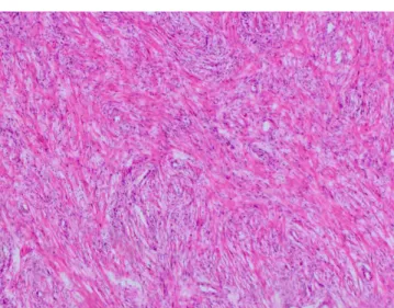
Microscopically the well demarcated lesion that was composed of spindle cells forming long, transecting bundles, was pushing, but not iniltrating the normal prostate tissue. Focal nuclear hy-perchromasia and pleomorphism were observed. Mitosis was rare. There was no necrosis or atypical mitotic igures. There were foci of mononuclear inlammatory response, including plasma cells, and myxoid degeneration (Figure 1). Vimentin, smooth muscle actin were positive, anaplastic lymphoma ki-nase -1 (ALK-1) was focal positive, S-100 and pancytokeratin were negative in immunohistochemical study. ALK positivity was conirmed with leurocein in-situ hybridization (FISH) with DAKO split signal FISH probe (Figure 2).
The case was diagnosed as inlammatory myoibroblastic tu-mor. The patient is under follow up, for 5 years and no recur-rence occurred.
Discussion
Inlammatory myoibroblastic tumors are rare lesions that can be seen in a variety of organs (e.g. lungs, heart, liver, vagina,
kidneys) [2, 3]. The ethiopathogenesis is uncertain. The term “inlammatory pseudotumor” was irst used by Umiker and Iven-son in 1954 for desricribing 4 cases in lung [2]. Compared with the respiratory system the term “inlammatory pseudotumor” is relatively new in genitourinary tract. First “reactive pseudo-sarcomatous response” in genitourinary tract was described in bladder in 1980 [4], and the irst IMT in prostate was described 4 years later by Haiz et al [5].
IMTs are spindle cell lesions that are believed to be originate from the myoibroblastic cells [6]. They may mimic malignant lesions both clinically and microscopically. In clinic, IMTs may show rapid grow and high PSA levels. Microscopically they may be confused with sarcomas and spindled carcinomas, which will lead to cystoprostatectomy and pelvic lymph node dissection. In our case the PSA levels were not suggestive of malignancy and the clinical symptoms of the patient were existing nearly for two years, without a rapid increase. The size of our lesion is larger than the previously reported IMTs of prostate [1, 7]. This slow pace of growth may explain the relatively larger size of the lesion. If the lesion showed rapid growth and caused more dramatic symptoms, the patient would have admitted to the hospital earlier and probably the lesion would be excised in an earlier phase. In microscopic examination there was focal nuclear pleomorphism and hyperchromasia. Mitosis was rare
Figure 1. Photomicrograph reveals transecting spindle cell bundles intermixed with mononuclear inlammatory iniltrate (HE x100)
| Journal of Clinical and Analytical Medicine
İnlamatuar Myoibroblastik Tümör / Inlammatory Myoibroblastic Tumor
3
and necrosis was absent. In sarcomas and spindled carcinomas, nuclear pleomorphism and hyperchromasia are more prominent with a high mitotic rate and invasive pattern. Foci of necrosis may also be present.
Postoperative spindle cell nodule (PSCN), and IMT were thought to be diferent entities, but now they are believed to be relect-ing the same lesion with some minor diferences, particularly in patient history. PSCN develops ater an instrumentation or operation, and tends to be smaller than IMT [1, 6]. Our patient had no history of a previous operation or instrumentation. Embryonal, ibroblastic, and smooth muscle nodules are the stromal nodules of benign prostatic hyperplasia (BPH) [1]. They are all well demarcated nodules and may contain pleomorphic spindle cells and hyperchromatic pleomorphic nuclei [8], like IMT. All BPH nodules are positive for S-100 [1]. In our case S-100 was negative. Although one may speculate that it may be possible that some IMT nodules were being reported as BPH nodules in the past, and the real incidence of IMT may be higher than the current data, ater nearly 30 years of its irst descrip-tion, there is still a small number of case of reports in the litera-ture regarding prostatic IMTs.
Immunohistochemical ALK-1 positivity may be seen in genito-urinary tract, most commonly in bladder, IMTs. In a study by Montgomery et al, FISH ALK results and immunohistochemical ALK results were showed to be compatible [7]. The ALK positiv-ity suggests that IMTs of genitourinary tract are true neoplastic processes rather than reactive ones. ALK positivity may not be a predictor of recurrence [7].
IMTs of prostate are rare lesions and their distinction from sar-comas and spindle cell carcinomas is crucial to prevent an over-treatment. Most IMTs of prostate are treated with transurethral resection, an interventional therapy that aims to resolve the symptoms rather than a total resection of the mass. Since IMTs are true neoplastic processes, close follow up of the patients with IMT diagnosis may be important.
References
1. Helpap B. Non-epithelial tumor-like leions of the prostate: a never ending diagnostic problem. Virchows Arch 2002;441(3):231-7.
2. Patnana M, Sevrukov AB, Elsayes KM, Viswanathan C, Lubner M, Menias CO. Inlammatory pseudotumor: the great mimicker. Am J Roentgenol 2012;198 (3):W217-27 doi: 10.2214/AJR.11.7288.
3. Atis G, Gurbuz C, Kiremit MC, Guner B, Zemheri E, Caskurlu T. Pseudosarcoma-tous ibromyxoid tumor of the prostate. ScientiicWorldJournal 2011;11:1027-30. doi: 10.1100/tsw.2011.87.
4. Roth JA. Reactive pseudosarcomatous response in urinary bladder. Urology 1980;16(6):635-7.
5. Haiz MA, Toker C, Sutula M. An atypical ibromyxoid tumor of the prostate. Cancer 1984;54(11):2500-4.
6. Bjerggaard JJ, Langkilde NC, Lundbeck F, Marcussen N. Pseudosarcomatous i-bromyxoid tumor of the prostate. Scand J Urol Nephrol 2003;37(1):85-87. 7. Montgomery EA, Shuster DD, Burkhart AL, Esteban JM, Signigroli A, Elwood L, et al. Inlammatory myoibroblastic tumors of the urinary tract: A clinicopathologic study of 46 cases, including a malignant example of inlammatory ibrosarcoma and a subset associated with high-grade urothelial carcinoma. Am J Surg Pathol 2006; 30(12):1502-12.