Lactose Intolerance in Infants with Gluten-Sensitive
Enteropathy: Frequency and Clinical Characteristics
Nedeljko Radlović1, Marija Mladenović2, Zoran Leković1, Dragana Ristić1, Momčilo Pavlović3, Zorica Stojšić4, Biljana Vuletić5, Vladimir Radlović1, Dejan Nikolić1, Jelena Djurdjević6, Milan Gajić71University Children’s Hospital, Belgrade, Serbia; 2Health Centre, Valjevo, Serbia;
3General Hospital, Subotica, Serbia;
4Institute for Pathology, School of Medicine, University of Belgrade, Belgrade, Serbia; 5Paediatric Hospital, Clinical Centre “Kragujevac”, Kragujevac, Serbia;
6Institution for Worker Protection “Railways of Serbia”, Belgrade, Serbia;
7Institute for Statistics, School of Medicine, University of Belgrade, Belgrade, Serbia
INTRODUCTION
Gastrointestinal lactose intolerance presents the most frequent nutritional disorder [1-3]. It usually develops as the result of the primary or secondary deficiency of lactase activity (hypolactasia) [2, 4]. Contrary to the primary, which presents as a developmental or genet-ically defined occurrence, the secondary form of lac-tose intolerance is caused by the damage of the small bowel mucosa [2-6]. As it disappears with the patient’s improvement this form of lactose intolerance is also called transitory. Numerous diseases followed by the morphological damage of the small bowel mucosa, such as viral enteritis, intestinal lambliasis, protein-sensitive enteropathy, severe malnutrition and oth-ers, lead to secondary lactose intolerance (SLI) [2, 6, 7]. The secondary disorder of lactose tolerance also occurs as a reaction to the use of antibiotics and gas-trointestinal prokinetics, as well as after gastroec-tomy and extensive small bowel resection [4, 8-10]. Clinical manifestation of lactose intolerance is, gen-erally speaking, most variable and depends, not only on the severity of enzymic deficit and on the degree
of its overload, but on the patient’s age and compen-satory capacity of the colon as well [4, 6, 7, 11-13].
One of the diseases that are relatively often fol-lowed by secondary hypoplasia is also gluten-sensi-tive enteropathy (GSE) [2, 4-6, 14-16]. According to the reports from the literature, in this disease a clin-ically manifest deficit of lactose activity, especially that of severe form, is relatively rare [2, 7, 14-18]. It mainly occurs in severe and neglected forms of the disease, and by its presence, it significantly contrib-utes both to the severity of diarrhoeal disorder and to the degree of undernourishment of the patient [6, 7, 16]. Having in mind all these facts and milk nutri-tional significance, it is clear that SLI presents a seri-ous problem (handicap) for children with GSE, and particularly those of the earliest age [7].
OBJECTIVE
The goal of the study was to assess the frequency of SLI in infants with GSE. In addition, we evaluated the relationship of this disorder with the duration and age
SUMMARY
Introduction Secondary lactose intolerance (SLI) belongs to the rarer manifestations of gluten-sensitive enteropathy (GSE). It occurs in more severe forms of the disease and its presence contributes significantly to the degree of its expression.
Objective The goal of the study was to determine the frequency of SLI in infants with clinically classic form of GSE, as well as its relationship with the duration, severity and age at the diagnosis of the basic disease and the degree of small bowel mu-cosa damage.
Methods The study was based on a sample of 42 infants, 30 female and 12 male, aged 7-12 months (x=9.98±1.69), with a cli-nically classic form of GSE. The diagnosis of GSE was established based on the characteristic pathohistological appearance of small bowel mucosa and clinical improvement of patients on gluten-free diet, while SLI on pathological lactose or milk tole-rance test. The assessment of basic disease severity was based on body mass divergence in relation to the standard value, as well as on Hb and serum iron levels, while the degree of small bowel mucosa damage was determined according to the mo-dified Marsh criteria.
Results SLI was verified in 8/42 or 19.05% of patients. In addition to the symptoms and clinical signs of GSE, all the patients with SLI also featured the problems characteristic of lactose tolerance disorders, i.e. watery diarrhoea, borborygmus and me-teorism occurring after milk meals. In addition, all had perianal erythema (6 with erosive changes), as well as destructive en-teropathy (5 subtotal and 3 total). The difference in the duration of the basic disease, age at diagnosis, as well as in the degree of body mass deviation from the standard value between the lactose-tolerant and lactose-intolerant infants was not found. In addition, no difference in Hb and serum iron levels or in the degree of small bowel mucosa damage was found between the two groups.
Conclusion Our findings indicate that SLI presents a relatively frequent occurrence in infants with clinically classic GSE, as well as that it occurs independently to the duration, severity and age at diagnosis of the basic disease and the degree of small bowel mucosa damage.
at GSE diagnosis, as well as with the basic clinical and lab-oratory nutritional parameters of patients, and the degree of small bowel mucosa damage.
METHODS
We retrospectively analyzed a sample of 42 infants, 30 female and 12 male, aged 7-12 months (X=9.98±1.60), with a clinically classic type of GSE, i.e. the type of the disease followed by chronic diarrhoea (>2 weeks) and disordered development. The diagnosis of GSE was based on the char-acteristic pathohistological appearance of the small bowel mucosa and clinical improvement of the patient on gluten-free diet [19]. The diagnosis was preceeded by a detailed illness history, a complete physical examination and rele-vant laboratory investigations.
All the patients with the history of watery, explosive and foamy stools after milk intake and/or the presence of perianal erythema with marked meteorism, underwent lactose tolerance test (LTT) or milk tolerance test (MTT). The confirmation of lactose intolerance was based on path-ological LTT or MTT findings, i.e. the presence of watery diarrhoea, meteorism, as well as positive Clini test find-ings (>0.5%) and a low stool Ph (<5.5) after the intake of 10% of lactose solution in the dosage of 2 g/kg body mass or 200-220 ml of highly adopted cow’s milk [6, 7, 20, 21]. None of the patients was on antibiotics, and none had gas-trointestinal infection or some other condition followed by lactose intolerance.
In addition, all the patients were investigated in detail for history data at the onset and duration of the basic disease, while during clinical examination in each a precise body length and body weight were measured and compared to the referent values for the corresponding age and gender [22]. The body length values were expressed in percen-tiles, and body weight deviations in relation to ideal val-ues in percentages. Hb and serum iron levels, as the lab-oratory parameters of the nutritional status, were deter-mined by standard methods from a blood sample taken in the morning before breakfast. The diagnostic criteria for anaemia was serum Hb level below 110 g/L, and for sideropenia serum iron concentration below 10.7 μmol/L [23-25]. Hb values ranging from 100-109 g/L indicated mild, from 70-99 g/L moderate, and below 70 g/L severe anaemia [23].
Small bowel mucosa samples were obtained by aspira-tion or endoscopic enterobiopsy. By the former method biopsy was performed from the initial part of the jeju-num or duodejeju-num, and by the latter from the postbul-bous (descending) part of the duodenum. Using aspiration enterobiopsy, we obtained two, and by endoscopic entero-biopsy three to five samples of the mucosa. Immediately after the biopsy and adequate orientation, each speci-men was stereomicroscopically analyzed in detail. After the stereomicroscopical evaluation and a precise descrip-tion, the mucosa specimens were immersed in a standard formalin solution and were then sent for a pathohisto-logical analysis. The classification of the degree of small bowel mucosa damage was made according to the modi-fied Marsh criteria, dividing it into inflammatory damage
of infiltrative (I), infiltrative-hyperplastic (II), destructive (III) and hypoplastic (IV) type [26, 27]. Depending on the degree of villous degeneration, destructive enteropathies were additionally differentiated into partial (IIIa), subto-tal (IIIb) and tosubto-tal (IIIc) [27, 28].
The difference between the lactose intolerant and tol-erant infants in the duration and age at GSE diagnosis, as well as in the degree of body weight deficit and Hb and serum iron levels were analyzed by the Student’s t-test, and in the severity of small bowel mucosa damage by the Mann-Whitney test.
RESULTS
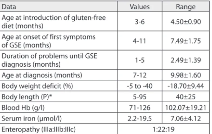
Basic data related to the whole group of patients are pre-sented on Table 1. Among the total 42 patients, 35 infants were on maternal milk; of these, only 4 were concurrently introduced to gluten containing diet. However, none of the infants was breast fed at the time of GSE diagnosis. Except for one infant whose body weight was below the low limits of the referent value for the corresponding age and gender, the remaining patients had normal longitu-dinal growth. All the infants had body weight deficit; in 19 (45.24%) it was above 20%. Anaemia was registered in 30 (71.43%) patients, of whom in 18 it was mild and in 12 moderate, while sideropenia was detected in 34 (80.95%). All the patients had enteropathy of the most severe degree, of whom in one only it was partial.
Of total 42 patients with the classic form of GSE, SLI was confirmed in 8 or 19.0%. Beside the symptoms and clinical signs of GSE, all SLI patients also had additional problems, i.e. watery diarrhoea, borborygmus and mete-orism after milk meal. In addition, all had perianal ery-thema, of whom with erosive changes in 6. None of the patients had gastrointestinal infection or any other path-ological conditions followed by lactose intolerance, and none showed allergy to cow’s milk proteins. Nutritional lactose restriction, with gluten-free diet, resulted in a rapid recovery of the patients followed by improvement in the consistency and number of stools, as well as in the loss of perianal erythema. In none of the infants, SLI concomi-tant with GSE did not last over 2-3 weeks after the intro-duction of the diet.
Table 1. Basic data in infants with GSE (n=42)
Data Values Range
Age at introduction of gluten-free
diet (months) 3-6 4.50±0.90
Age at onset of first symptoms
of GSE (months) 4-11 7.49±1.75
Duration of problems until GSE
diagnosis (months) 1-5 2.49±1.39
Age at diagnosis (months) 7-12 9.98±1.60 Body weight deficit (%) -5 to -40 -18.70±9.44
Body length (P)* 5-95 40±25
Blood Hb (g/l) 71-126 102.07±19.21
Serum iron (μmol/l) 2.2-19.5 7.06±4.12
Enteropathy (IIIa:IIIb:IIIc) 1:22:19
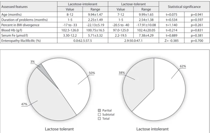
Among the features set in the study objectives, differ-ences between the lactose intolerant and lactose tolerant infants are presented on Table 2 and Figure 1. As evident, there were no significant differences between these two groups of patients, either in the basic disease duration, age at diagnosis, or in the deficit of body weight, and Hb and serum iron levels. In addition, there was also no significant difference in the severity of small bowel mucosa damage.
DISCUSSION
Lactose is the basic milk carbon hydrate in most mam-mals [29]. It consists of glucose and galactose molecules interlinked by the β-1.4 glucoside configuration. In addi-tion to energetic significance, lactose stimulates the resorp-tion of calcium, magnesium and iron, as well as the col-onization of the large bowel with Bifidobacterium and Lactobacillus bacteria [2, 30]. Lactose hydrolysis, which presents a precondition for its absorption, is promoted by lactase (β-galactosidase), a specific hydrolase that is linked with its C-terminal ending to the luminal side of the eryth-rocyte membrane in the proximal small bowel segment [2, 7, 31]. After being released, glucose and galactose mole-cules are by active co-transport with sodium transferred into the enterocyte, which then exits it to be easily diffused throughout the portal bloodstream. By phosphorization processes, transfer to uridine-diphosphate and epimeriza-tion occurring in the liver under the activity of galacto-kinase, galactose 1-phosphate uridyl transferase and uri-dine diphosphate-4-epimerase, galactose is transformed
into glucose [33, 34]. Therefore, according to the level of development lactose tolerance disorders are classified into two groups of clinical entities, of which the former occur due to hypolactase, gastrectomy or glucose and galactose malabsorption, and the latter due to the deficit of galac-tokinase, galactose 1-phosphate uridyl transferase or uri-dine diphosphate-4-epimerase [2, 35]. Except for the defi-ciency of lactase activity, other causes of lactose intoler-ance are rare [2, 35].
GSE belongs to the diseases often followed by hypolacta-sia [2, 4, 6, 14-18]. The deficiency of lactase activity occurs as the result of small bowel mucosa inflammation, i.e. the reduction of its functional surface and epithelial immatu-rity [36]. Although the changes are most prominent in the small bowel segment, where also lactase activity is highest owing to its remaining fraction and compensatory role of the colon, clinically manifest hypolactasia is relatively rare and is mostly seen in the severe forms of the disease [2, 4, 7, 14, 17, 18, 37, 38]. In the group of our SLI patients, it was disclosed in 8/42 or 19.05%. All had a severe form of GSE and destructive small bowel mucosa damage; in 5 subto-tal and in 3 tosubto-tal. In addition, the patients were of infan-tile age, which is characterized by a relatively high lactose overload, physiologically more vivid peristalsis and a lower compensatory capacity of the colon [4, 6, 7].
Clinical features of lactose intolerance are quite typical. Problems occur immediately after milk meals and depend, not only on the enzymatic deficiency and on the degree of its overload, but also on the patient’s age, as well as the compensatory capacity of the colon [4, 37-39]. More severe forms of the disorder, particularly in infants and small Table 2. Difference in age, duration of problems, clinical and laboratory parameters of nutritional status and the degree of small bowel mu-cosa damage between the lactose-intolerant and lactose-tolerant infants with GSE (n=42)
Assessed features Lacotose-intolerant Lactose-tolerant Statistical significance
Value Range Value Range
Age (months) 8-12 9.94±1.47 7-12 9.99±1.65 t=0.075 p=0.941
Duration of problems (months) 1-5 2.25±1.49 1-5 2.54±1.38 t=0.534 p=0.597
Percent in BW divergence -17 to -33 -22.13±5.19 -20.5 to -40 -17.91±10.08 t=1.140 p=0.261
Blood Hb (g/l) 102.5-126.0 100.75±16.5 97.0-125.0 102.4±20.05 t=0.214 p=0.831
Serum Fe (μmol/l) 3.30-12.2 5.71±3.32 2.2-19.5 7.38±4.29 t=0.889 p=0.381
Enteropathy IIIa:IIIb:IIIc (%) 0.0:62.5:37.5 2.9:50.0:47.1 Z= -0.385 p=0.700
Graph 1. Distribution according to the severity of enteropathy in lactose-tolerant and lactose-intolerant infants with GSE (n=42) 3%
50%
47%
WĂƌƟĂů ^ƵďƚŽƚĂů dŽƚĂů
62%
38%
children, are characterized by osmotic diarrhoea, period-ically of such intensity that, not only does it disturb water and electrolyte balance, but also the nutritional status of the child, while in milder forms and at older age the basic symptomatology involves abdominal pains of colic type, meteorism and increased flatulence [6, 7]. All our patients with SLI, beside the symptoms and clinical signs of GSE, also had the problems characteristic of lactose maldiges-tion, i.e. watery diarrhoea, borborygmus and meteorism after milk meals. In addition, in all we disclosed perianal erythema associated with erosive changes in 6 of them.
Beside a strict diet, the treatment of SLI concomitant with GSE also involves a contemporary elimination of lactose in the patient’s nutrition [3, 6, 7, 40-42]. With this goal, the infant on artificial diet is fed on some of milk lac-tose-free formulas, and the older child on yogurt or some other fermented dairy product (sour milk, kefir yogurt and cheese) [4, 6, 43-45]. As all our patients with SLI were infants, beside strict gluten-free diet, all were fed on lactose-free cow’s milk formulas. The application of these dietetic-therapeutic measures resulted in the decreased number and improvement of stool consistency, withdrawal of peri-anal erythema and increase in the patients’ body weight. In our sample of patients, lactose elimination from meals was necessary to be used for only 2-3 weeks.
However, the comparison of the basic characteristics of GSE that could influence the clinical expression of
hypo-lactasia did not indicate the presence of significant dif-ferences. It showed that in this relation the studied sam-ple was homogenous, but concurrently a question also emerges as to the basic clinical expression of hypolacta-sia in 8 of 42 patients. As none of these infants, except for GSE and associated malnutrition, had no other cause of lactose intolerance, it can be concluded that the explana-tion for this could be found in different extensities, i.e. in the degree of small bowel mucosa involvement as a whole, as well as in the individual variations of the compensa-tory capacities of the colon and the level of lactose activ-ity [4, 6, 33, 35, 46].
CONCLUSION
The results of our study indicated that there was a relatively high incidence of SLI in infants with a classic form of GSE. Beside symptoms and clinical signs of such form of GSE, all the infants with SLI had problems characteristic of lactose intolerance. As the sample of subjects was quite homoge-nous, both regarding the age and the severity of the basic disease, the presence of lactose intolerance in one group of patients could be explained by the difference in the exten-siveness of small bowel mucosa disorder, as well as in the individual variations in the compensatory capacity of the colon and the level of lactose activity.
REFERENCES
1. Auricchio S. Pathogenetic mechanisms in coeliac disease. In: Bianchi-Porro G, Farthing MJG, editors. New Horizons in
Gastrointestinal and Liver Disease: Mechanisms and Management. Paris: JL Eurotext; 1999. p. 61-7.
2. Hack S, Bergwerk A. In: Ekvall SW, Ekvall VK, editors. Pediatric Nutrition in Chronic Diseases and Developmental Disorders. Oxford: University Press; 2005. p. 340-5.
3. Naim HY, Zimmer KP. Genetically determined disaccharidase deficiency. In: Kleinman Re, Sanderson IR, Goulet O, Sherman PM, Mieli-Vergani G, Sheinder BL, editors. Walker’s Pediatric Gastrointestinal Disease. Hamilton: BC Decker Inc; 2008. p. 273-87. 4. Tuula VH, Marteau P, Korpela R. Lactose intolerance. J Am Coll Nutr.
2000;19:165-75.
5. Sahi T. Genetics and epidemiology of adult type hypolactasia. Scand J Gastroenterol. 1994;(Suppl 202):7-20.
6. Amarican Academy of Pediatrics, Committee on Nutrition. Lactose intolerance in infants, children, and adolescents. Pediatrics. 2006;118:1279-86.
7. Walker-Smith JA. Lactose intolerance. In: Gracey M, Walker-Smith JA, editors. Diarrheal Disease. Philadelphia: Lippincott-Raven Publ; 1997. p. 171-89.
8. Caron F, Ducrotte P, Lerebours E, Colin R, Humbert G, Denis P. Effects of amoxicillin-clavulonate combination on the motility of the small intestine in human being. Antimicrob Agents Chemother. 1991;35:1085-8.
9. Alvarez-Elcoron S, Enzler M. The macrolides: erythromycin, clarithromycin, and azithromycin. Mayo Clin Proc. 1999;74:613-34. 10. Tonini M. Recent advances in the pharmacology of gastrointestinal
prokinetics. Pharm Res. 1996;33:217-26.
11. Suarez FL, Savaiano D, Arbisi P, Levitt MD. Tolerance to the daily ingestion of two cups of milk by individuals claming lactose intolerance. Am J Clin Nutr. 1997;65:1502-6.
12. Labayen I, Forga L, Gonzalez A, Lenoir-Wijnkoop R, Nutr R. Relationship between digestion, gastrointestinal transit time and symptoms in lactose malabsorbers after dairy consumption. Aliment Pharmacol Ther. 2001;15:543-9.
13. Bhatnagar S, Aggarwal R. Lactose intolerance. Br Med J. 2007;334:1331-2.
14. Carazza GR, Strocchi A, Gasbarrini G. Fasting breath hydrogen in celiac disease. Gastroenterology. 1987;93:53-8.
15. Farrell RJ, Kelly CP. Celiac sprue. N Engl J Med. 2002;346:180-8. 16. Weir DC, Kelly C. Celiac disease. In: Duggan C, Watkins JB, Walker AW,
editors. Nutrition in Pediatrics. Hamilton: BC Decker Inc; 2008. p. 561-8. 17. Bodè S, Gudmand-Høyer E. Incidence and clinical significance of
lactose malabsorption in adult coeliac disease. Scand J Gastroenterol. 1988;23:484-8.
18. Roggero P, Ceccatelli MP, Volpe C, Donattini T, Giuliani MG, Lambri A, et al. Extent of lactose absorption in children with active celiac disease. J Pediatr Gastroenterol Nutr. 1989;9(3):240-4.
19. Walker-Smith JA, Guandalini S, Schmitz J, Shmerling DH, Visakorpi JK. Revised criteria for diagnosis of coeliac disease. Arch Dis Child. 1990;65:909-11.
20. Arola H. Diagnosis of hypolactasia and lactose malabsorption. Scand J Gastroenterol. 1994;29(Suppl 202):26-35.
21. Kahana DD, Ulshen MH, Martin MG. Carbohydrate absorption and malabsorption. In: Duggan C, Watkins JB, Walker AW, editors. Nutrition in Pediatrics. Hamilton: BC Decker Inc; 2008. p. 685-99. 22. Needlman RD. Growth and development. In: Bermasn RE, Kliegman
RM, Arvin AM, editors. Nelson Textbook of Pediatrics. Philadelphia: WB Saunders Comp; 1996. p. 30-72.
23. DeMaeyer ME, Dallman P, Gurney MJ, Hallberg L, Sood KS, Srikantia GS. Preventing and controlling iron deficiency anaemia through promary health care. A guide for health administrations and programme managers. Geneva: World Health Organization; 1989. 24. International Nutritional Anaemia Consultative Group, World
Health Organization, United Nations Children’s Found. Guidelines for the use of iron supplements to prevent and treat iron deficiency anaemia. Washington DC: ILSI Press; 1998.
25. Belton NR. Biochemical and physiological tables and reference ranges for laboratory tests. In: McIntosh N, Helms PJ, Smyth RL, editors. Forfar&Arneil’s Textbook of Pediatrics. Edinburgh: Churchill Livingstone; 2002. p. 1879-916.
27. Report of a working group of the United European
Gastroenterology Week in Amsterdam, 2001. When is a coeliac a coeliac? Eur J Gastroenterol Hepatol. 2001;13:1123-8.
28. Rostami K, Kerckhaert J, Tiemessen R, von Blomberg ME, Meijer JWR, Mudler CJJ. Sensitivity of anti-endomisium and antigliadin antibodies in untreated celiac disease: disappointing in clinical practice. Am J Gastroenterology. 1999;94:888-94.
29. Lukas A, Zlotkin S. Nutritional requirements. In: Lukas A, Zlotkin S, editors. Infant Nutrition. Oxford: Health Press; 2003. p. 14-22. 30. Schuette SA, Knowles JB, Ford HE. Effect of lactose or its
component sugar on jejunal calcium absorption in adult man. Am J Clin Nutr. 1989;50:1084-7.
31. Swagerty DL, Walling AD, Klein RM. Lactose intolerance. Am Fam Physican. 2002;65:1845-55.
32. Schultz SG, Zalusky R. Transport in isolated rabbit ileum: II Interaction between active sodium and active sugar transport. J Gen Physiol. 1964;47:1043.
33. Arola H, Tamm A. Mrtabilism of lactose in the human body. Scand J Gastroenterol. 1994;29(Suppl 202):21-5.
34. Horton RH, Moran LA, Scrimgeour KG, Perry MD. Glycolisis. In: Horton RH, Moran LA, Scrimgeour KG, Perry MD, editors. Principles of Biochemistry. London: Pearson Educat; 2006. p.327-56. 35. van Calcar S, Wolff J. Galactosemia. In: Ekvall SW, Ekvall VK, editors.
Pediatric Nutrition in Chronic Diseases and Developmental Disorders. Oxford: University Press; 2005. p. 335-9.
36. Walker-Smith JA. Celiac disease. In: Walker AW, Durie PR, Hamilton RJ, Walker-Smith JA, Watkins JB, editors. Pediatric Gastrointestinal Disease. Philadelphia: BC Decker Inc; 2000. p. 727-46.
37. Hertzler SR, Savaiano DA. Colonic adaptation to daily lactose feeding in lactose maldigesters reduces lactose intolerance. Am J Clin Nutr. 1996;64:232-6.
38. Vesa TH, Korpela RA, Sahi T. Tolerance to small amounts of lactose. Am J Clin Nutr. 1996;64:197-201.
39. Hertzler SR, Savaiano DA. Colonic adaptation to daily lactose feeding in lactose maldigesters reduces lactose intolerance. Am J Clin Nutr. 1996;64:232-6.
40. Caballero B, Solomons NW. Lactose-reduced formulas for the treatment of persistent diarrhea. Pediatrics. 1990;86:645-6. 41. Ojetti V, Gabrielli M, Migneco A, Lauritano C, Zocco MA, Scarpellini
E, et al. Regresion of lactose malabsorption in coeliac patients after receiving a gluten-free diet. Scand J Gastroenterol.
2008;43(2):174-7.
42. Radlović N. Celiac disease in children – modern diagnostic approach. Srp Arh Celok Lek. 2008; 136(Suppl 2):152-7.
43. Heubi J, Karasov R, Reisinger K, Blatter M, Rosenberg L, Vanderhoof J, et al. Randomized multicentar trial documenting and safety of a lactose-free and lactose-containing formula for term infants. J Am Diet Assoc. 2000;100:212-7.
44. Boudraa G, Benbouabdellah M, Hachelaf W, Boisset M, Desjeux JF, Touhami M. Effect of feeding yogurt versus milk in children with acute diarrhea and carbohydrate malabsorption. J Pediatr Gastroenterol Nutr. 2001;33:307-13.
45. Labayen I, Forga L, Gonzalez A, Lenoir-Wijnkoop I, Nutr R, Martinez JA. Relationship between lactose digestion, gastrointestinal transit time and symptoms in lactose malabsorbers after dairy
consumption. Aliment Pharmacol Ther. 2001;15:543-9. 46. Gupta SK, Chong SK, Fitzgerald JF. Disaccharidase activities in
children: normal values and comparison based on symptoms and histological changes. J Pediatr Nutr. 1999;28:246-51.
Nedeljko RADLOVIĆ
Univerzitetska dečja klinika, Tiršova 10, 11000 Beograd, Srbija
Tel.: +381 (0)11 2060 697; Email: [email protected]
KRATAK SADRŽAJ
Uvod Se kun dar na in to le ran ci ja lak to ze (SIL) je ret ka ma-ni fe sta ci ja glu ten-sen zi tiv ne en te ro pa ti je (GSE). Ja vqa se u te žim ob li ci ma bo le sti i bit no do pri no si ste pe nu we nog is po qa va wa.
Ciq rada Ciq ra da je bio da se utvr di uče sta lost SIL kod odoj ča di s kli nič ki kla sič nom GSE, kao i wen od nos sa tra ja-wem, te ži nom i uz ra stom di jag no sti ko va wa osnov ne bo le sti i ste pe nom ošte će wa slu zni ce tan kog cre va.
Metode rada Is tra ži va we je ob u hva ti lo 42 odoj če ta (30 žen skog i 12 mu škog po la), uz ra sta od se dam do 12 me se ci (pro seč no 9,98±1,69 me se ci), s kli nič ki kla sič nim ob li kom GSE. Di jag no za GSE je po sta vqe na na osno vu ti pič nog pa to hi-sto lo škog iz gle da slu zni ce tan kog cre va i re zul ta ta kli-nič kog opo rav ka bo le sni ka na di je ti bez glu te na, a di jag no-za SIL na osno vu pa to lo škog na la no-za te sta ko jim se is pi ti-va lo pod no še we lak to ze ili mle ka. Pro ce na te ži ne osnov-ne bo le sti za sni va la se na od stu pa wu te le sosnov-ne ma se u od no su na stan dard nu vred nost, kao i na ni vo i ma he mo glo bi na i gvo-žđa u kr vi, dok su za od re đi va we ste pe na ošte će wa slu zni-ce tan kog cre va ko ri šće ni mo di fi ko va ni Mar šo vi (Marsh) kri te ri ju mi.
Rezultati SIL je po tvr đe na kod osam is pi ta ni ka (19,05%). Osim simp to ma i kli nič kih zna ko va GSE, kod svih bo le sni-ka sa SIL su se is po qa va le i smet we ti pič ne za po re me ćaj pod no še wa lak to ze: vo de na di ja re ja, bor bo rig mi i me te o ri-zam po sle obro ka mle ka. Ta ko đe, kod svih su uoče ni pe ri a nal-ni eri tem (kod šest s ero ziv nal-nim pro me na ma) i de struk tiv-na en te ro pa ti ja (kod pet sup to tal tiv-na, a kod tri to tal tiv-na). Raz-li ka u tra ja wu osnov ne bo le sti, uz ra stu u ko jem je po sta vqe-na di jag no za i ste pe nu od stu pa wa te le sne te ži ne u od no su vqe-na stan dard nu vred nost iz me đu odoj ča di ko ja pod no se lak to zu i one ko ja je ne pod no se ni je za be le že na. Ta ko đe, iz me đu ove dve gru pe is pi ta ni ka ni je bi lo raz li ke ni u ni vo i ma he mo-glo bi na i gvo žđa u kr vi, ni ti u ste pe nu ošte će wa slu zni ce tan kog cre va.
Zakqučak Re zul ta ti ovog is tra ži va wa po ka zu ju da je SIL re-la tiv no če sta po ja va kod odoj ča di s kli nič ki kla sič nom GSE, te da se ja vqa ne za vi sno od tra ja wa, te ži ne i uz ra sta di jag-no sti ko va wa osjag-nov ne bo le sti i ste pe na ošte će wa slu zni ce tan kog cre va.
Kqučne reči: intolerancija laktoze; odojčad; gluten-senzi-tivna enteropatija
еку д
толе
ј
л кто е код одој д
луте
-
е
т
о
е те оп т јо
: у е т ло т кл
ке одл ке
Недељко Радловић1, Марија Младеновић2, Зоран Лековић1, Драгана Ристић1, Момчило Павловић3, Зорица Стојшић4, Биљана Вулетић5, Владимир Радловић1, Дејан Николић1, Јелена Ђурђевић6, Милан Гајић7
1Универзитетска дечја клиника, Београд, Србија; 2Здравствени центар, Ваљево, Србија;
3Општа болница, Суботица, Србија;
4Институт за патологију, Медицински факултет, Универзитет у Београду, Београд, Србија; 5Педијатријска клиника, Клинички центар „Крагујевац”, Крагујевац, Србија;
6Завод за заштиту радника „Железница Србије”, Београд, Србија;