Ferritin Is Required in Multiple Tissues during
Drosophila melanogaster
Development
Nicanor González-Morales☯, Miguel Ángel Mendoza-Ortíz☯, Liisa M. Blowes¤a,
Fanis Missirlis¤b, Juan R. Riesgo-Escovar*
Departamento de Neurobiología del Desarrollo y Neurofisiología, Instituto de Neurobiología, Universidad Nacional Autónoma de México, Campus UNAM Juriquilla, Querétaro, 76230, México
☯These authors contributed equally to this work.
¤a Current address: School of Biological and Chemical Sciences, Queen Mary University of London, 327 Mile End Road, London, E1 4NS, United Kingdom
¤b Current address: Departamento de Fisiología, Biofísica y Neurociencias, CINVESTAV-IPN, Zacatenco, 07360, México
Abstract
InDrosophila melanogaster, iron is stored in the cellular endomembrane system inside a protein cage formed by 24 ferritin subunits of two types (Fer1HCH and Fer2LCH) in a 1:1 stoichiometry. In larvae, ferritin accumulates in the midgut, hemolymph, garland, pericar-dial cells and in the nervous system. Here we present analyses of embryonic phenotypes for mutations inFer1HCH,Fer2LCHand in both genes simultaneously. Mutations in either gene or deletion of both genes results in a similar set of cuticular embryonic phenotypes, ranging from non-deposition of cuticle to defects associated with germ band retraction, dorsal closure and head involution. A fraction of ferritin mutants have embryonic nervous systems with ventral nerve cord disruptions, misguided axonal projections and brain malformations. Ferritin mutants die with ectopic apoptotic events. Furthermore, we show that ferritin maternal contribution, which varies reflecting the mother’s iron stores, is used in early development. We also evaluated phenotypes arising from the blockage of COPII transport from the endoplasmic reticulum to the Golgi apparatus, feeding the secretory pathway, plus analysis of ectopically expressed and fluorescently marked Fer1HCH and Fer2LCH. Overall, our results are consistent with insect ferritin combining three functions: iron storage, intercellular iron transport, and protection from iron-induced oxidative stress. These functions are required in multiple tissues duringDrosophilaembryonic
development.
Introduction
Iron is the most abundant transition metal on earth, commonly found at active sites of enzymes in the form of heme or iron-sulfur clusters, or as mono-nuclear or di-nuclear iron [1]. Because of the high reactivity between iron and oxygen, iron has become a key player in aerobic metabolism. Excessive iron catalyzes noxious oxidative stress. Therefore, iron concentration
a11111
OPEN ACCESS
Citation:González-Morales N, Mendoza-Ortíz MÁ, Blowes LM, Missirlis F, Riesgo-Escovar JR (2015) Ferritin Is Required in Multiple Tissues during
Drosophila melanogasterDevelopment. PLoS ONE 10(7): e0133499. doi:10.1371/journal.pone.0133499
Editor:Efthimios M. C. Skoulakis, Alexander Fleming Biomedical Sciences Research Center, GREECE
Received:April 22, 2015
Accepted:June 28, 2015
Published:July 20, 2015
Copyright:© 2015 González-Morales et al. This is an open access article distributed under the terms of theCreative Commons Attribution License, which permits unrestricted use, distribution, and reproduction in any medium, provided the original author and source are credited.
Data Availability Statement:All relevant data are within the paper and its Supporting Information files.
within subcellular compartments and extracellular fluids is tightly regulated. The Divalent Metal Transporter 1 (DMT1) is responsible for cellular iron uptake [2,3].
Ferritin participates in iron homeostasis as the main iron storage complex in both prokary-otes and eukaryprokary-otes [4,5]. Ferritin major form (holoferritin) is cytosolic and consists of 24 H and L protein chains subunits that assembles into a cage-like structure. This complex seques-ters up to 4,500 iron atoms in its interior. The H chains contain a ferroxidase center necessary for iron internalization while the L chains contain acidic groups exposed in the interior surface of the holoferritin complex facilitating iron mineralization [6]. Ferritin genes are regulated dur-ing translation by the iron regulatory protein (IRP) binddur-ing to an iron responsive element (IRE) in the ferritins mRNAs 5' untranslated region [7–9]. The discovery of ferritin receptors Scara5 [10] and Tim2 [11] in mice has led to the idea that ferritin might be involved in iron transport [12,13]; however, this idea remains controversial [14].
In insects, ferritin shells have an H12L12 organization due to inter- and intra- subunit disul-fide-bonds that ensure protein folding and assembly [15]. Intracellular ferritin in most insects is directed to the endoplasmic reticulum and the Golgi apparatus [16,17]. TheDrosophila mela-nogastergenome encodes three ferritin genes:Ferritin 1 heavy chain homologue (Fer1HCH) andFerritin 2 light chain homologue (Fer2LCH)that together produce the major ferritin com-plex [18–20].Ferritin 3 heavy chain homologue (Fer3HCH)encodes the mitochondrial ferritin, predominantly expressed in testis [16,21].
TheDrosophilaIron Regulatory Protein-1A (IRP-1A), in its iron-sulfur cluster depleted modality, binds IREs thereby regulating the translation of a subset ofFer1HCHmRNA tran-scripts [22]. The soleDrosophilaDMT1 homolog [23] was originally isolated as a gustatory mutant namedMalvolio (Mvl)[24,25].
Previous work has shown that ferritin is required for embryonic and larval development [17,26] and that the ferroxidase center of the H chain is essential [17], but the specific pheno-types of the ferritin mutants have not been studied. It has been proposed thatDrosophila ferri-tin might also serve a role as an extracellular source of iron [10,26–28], but functional analyses supporting this exist only for the secreted ferritin of ticks [29,30].
Here we analyzeFer1HCHandFer2LCHmutant embryonic phenotypes. We show that key functions of the ferritin subunits are likely mediated through the ferritin complex, as single mutant phenotypes are indistinguishable from double mutants during embryogenesis. We also show that ferritin mutant phenotypes can be enhanced when embryos have reduced or are deprived of ferritin maternal contribution, by limiting iron uptake in parental diets, or by induction of germ line clones, respectively. Ferritin mutant phenotypes encompass a wide range of cuticular phenotypes, implying widespread embryonic functions. A strong central ner-vous system (CNS) defect results from mutations in ferritin genes. We show that this pheno-type is accompanied by ectopic apoptosis. Last, we show that blocking the intracellular secretory pathway during embryogenesis results in the mislocalisation of ferritin, and that ectopically expressed and marked ferritin subunits are in tissues and organs where they were not synthesized, also implying transport. We hypothesize that failure of ferritin transport con-tributes to mutant phenotypes and lethality.
Results
Pleiotropic phenotypes of ferritin mutants in embryonic cuticle
preparations
Nüsslein-Volhard and Wieschaus examinedDrosophilaembryonic cuticular defects exten-sively (affecting in principle the ectodermally derived tegument and cuticle deposition) and showed it to be a useful and comprehensive way of analyzing genes required during
laboratory budget to J.R.R.E. The funders had no role in study design, data collection and analysis, decision to publish, or preparation of the manuscript.
embryogenesis, since many times mutations affecting the cuticle also affect other embryonic tissues and cell types [31]. We analyzed cuticles of previously described transposon-induced loss-of-function ferritin alleles:Fer1HCH451andFer2LCH35[17,26]. We also used and com-pared them with a null mutation for both ferritin genesDf(3R)Fer[32], a GFP-trap allele,
Fer1HCHG188[17], and a new allele,Fer2LCHΔ17, that fails to complementFer1HCH451,
Fer1HCHG188, andFer2LCH35(seeMaterials and Methods). All of these alleles are lethal. A significant number of ferritin mutant embryos die during embryogenesis (36% for
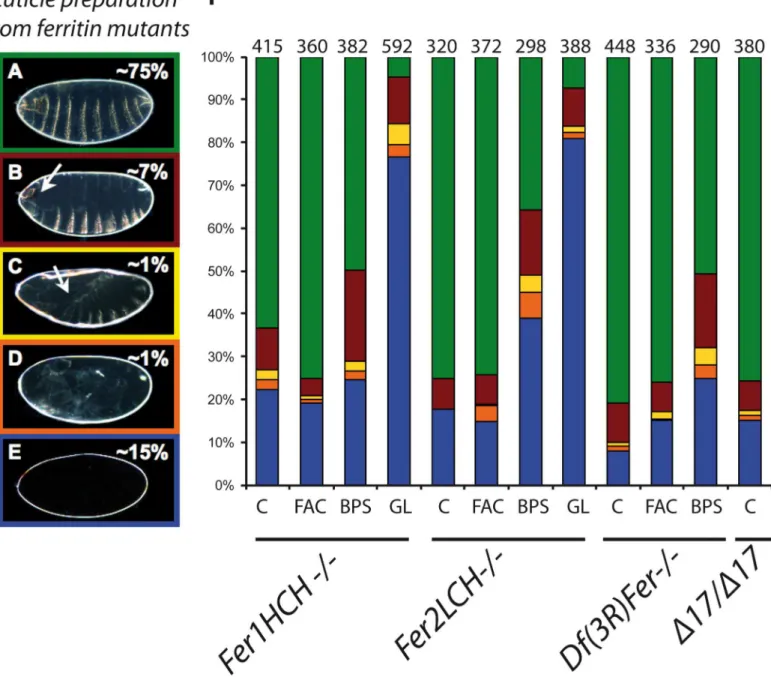
Fer2LCH35, 43% forFer1HCH451, and 90% forDf(3R)Fer; in our hands in these experiments in control flies only 1% of embryos die during embryogenesis. Differing numbers of mutant cuticles may just reflect level of lack-of-function attained in the different mutant genotypes, as opposed to qualitative differences, since they all exhibit exactly the same mutant phenotypes). Cuticle preparations of zygotic ferritin homozygous mutants are mostly wildtype (approxi-mately 75%), but a fraction (overall 25%) has no cuticle deposition, or cuticular defects (a small fraction have germ band extension and retraction, dorsal closure, and head involution defects, among others;Fig 1). Quantification showed that the majority of mutant embryos presented either a normal cuticle (~75%) or no cuticle at all (~15%). A significant finding was that the fer-ritin mutant genotypes tested showed similar percentages of mutant phenotypes. We interpret this to mean that individual ferritin subunits only function in concert (as the ferritin complex, holoferritin) and do not have vital subunit-specific functions. These diverse phenotypes also suggest that ferritin is required in several tissues during development, consistent with differen-tial iron needs in cells and tissues. Notably, a number of iron sulfur cluster proteins are induced during the final stages of embryogenesis to support glycolysis in an aerobic environment [33]. As cuticle deposition occurs during the last stages of embryonic development, defects in cuticle deposition may have a late cause. On the other hand, mutant cuticles pinpoint defects mani-fested earlier during development: germband extension-retraction (stages 9–12), dorsal closure (stages 13–15), and head involution (stages 14–16). Overall, we found defects from mid-devel-opment (stage 9) onwards. All these defects were not found in control stocks.
Ferritin maternal contribution is utilized during early embryonic
development
Ferritin is maternally contributed [17]. We therefore wondered if our analysis of zygotic mutants would miss early ferritin requirements (even obligate maternally-encoded ferritin requirements) fulfilled by this maternal contribution. To analyze how maternal ferritin func-tioned during embryogenesis we followed two strategies: a) curtailed iron availability in moth-ers, where some maternally deposited ferritin mRNAs and proteins would be available in the egg, but iron would be limited, and b) generating female germ line clones without wild type copies of the two main ferritin genes.
A way of reducing ferritin expression and iron availability in adults is to add an iron-specific chelator in the diet (S1 Fig)[16,34]. We hypothesized that reduced overall iron levels would result in decreased ferritin (and iron) maternal contribution and a more severe embryonic phe-notype. Homozygous mutant embryos derived from heterozygous adults fed with 200μM
augmented from ~2% to ~4%, and embryos failing to complete dorsal closure increased from ~1% to ~4% (Fig 1F; BPS columns). At the same time, the percentage of“wild type”cuticles decreased. Using cuticle preparations, this evidence points to iron levels being critical for all ferritin embryonic functions, and BPS treatment as being a way of generating more extreme lack-of-function conditions. No new mutant phenotypes, different from non-iron reduced zygotic mutants, were detected in these embryos, supporting that ferritin and iron deficits are Fig 1. Ferritin mutants result in a variety of cuticle phenotypes quantified by different colors.Examples are shown of (A) wild type cuticle (green), (B) head involution defects (red), (C) dorsal closure defects (yellow), (D) germ band retraction defects (orange), and (E) no cuticle deposition (blue). (F) Percentages of cuticular phenotypes of ferritin mutants. An enhancement of the earlier phenotypes was seen in mutant embryos whose mothers were fed BPS, which was dramatic in embryos derived from ferritin mutant germline clones. C: normal diet, FAC: high iron diet, BPS: low iron diet, GL: germline clones, n: number of embryos examined per genotype. An asterisk denotes statistical difference compared to the control lane at p<0.001.
phenotypically coincidental. Once again, the changes in phenotypic classes abundances were consistently similar inFer1HCH451,Fer2LCH35, andDf(3R)Fer.
On the other hand, feeding extra iron to adults did not result in a rescue of the zygotic embryonic phenotype of the mutant offspring (Fig 1F, FAC), even though total levels of ferritin in mothers increased (S1 Fig). This shows that maternal ferritin contribution is not sufficient to fully rescue embryogenesis in ferritin homozygous mutants, implying an obligate role for zygotically expressed ferritin in embryonic development.
To further explore whether maternal ferritin only partially rescues zygotic ferritin mutants, especially during early development, we generated homozygous ferritin mutant germ line clones. Most germ line clones had no cuticle or bore cuticles with defects (95% of mutant embryos). The‘no cuticle’phenotype percentage changed spectacularly from ~15% to ~80% (Fig 1F). Again, no new mutant phenotypes were seen. This augmentation of phentoypes is in agreement with the BPS experiment above, consistent with early ferritin/iron requirements during embryogenesis. A wild type copy of zygotic ferritin can rescue a small fraction of mutant embryos that were maternally deprived of ferritin, implying no obligate function for maternally supplied ferritin.
Heterozygous embryos without maternal contribution develop normally into adults without defects. Taken together, these results show that maternally supplied ferritin is employed early during development, but can be functionally replaced by zygotically expressed ferritin. We hypothesize that this rescue is due to zygotic ferritin genes being overexpressed in order to compensate for lack of maternal ferritin, sufficient to handle iron needs during embryonic development. It still remains to be seen how iron is maternally deployed in the oocyte when there is no maternal Fer1HCH and Fer2LCH available, as ferritin complex formed from these two proteins is the main iron storage complex in the fly.
The diversity of cuticular phenotypes, and their increase in conditions where iron availabil-ity is reduced and/or ferritin is deprived, leads to a common‘syndrome’of mutant cuticles, where lack of cuticle deposition becomes particularly abundant. The different mutant embry-onic cuticular phenotypes, including their increase in maternally reduced or deprived embryos, are consistent with ferritin requirements several times during development.
Central Nervous System (CNS) phenotypes of ferritin mutants
The majority of zygotic ferritin mutant embryos derived from heterozygous mothers die with an apparently normal cuticle. Since not all functional requirements in embryonic lethal muta-tions are reflected as cuticular defects, we also sought to study other tissues and organs. We sought to study an internal organ or tissue that might be affected by lack of ferritin, and con-tribute to the death of the mutant embryos, an organ or tissue whose defects which might not necessarily reflect in the cuticle. Since ferritin transcripts are first detected in the developing neuroectoderm at stages 7–8 and the protein becomes concentrated in the CNS of first instar larvae [21,35], we asked whether the CNS developed normally in ferritin mutants.
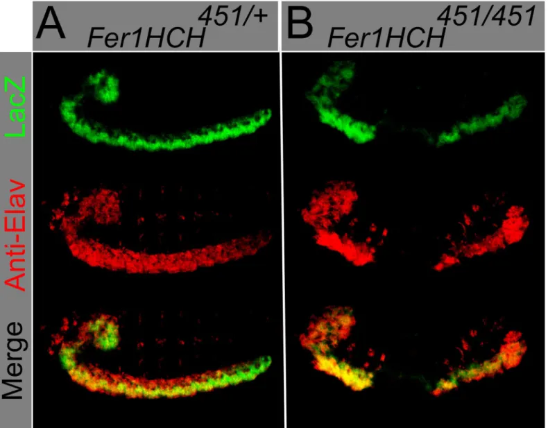
strongly expressed in the neuroectoderm (Fig 2). Co-localization between the neuronal marker Elav andβ-galactosidase was also observed in homozygous mutant embryos that have a dis-rupted CNS (Fig 2). The earliest stage we see defects with Elav antibody are stage 11–12, some time after nervous system ferritin expression is seen at stages 7–8.
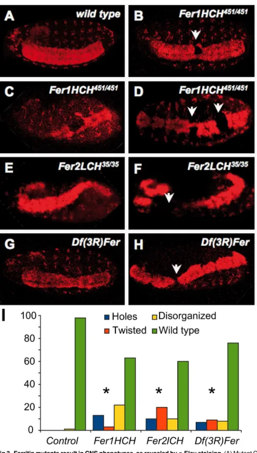
We then stained embryos with antibodies against several neuronal markers. Anti-Elav, which marks neuronal nuclei, showed that some ferritin mutants harbor holes in the abdomi-nal CNS segments (Figs2and3). More severe phenotypes were also present, albeit in fewer embryos, including aberrant condensation of the CNS, twisted CNS, and loss of parts of the brain and peripheral nervous system (PNS). Importantly, and consistent with our analysis of the cuticle phenotypes discussed above, these phenotypes were observed with both ferritin alleles and with the 2.2 kb genomic deletion that specifically deletes bothFer1HCHand
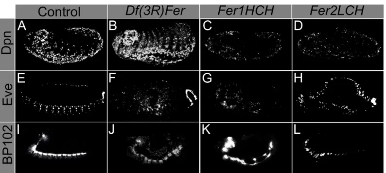
Fer2LCH(Fig 3B-3H, distribution of defects in 3I, compared with the control 3A). These phe-notypes were significantly different from control embryos (Fig 3I). The developing CNS con-sists of at least four types of cells: neuroblasts, ganglion mother cells, neurons, and glia. Neuroblasts are CNS precursor cells and give rise to ganglion mother cells; and these, in turn, give rise to neurons and glia [36]. In order to test whether neuroblasts and ganglion mother cells were affected in ferritin mutant embryos, we performed antibody staining against Dead-pan (Dpn) and Evenskipped (Eve) [37,38]. Anti-Deadpan staining, which marks all neuro-blasts, shows that CNS defects are already present within neuroblast cell lineages in at least some mutant embryos (Fig 4A-4D). As expected for early CNS defects, mutant embryos can also be found where Eve positive ganglion mother cells are affected (Fig 3E-3H). CNS defects are detected from the time neuroblast cell lineages are specified in some mutant embryos, at stages 7–8, precisely during germband extension-retraction, when the earliest cuticle defects are seen. These early defects may result in more maturecontorted and aberrant nervous sys-tems, as seen with anti-Elav.
In order to examine other aspects of later embryonic CNS stages (besides anti-Elav), we used anti- BP102 antibody to study condensed CNS and axonal trajectories in ferritin mutant embryos. Anti-BP102 stains axons and CNS. We studied the condensed ventral nerve cord and axons. Anti-BP102 staining revealed that in some mutant embryos the CNS ventral nerve cord is aberrant and contorted, and that axons are misguided (Fig 3I-3L). Altogether, CNS defects occur throughout CNS development.
Ectopic apoptotic activation in ferritin mutants
The
Drosophila
DMT1 homolog,
Malvolio
, is upregulated in ferritin
mutants
Mutations inMvlresult in reduced iron within intestinal iron storage cells, but also in the whole body [46].Mvlmutants can suppress both intestinal iron accumulation resulting from ferritin [26] and from Multicopper Oxidase-1 (MCO1) misregulation [47]. A recent paper by the same group shows that the likely reductase for this enzyme is ascorbate; its influence on iron homeostasis is conserved in mosquitoes [48]. In view of the above findings, we used
Mvl97f, a P-element insertion mutant carrying aLacZreporter for gene activity that also leads to reducedMvlexpression [24,46] to ask whetherMvland ferritin genes interact.
In control embryos, LacZ showed a very restricted pattern ofMvlexpression consistent with previous studies [21,24]. However, in a mutant ferritin background,Mvl- driven LacZ is Fig 2.Fer1HCH451lacZ
enhancer trap is expressed in the embryonic CNS.Using an antibody against Anti- Beta-Galactosidase (green) and an antibody against the neuronal marker Elav (red), colocalization is observed in (A) heterozygousFer1HCH451/+and (B) homozygousFer1HCH451embryos. Ferritin
homozygous mutant embryo has abnormally shaped and separated nervous system.
Fig 3. Ferritin mutants result in CNS phenotypes, as revealed byɑ-Elav staining.(A) Mutant CNS (B-H)
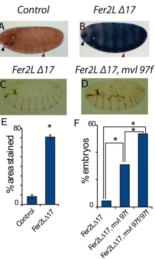
upregulated, its levels increasing as development proceeds (Fig 6A, 6B and 6E). We hypothesize than in the absence of functional ferritin, iron depleted cells upregulateMvlin an effort to curve iron depletion. Quantitation showed a significant difference of expression (Fig 6E). Sig-nificantly, the addition of one copy of theMvl97fhypomorphic allele into a ferritin-depleted embryo resulted significantly in the appearance of necrotic patches, which were rarely present inMvl97for in ferritin homozygous mutants alone (Fig 6C, 6D and 6F).
Ferritin expression, localization and trafficking during development
Ferritin might participate in an iron import pathway [26]. The above interaction between DMT1 and ferritin is reminiscent of recent findings in mammals. Indeed, the mammalian fer-ritin receptor SCARA5 is upregulated in the absence of the transferrin receptor [10].In order to study this proposed transport function of ferritin, we used theFer1HCHG188
mutant allele.Fer1HCHG188is a mutantFer1HCHallele, shown to generate a chimeric GFP-Fer1HCH protein that faithfully mimics the endogenous Fer1HCH pattern in heterozy-gous condition [17,32,35,49]. The GFP tag in homozygosis is thought to block the correct func-tion of ferritin, because in homozygotes all the H-subunits carry a GFP tag, and embryonic development fails [17].Fer1HCHG188/+flies are viable as heterozygotes, showing a dominant mild effect: a small reduction in iron accumulation within ferritin [34].
During stages 16–17 of embryonic development GFP tagged Fer1HCH is present in hemo-cytes inFer1HCHG188heterozygotes (Fig 7A). Hemocytes are large cells that are loosely
defects in different ferritin mutants that present CNS defects; statistical difference compared to control following a Chi squared test, at p<0.0001, and is denoted by an asterisk (n = 32, 10, and 138 embryos, respectively).
doi:10.1371/journal.pone.0133499.g003
Fig 4. Neuroblast and ganglion mother cells populations and neuronal axons derived from embryonic neurons are affected in ferritin mutants.
Neuroblast populations, as marked by Dpn (A, control) are affected in ferritin mutants (B-D). Ganglion mother cells (E, control) are affected in ferritin mutants (F-H). Axons emanating from the brain and ventral nerve cord have a stereotyped pattern in normal development (I). In ferritin mutants, the axons form but are disorganized (J-L).
associated with peripheral tissues circulating in the hemolymph, where they function as both phagocytic and immune cells [50]. There is no report of ferritin mRNA expression in hemo-cytes. In order to confirm that the large, ferritin-accumulating cells were actually hemocytes we used theCg-Gal4line to drive expression, exclusively in hemocytes, of a nuclearRFP in
Fer1HCHG188/+embryos. GFP-Fer1HCH is present in the same cells as Cg-nRFP (Fig 7A). Ferritin mRNAs have been located in the fat body [17]. In a similar experiment as above, we usedFer1HCHG188/+and tagged the fat body with FB-nRFP, a fat body specific Gal4 line.
Fer1HCHG188GFP and FB-nRFP are not coincidental (Fig 7B). If the ferritin mRNAs are translated in the fat body, they are either degraded quickly or transported elsewhere. A likely target candidate for transport is the hemocytes, where conversely there are no ferritin mRNAs; rather ferritin protein is present.
We noticed that homozygous mutantFer1HCHG188embryos showed a clumped and con-centrated accumulation of GFP-Fer1HCH than similarly staged heterozygousFer1HCHG188
embryos (Fig 7C and 7D). HomozygousFer1HCHG188embryos have a lower and restricted GFP-Fer1HCH expression; this is mainly seen in the intestinal region (Fig 7D), suggesting either degradation of the mutant protein or that mutant, non-functional heteropolymers (com-posed exclusively of GFP- Ferritin1HCH and Ferritin 2LCH subunits), are not trafficked. The intestine could also be a possible source of transported ferritin, as iron from the diet is accumu-lated there and both ferritins mRNAs and proteins are present there (Fig 7C). Fewer hemocytes haveFer1HCHG188–derived GFP in the mutant (Fig 7E and 7F), again consistent with ferritin
transport from intestinal and / or fat body sources.
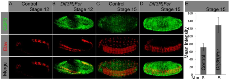
To test further whether ferritin is delivered from other embryonic tissues, we blocked the intracellular secretory pathway by means of a lethal mutation in Sec23. sec23j13C 8is a P-ele-ment insertion in the 5’UTR ofsec23, expected to eliminate or severely attenuate gene function [51]. If ferritin is indeed transported during embryogenesis, blocking the secretory pathway will impede its exit from the cells where it is originally transcribed. Ferritin was detected clumped insec23j13C8/j13C8mutants (compareFig 7H to 7G); GFP-Fer1HCH aggregates were detected mainly around the midgut (Fig 7H) in a similar expression pattern as that Fig 5. Ferritin mutants cause apoptosis in the CNS and other tissues.(A-D) Whole embryos were stained with anα-CSP3act marking apoptotic cells
(green), and anα-Elav marking neurons (red). Ectopic apoptosis was observed in ferritin mutants from stage 12 onwards; at this stage it was mostly restricted
to the neurogenic region (B). At stage 15 apoptosis covers most mutant embryonic tissues (D). Quantification of the mean intensity value on CSP3act staining in control and ferritin mutants at stage 15 show a significant difference, with higher levels of staining in mutant embryos (n = 5; T-test, p = 0.0113).
Fig 6. Ferritin genes interact genetically with the DMT1 homologMvl.(A) TheMvl97f-LacZline shows a spatially restricted expression pattern forMvl, mainly in the head region, the brain, and a segmentally repeated pattern. (B) In a ferritin homozygous mutant background,Mvl97f-LacZexpression increases. Black
observed in homozygousFer1HCHG188mutants. This is consistent with ferritin trafficking during embryonic development.
Thus, in embryos impaired in the secretory pathway some cells fail to accumulate ferritin, suggesting that in wild type embryos ferritin may be actively secreted into the hemolymph. We then made use of two constructs, UAS-mcherry::Fer2LCH and UAS-GFP::Fer1HCH (Gambis, Steller & Mollereau, personal communication), and expressed them using the hemocyte Cg-Gal4line. We found that embryonic expression of the ectopic, tagged ferritin subunits was largely, although not absolutely, coincidental, and that expression was seen in cells other than hemocytes (S4 Fig) in late embryos.
We also used the same fluorescently tagged ferritin constructs to see whether these tagged ferritin proteins could also be expressed and transported in larvae. We usedFer2LCH-Gal4to drive expression (S5A and S5B Fig). We found expression of both constructs in the midgut, as expected (due toFer2LCH-Gal4), but also in Garland cells, where noFer2LCH-Gal4expression is seen, implying ferritin transport. We also used two other drivers:elav-Gal4for expression in neurons (S5C Fig) andrepo-Gal4for expression in glial cells (S5D Fig). In both cases, fluores-cence is also seen in Garland cells, and again, implying ferritin transport. Garland cells are nephrocytes, part of the larval fly hemolymph filtration system [52], and may come in contact and accumulate ferritin circulating in the hemolymph.
Taken together, all these data point that ferritin traffics between tissues, and that hemocytes and hemolymph may play key roles in this trafficking process, conveying ferritin from one tis-sue to another. Such a role for hemocytes has been suggested in the context of tistis-sue communi-cation in the innate immune response [53].
Discussion
Insect embryos must course through development with limited amounts of iron, provided dur-ing oogenesis by the mother, in partviaferritin. It seems reasonable to assume that tissues developing at different rates present different iron requirements, and therefore iron transport must be of vital importance for normal development. This may also mean that ferritin is used for trafficking from storage sites, as development proceeds, to tissues and cells where it is required, as an iron conveyor.
Here we show that ferritin mutants die during embryogenesis with a wide range of pheno-types. Ferritin mutants first show cuticular defects after gastrulation, during germband exten-sion-retraction and the formation of the nervous system, if we classify lack of cuticle formation only as a late occurring defect. Around stage 12, at about the initiation of dorsal closure, there is already ectopic apoptosis, and it becomes more prevalent and intense as embryonic develop-ment continues. Judging from the generalization of apoptosis, not all portions of the embryo are affected in a similar manner, as ectopic apoptotic signal is first seen in the neurogenic region, before becoming generalized. Most zygotic mutants do from cuticle, implying that even at a stage where there is prevalent apoptosis, some cells survive (differentiating epidermis cells) enough to secrete cuticle at the end of embryogenesis. In contrast, the CNS is affected from the start.
If iron provided maternally by ferritin is reduced or completely missing, the same pheno-types are present, but particularly, embryos not forming cuticle become prevalent. This implies
background resulted in the appearance of necrotic patches in the cuticle (C, D). (E) Quantification of the total area covered by LacZ staining in control and ferritin mutant backgrounds, (p<0.0001; T-test, asterisk). (F) Quantification of the number of embryos showing necrotic patches with one or twoMvl97falleles; statistical difference using a Chi squared test with p<0.0001 is shown by an asterisk.
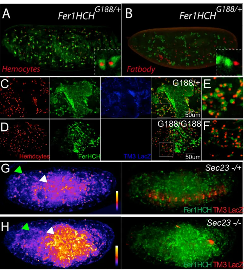
Fig 7. Marked ferritin accumulation in embryos with different genetic backgrounds.Fer1HCHprotein was visualized in all embryos using the
Fer1HCHG188GFP trap line (green). In stage 17Fer1HCHG188/+embryos that successfully complete development, ferritin accumulates mainly in the midgut
and hemocytes. (A) The GFP-Fer1HCH signal is found in hemocytes marked bycgdriving nuclear RFP, but not (B) in the fat bodies marked by the fat body driver FB driving nuclear RFP. (C-F)Fer1HCHG188homozygous embryos, which die like other ferritin mutants, also show intestinal ectopically localized
that the‘no cuticle’phenotype stems from a very early ferritin requirement manifested late by lack of cuticle deposition. Ferritin damage ends in ectopic apoptosis, eventually generalized throughout the embryo.
We also provide evidence of ferritin trafficking. Blocking the secretory pathway during development causes abnormal ferritin distribution, suggesting an additional iron-trafficking role for ferritin. Our ectopic studies point in the same way, at least for the ectopically produced ferritin subunits. This may be responsible for differences in ferritin mRNA and protein expres-sion. Ferritin mutations also cause an up-regulation ofMvl, probably as a means to stimulate iron uptake from iron deprived cells.
Tang and Zhou demonstrated the importance of ferritin expression in the larval intestine [26]. Our studies in larval intestine are consistent with this, and suggest that ferritin expression in the gut is a starting point for ferritin larval function.
Our results also point to both ferritin subunits being jointly required in the embryonic CNS. In adult flies, in contrast, both genes might also have different functions. RNA interference in subsets of neurons againstFer2LCHbut not againstFer1HCHdisrupted circadian rhythms [54]. Furthermore, some cell types, including commonly used cell culture lines [55,56], only expressFer1HCHand notFer2LCH. Overexpression of eitherFer1HCHorFer2LCH, or both subunits simultaneously inDrosophilaglia [43] or neurons [53,57] resulted in qualitatively dif-ferent responses. We also find that embryonic ectopic expression of both ferritin subunits leads to some cells differentially expressing one of the two genes. Use of theelav-Gal4driver to silence eitherFer1HCHorFer2LCHresulted in viable adults with perturbed circadian behavior [54] and apparent neurodegeneration [26]. RNA interference is known to cause reduced expression but not complete silencing of its targets, which may explain why ferritin RNAi flies survived to adulthood. In addition and consistent with our early CNS defects results, overex-pression of ferritin subunits in this last study withelav-Gal4failed to rescue their respective mutants [26], implying an early ferritin neuronal requirement, before theelav-Gal4transgene is active, as seen in our staining experiments with early CNS markers. Furthermore, disrupting ferritin levels (either by reductions or ectopic expression may provoke disequilibrium of ferri-tin proteins and explain the differing results observed.
We conclude that both ferritins are normally required, but that sometimes regulation in vivo is effected primarily via Fer2LCH. Thus, the question of how different cell types regulate in vivo the two ferritin genes and subunits and whether they always act in concert requires fur-ther investigation.
Materials and Methods
Fly stocks
All flies wereDrosophila melanogasterMeigen. As a control strainy,wflies were used.
Fer1HCH451andFer2LCH35are P(ry[+t7.2] = PZ) insertion alleles generated during a large-scale mutagenesis screen [58], and have been partially characterized elsewhere [17]. They were obtained from the Bloomington Drosophila Stock Center (BDSC); stock numbers #11497 and #11483, respectively.Fer2LCHΔ17was generated from an imprecise excision of Fer2L-CHEP1059(described in Flybase) and interferes with expression of both genes, as confirmed
pathway using a homozygous mutantsec23j13C8background leads to a decrease of ferritin levels in the hemocytes (green arrowheads),; and ectopic
accumulation in the intestine (white arrowheads). Intensity of GFP::Fer1HCH flurescence is shown in the left side of panels G and H using a fire scale. The fire scales are shown in the lower right side of each, with warmer colors (red-orange-yellow-white) denoting higher intensity of fluorescence, and colder colors (purple-blue) denoting lower levels of fluorescence for the green channel. Note higher fluorescence levels in thesec23homozygous mutant embryo (H). A merged image showing GFP::Fer1HCH fluorescence and control RFP staining (G) due to the balancer chromosome is shown to the right side of each panel.
by complementation crosses.Df(3R)Ferwas a gift from Alexis Gambis, Bertrand Mollereau, and Hermann Steller and is a 2.2 kb deletion disrupting specificallyFer1HCHandFer2LCH
[34]. To generate germline clones,Fer1HCH451andFer2LCH35were recombined unto FRT82 containing chromosomes [59].Fer1HCHG188is a protein trap line and has been extensively described elsewhere [17,60].Mvl97fis an homozygous viableP(lacW)insertion obtained from the (BDSC stock #5151) (Rodriguez, et al. 1995).Sec23j13c8mutant is aP(lacW)insertion within the 5'UTR ofSec23[51]; BDSC stock #10218.Cg-Gal4(BDSC stock #7011) was used to drive expression in the hemocytes,drm-Gal4(BDSC stock #7098) in embryonic gut and scat-tered cells around the epidermis,FB-Gal4in the fat body (P{GAL4}fat; Flybase ID;
FBti0013267) [16,61,62]. UAS-mcherry::Fer2LCH and UAS-GFP::Fer1HCH were a gift from Alexis Gambis, Hermann Steller and Bertrand Mollereau (Gambis, Steller, and Mollereau, per-sonal communication), and will be described elsewhere. In cases where recombinant or double balanced stocks were needed they were generated following conventional crossing schemes.
Iron diets
Flies were raised for 3 successive generations on standard medium supplemented with 200μM
Bathophenanthrolinedisulfonic acid disodium salt (SIGMA #B1375) referred to as BPS in the text or with 1 mM ammonium iron (III) citrate (SIGMA #F5859) referred to as FAC. Adults were used for embryo collections. Protein extracts from female adults were also analyzed by non-reducing SDS- PAGE, confirming the differential accumulation of ferritin in flies raised on the respective diets.
Immunohistochemistry and confocal imaging
Following dechorionation with a commercial bleach solution, embryos from overnight collec-tions were devitellinized and fixed in a 1:1 mixture of heptane and 36% formaldehyde for 5 minutes and then washed in methanol. Embryos were then stored at -20 C or rehydrated, and used for staining. Primary antibodies used were: ratα-Elav 1:100, mouseα-BP102 1:100, mouseα-Eve 1:100 (Developmental Studies Hybridoma Bank, Indiana, USA);α-activated Cas-pase 3 1:100 (Cell Signaling, USA); and ratα-Deadpan 1:2, a gift from Cheng-Yu Lee. Second-ary antibodies used were: Alexa flour 546α-rat 1:100 (Santa Cruz Biotechnology, USA), Cy5 α-mouse 1:1000, Cy3α-mouse 1:1000, FITCα-rabbit 1:1000 (Zymax, USA). Signal from α-dead-pan staining was increased with the ABC kit from Vectastain (USA). A 510 Meta and 780 Duo confocal microscopes (Zeis, Germany) were used for fluorescent imaging, and images were processed with Zeiss software and ImageJ. Homozygous mutant embryos were selected by lack of TM3GFP of TM3LacZ. Accumulation of GFP::Fer1HCH inSec23mutants was evaluated using the fire LUT of ImageJ.
Cuticle preparations and X-Gal staining
Supporting Information
S1 Fig. Ferritin expression can be modulated by iron availability.Ferritin complexes were revealed by Coomasie staining and identified by size, when either flies were fed Bathophenan-throline Sulfate (BPS) to reduce iron availability (BPS is an iron chelator), or fed extra iron (FAC).
(TIF)
S2 Fig.Fer1HCH451is a functional ferritin enhancer trap line.β-Galactosidase expression is
normally restricted to the iron region in the larval midgut, but expression is enhanced in the anterior midgut (AMG) when iron fed, as occurs in wild type larve (Mehta et al. 2009). (TIF)
S3 Fig. Apoptosis in ferritin mutants.Apoptosis revealed with anα-active caspase 3 antibody (CSP3act) marking apoptotic cells (green), and anα-Elav marking neurons (red) in
Fer1HCH451/451(A) andFer2LCH35/35(B) mutant embryos. (TIF)
S4 Fig. Embryonic ectopic expression of fluorescence-tagged ferritin subunits reveals embryonic dynamic ferritin transport of tagged ferritins.(A) Control embryo where embry-onic hemocytes are revealed by theCg-Gal4line and UAS-RFP (red fluorescent protein). Cg-Gal4drives RFP expression specifically in hemocytes. (B-D) Overexpression of both ferritin tagged subunits in the hemocytes results in ferritin accumulation in these cells (asterisk), but also in other tissues (arrowhead). UAS-mcherry::Fer2LCH (UAS-cherry:Fer2LCH) tagged fer-ritin expression is in (B), and UAS-GFP::Fer1HCH (UAS-GFP:Fer1HCH) is in (C). A merged image in shown in D. The white arrowhead points to embryonic tissues harboring fluorescence other than hemocytes. The asterisk marks hemocytes. (E-G) Higher magnification images of a similar embryo revealing partial co-localization of tagged ferritin subunits driven byCg-Gal4, where cells accumulating one subunit but not the other are seen (dashed circles). UAS-mcherry::Fer2LCH expression is shown in (E), UAS-GFP::Fer1HCH is shown in (F), and a merged image is shown in G. The tagged ferritin lines will be described elsewhere (Gambis, Steller, and Mollereau, personal communication).
(TIF)
S5 Fig. Ectopic expression of tagged ferritin subunits reveals dynamic ferritin transport in larvae.(A) Ectopic expression of RFP::Fer2LCH using aFer2LCH-Gal4line results in ferritin accumulation in the anterior midgut and in Garland cells (dashed line). UAS-GFP was used as a transcription marker forFer2LCH-Gal4activity (A’) and is detected in the anterior midgut but not in the Garland cells. Merged image in (A”). (B) Likewise, ectopic expression of GFP:: Fer1HCH usingFer2LCH-Gal4also results in GFP::Fer1HCH accumulation in the anterior midgut and in Garland cells (dashed line). Nuclear RFP (B’) was used as a transcription marker forFer2LCH-Gal4and is detected in the anterior midgut but not in the Garland cells. Merged image in (B”). (C) Ectopic expression of tagged ferritin subunits in neurons viaelav-Gal4
results in theiraccumulation in Garland cells, implying GFP::Fer1HCH (C) and RFP::Fer2LCH (C’) transport from neurons to Garland cells. Merged image in (C”) also shows localization of Garland cells around digestive tract (combined fluorescence and transmitted light image). (D) Glial expression via therepo-Gal4 driver of both tagged ferritin subunits (GFP::Fer1HCH (D) and RFP::Fer2LCH (D’) results in tagged their accumulation in a discrete portion of midgut enterocytes. Merged image in (D”).
Acknowledgments
We thank María Teresa Peña-Rangel for technical assistance during the course of this project, Nydia Hernández-Rios for assistance with the use of the confocal microscope. We also acknowledge Cheng- Yu Lee for sending theɑ-deadpan antibody and Alexis Gambis,
Her-mann Steller, and Bertrand Mollereaufor sharing theDf(3R)Fer, and the UAS-mcherry:: Fer2LCH and UAS-GFP::Fer1HCH fly stocks prior to publication. This work is part of the PhD dissertation of Miguel Ángel Mendoza-Ortíz, who is a PhD student in the Programa de Doctorado en Ciencias Biológicas, Universidad Nacional Autónoma de México (UNAM), and received a PhD fellowship from Consejo Nacional de Ciencia y Tecnología (CONACYT) [164428].
Author Contributions
Conceived and designed the experiments: NGM MÁMO FM JRRE. Performed the experi-ments: NGM MÁMO LMB FM JRRE. Analyzed the data: NGM MÁMO FM JRRE. Contrib-uted reagents/materials/analysis tools: FM JRRE. Wrote the paper: NGM FM JRRE.
References
1. Sheftel AD, Mason AB, Ponka P. The long history of iron in the Universe and in health and disease. Bio-chim Biophys Acta. 2012; 1820: 161–87. doi:S0304-4165(11)00185-1 [pii] doi:10.1016/j.bbagen.2011. 08.002PMID:21856378
2. Gunshin H, Mackenzie B, Berger UV, Gunshin Y, Romero MF, Boron WF, et al. Cloning and characteri-zation of a mammalian proton-coupled metal-ion transporter. Nature. 1997; 388: 482–8. doi:10.1038/ 41343PMID:9242408
3. De Domenico I, McVey Ward D, Kaplan J. Regulation of iron acquisition and storage: consequences for iron-linked disorders. Nat Rev Mol Cell Biol. 2008; 9: 72–81. doi:nrm2295 [pii] doi:10.1038/nrm2295 PMID:17987043
4. Harrison PM, Arosio P. The ferritins: molecular properties, iron storage function and cellular regulation. Biochim Biophys Acta. 1996; 1275: 161–203. PMID:8695634
5. Hentze MW, Muckenthaler MU, Andrews NC. Balancing acts: molecular control of mammalian iron metabolism. Cell. 2004; 117: 285–97. doi:S0092867404003435 [pii] PMID:15109490
6. Santambrogio P, Levi S, Cozzi A, Rovida E, Albertini A, Arosio P. Production and characterization of recombinant heteropolymers of human ferritin H and L chains. J Biol Chem. 1993; 268: 12744–8. PMID:8509409
7. Pantopoulos K, Porwal SK, Tartakoff A, Devireddy L. Mechanisms of mammalian iron homeostasis. Biochemistry (Mosc). 2012; 51: 5705–24. doi:10.1021/bi300752r
8. Pantopoulos K. Iron metabolism and the IRE/IRP regulatory system: an update. Ann N Acad Sci. 2004; 1012: 1–13.
9. Piccinelli P, Samuelsson T. Evolution of the iron-responsive element. RNA. 2007; 13: 952–66. doi: rna.464807 [pii] doi:10.1261/rna.464807PMID:17513696
10. Li JY, Paragas N, Ned RM, Qiu A, Viltard M, Leete T, et al. Scara5 is a ferritin receptor mediating non-transferrin iron delivery. Dev Cell. 2009; 16: 35–46. doi:S1534-5807(08)00512-1 [pii] doi:10.1016/j. devcel.2008.12.002PMID:19154717
11. Todorich B, Zhang X, Slagle-Webb B, Seaman WE, Connor JR. Tim-2 is the receptor for H-ferritin on oligodendrocytes. J Neurochem. 2008; 107: 1495–505. doi:JNC5678 [pii] doi:10.1111/j.1471-4159. 2008.05678.xPMID:19014383
12. Fisher J, Devraj K, Ingram J, Slagle-Webb B, Madhankumar AB, Liu X, et al. Ferritin: a novel mecha-nism for delivery of iron to the brain and other organs. Am J Physiol Cell Physiol. 2007; 293: C641–9. doi:00599.2006 [pii] doi:10.1152/ajpcell.00599.2006PMID:17459943
13. Meyron-Holtz EG, Moshe-Belizowski S, Cohen LA. A possible role for secreted ferritin in tissue iron dis-tribution. J Neural Transm. 2011; 118: 337–47. doi:10.1007/s00702-011-0582-0PMID:21298454
15. Hamburger AE, West AP, Hamburger ZA, Hamburger P, Bjorkman PJ. Crystal structure of a secreted insect ferritin reveals a symmetrical arrangement of heavy and light chains. J Mol Biol. 2005; 349: 558–
69. doi:S0022-2836(05)00375-X [pii] doi:10.1016/j.jmb.2005.03.074PMID:15896348
16. Missirlis F, Holmberg S, Georgieva T, Dunkov BC, Rouault TA, Law JH. Characterization of mitochon-drial ferritin in Drosophila. Proc Natl Acad Sci U A. 2006; 103: 5893–8. doi:0601471103 [pii] doi:10. 1073/pnas.0601471103
17. Missirlis F, Kosmidis S, Brody T, Mavrakis M, Holmberg S, Odenwald WF, et al. Homeostatic mecha-nisms for iron storage revealed by genetic manipulations and live imaging of Drosophila ferritin. Genet-ics. 2007; 177: 89–100. doi:genetics.107.075150 [pii] doi:10.1534/genetics.107.075150PMID: 17603097
18. Georgieva T, Dunkov BC, Harizanova N, Ralchev K, Law JH. Iron availability dramatically alters the dis-tribution of ferritin subunit messages in Drosophila melanogaster. Proc Natl Acad Sci U A. 1999; 96: 2716–21.
19. Georgieva T, Dunkov BC, Dimov S, Ralchev K, Law JH. Drosophila melanogaster ferritin: cDNA encod-ing a light chain homologue, temporal and tissue specific expression of both subunit types. Insect Bio-chem Mol Biol. 2002; 32: 295–302. doi:S096517480100090X [pii] PMID:11804801
20. Mandilaras K, Pathmanathan T, Missirlis F. Iron absorption in Drosophila melanogaster. Nutrients. 2013; 5: 1622–47. doi:nu5051622 [pii] doi:10.3390/nu5051622PMID:23686013
21. Kumar S, Konikoff C, Van Emden B, Busick C, Davis KT, Ji S, et al. FlyExpress: visual mining of spatio-temporal patterns for genes and publications in Drosophila embryogenesis. Bioinformatics. 2011; 27: 3319–20. doi:btr567 [pii] doi:10.1093/bioinformatics/btr567PMID:21994220
22. Lind MI, Missirlis F, Melefors O, Uhrigshardt H, Kirby K, Phillips JP, et al. Of two cytosolic aconitases expressed in Drosophila, only one functions as an iron-regulatory protein. J Biol Chem. 2006; 281: 18707–14. doi:M603354200 [pii] doi:10.1074/jbc.M603354200PMID:16679315
23. Folwell JL, Barton CH, Shepherd D. Immunolocalisation of the D. melanogaster Nramp homologue Malvolio to gut and Malpighian tubules provides evidence that Malvolio and Nramp2 are orthologous. J Exp Biol. 2006; 209: 1988–95. doi:209/10/1988 [pii] doi:10.1242/jeb.02193PMID:16651563
24. Rodrigues V, Cheah PY, Ray K, Chia W. malvolio, the Drosophila homologue of mouse NRAMP-1 (Bcg), is expressed in macrophages and in the nervous system and is required for normal taste behav-iour. EMBO J. 1995; 14: 3007–20. PMID:7621816
25. D’Souza J, Cheah PY, Gros P, Chia W, Rodrigues V. Functional complementation of the malvolio muta-tion in the taste pathway of Drosophila melanogaster by the human natural resistance-associated mac-rophage protein 1 (Nramp-1). J Exp Biol. 1999; 202: 1909–15. PMID:10377272
26. Tang X, Zhou B. Ferritin is the key to dietary iron absorption and tissue iron detoxification in Drosophila melanogaster. FASEB J. 2013; 27: 288–98. doi:fj.12-213595 [pii] doi:10.1096/fj.12-213595PMID: 23064556
27. Nichol H, Law JH, Winzerling JJ. Iron metabolism in insects. Annu Rev Entomol. 2002; 47: 535–559. doi:10.1146/annurev.ento.47.091201.145237PMID:11729084
28. Zhou G, Kohlhepp P, Geiser D, Frasquillo MC, Vazquez-Moreno L, Winzerling JJ. Fate of blood meal iron in mosquitoes. J Insect Physiol. 2007; 53: 1169–78. doi:S0022-1910(07)00145-X [pii] doi:10.1016/ j.jinsphys.2007.06.009PMID:17689557
29. Hajdusek O, Sojka D, Kopacek P, Buresova V, Franta Z, Sauman I, et al. Knockdown of proteins involved in iron metabolism limits tick reproduction and development. Proc Natl Acad Sci U A. 2009; 106: 1033–8. doi:0807961106 [pii] doi:10.1073/pnas.0807961106
30. Galay RL, Aung KM, Umemiya-Shirafuji R, Maeda H, Matsuo T, Kawaguchi H, et al. Multiple ferritins are vital to successful blood feeding and reproduction of the hard tick Haemaphysalis longicornis. J Exp Biol. 2013; 216: 1905–15. doi:jeb.081240 [pii] doi:10.1242/jeb.081240PMID:23393286
31. Nüsslein-Volhard C, Wieschaus E. Mutations affecting segment number and polarity in Drosophila. Nature. 1980; 287: 795–801. PMID:6776413
32. Gutierrez L, Sabaratnam N, Aktar R, Bettedi L, Mandilaras K, Missirlis F. Zinc accumulation in heterozy-gous mutants of fumble, the pantothenate kinase homologue of Drosophila. FEBS Lett. 2010; 584: 2942–6. doi:S0014-5793(10)00422-9 [pii] doi:10.1016/j.febslet.2010.05.029PMID:20493851
33. Tennessen JM, Bertagnolli NM, Evans J, Sieber MH, Cox J, Thummel CS. Coordinated metabolic tran-sitions during Drosophila embryogenesis and the onset of aerobic glycolysis. G3 Bethesda Md. 2014; 4: 839–850. doi:10.1534/g3.114.010652
35. Mehta A, Deshpande A, Bettedi L, Missirlis F. Ferritin accumulation under iron scarcity in Drosophila iron cells. Biochimie. 2009; 91: 1331–4. doi:S0300-9084(09)00129-1 [pii] doi:10.1016/j.biochi.2009.05. 003PMID:19465081
36. Biffar L, Stollewerk A. Conservation and evolutionary modifications of neuroblast expression patterns in insects. Dev Biol. 2014; 388: 103–116. doi:10.1016/j.ydbio.2014.01.028PMID:24525296
37. Boone JQ, Doe CQ. Identification of Drosophila type II neuroblast lineages containing transit amplifying ganglion mother cells. Dev Neurobiol. 2008; 68: 1185–95. doi:10.1002/dneu.20648PMID:18548484
38. Kohwi M, Lupton JR, Lai SL, Miller MR, Doe CQ. Developmentally regulated subnuclear genome reor-ganization restricts neural progenitor competence in Drosophila. Cell. 2013; 152: 97–108. doi:S0092-8674(12)01437-7 [pii] doi:10.1016/j.cell.2012.11.049PMID:23332748
39. Thompson K, Menzies S, Muckenthaler M, Torti FM, Wood T, Torti SV, et al. Mouse brains deficient in H-ferritin have normal iron concentration but a protein profile of iron deficiency and increased evidence of oxidative stress. J Neurosci Res. 2003; 71: 46–63. doi:10.1002/jnr.10463PMID:12478613
40. Pham CG, Bubici C, Zazzeroni F, Papa S, Jones J, Alvarez K, et al. Ferritin heavy chain upregulation by NF-kappaB inhibits TNFalpha-induced apoptosis by suppressing reactive oxygen species. Cell. 2004; 119: 529–42. doi:S0092867404009985 [pii] doi:10.1016/j.cell.2004.10.017PMID:15537542
41. Rouault TA. The role of iron regulatory proteins in mammalian iron homeostasis and disease. Nat Chem Biol. 2006; 2: 406–14. doi:nchembio807 [pii] doi:10.1038/nchembio807PMID:16850017
42. Gambis A, Dourlen P, Steller H, Mollereau B. Two-color in vivo imaging of photoreceptor apoptosis and development in Drosophila. Dev Biol. 2011; 351: 128–34. doi:S0012-1606(10)01296-0 [pii] doi:10. 1016/j.ydbio.2010.12.040PMID:21215264
43. Kosmidis S, Botella JA, Mandilaras K, Schneuwly S, Skoulakis EM, Rouault TA, et al. Ferritin overex-pression in Drosophila glia leads to iron deposition in the optic lobes and late-onset behavioral defects. Neurobiol Dis. 2011; 43: 213–9. doi:S0969-9961(11)00095-7 [pii] doi:10.1016/j.nbd.2011.03.013 PMID:21440626
44. Dixon SJ, Lemberg KM, Lamprecht MR, Skouta R, Zaitsev EM, Gleason CE, et al. Ferroptosis: an iron-dependent form of nonapoptotic cell death. Cell. 2012; 149: 1060–1072. doi:10.1016/j.cell.2012.03. 042PMID:22632970
45. Denton D, Mills K, Kumar S. Methods and protocols for studying cell death in Drosophila. Methods Enzym. 2008; 446: 17–37. doi:S0076-6879(08)01602-9 [pii] doi:10.1016/S0076-6879(08)01602-9
46. Bettedi L, Aslam MF, Szular J, Mandilaras K, Missirlis F. Iron depletion in the intestines of Malvolio mutant flies does not occur in the absence of a multicopper oxidase. J Exp Biol. 2011; 214: 971–8. doi:214/6/971 [pii] doi:10.1242/jeb.051664PMID:21346125
47. Lang M, Braun CL, Kanost MR, Gorman MJ. Multicopper oxidase-1 is a ferroxidase essential for iron homeostasis in Drosophila melanogaster. Proc Natl Acad Sci U A. 2012; 109: 13337–42.
doi:1208703109 [pii] doi:10.1073/pnas.1208703109
48. Peng Z, Dittmer NT, Lang M, Brummett LM, Braun CL, Davis LC, et al. Multicopper oxidase-1 orthologs from diverse insect species have ascorbate oxidase activity. Insect Biochem Mol Biol. 2015; 59: 58–71. doi:10.1016/j.ibmb.2015.02.005PMID:25701385
49. Uhrigshardt H, Rouault TA, Missirlis F. Insertion mutants in Drosophila melanogaster Hsc20 halt larval growth and lead to reduced iron-sulfur cluster enzyme activities and impaired iron homeostasis. J Biol Inorg Chem. 2013; 18: 441–9. doi:10.1007/s00775-013-0988-2PMID:23444034
50. Evans IR, Wood W. Drosophila embryonic hemocytes. Curr Biol CB. 2011; 21: R173–174. doi:10. 1016/j.cub.2011.01.061PMID:21377090
51. Abrams EW, Andrew DJ. CrebA regulates secretory activity in the Drosophila salivary gland and epider-mis. Development. 2005; 132: 2743–58. doi:dev.01863 [pii] doi:10.1242/dev.01863PMID:15901661
52. Na J, Cagan R. The Drosophila nephrocyte: back on stage. J Am Soc Nephrol. 2013; 24: 161–163, doi:
10.1681/ASN.2012121227PMID:23334393
53. Wu Z, Du Y, Xue H, Wu Y, Zhou B. Aluminum induces neurodegeneration and its toxicity arises from increased iron accumulation and reactive oxygen species (ROS) production. Neurobiol Aging. 2012; 33: 199 e1–12. doi:S0197-4580(10)00283-6 [pii] doi:10.1016/j.neurobiolaging.2010.06.018PMID: 20674094
54. Mandilaras K, Missirlis F. Genes for iron metabolism influence circadian rhythms in Drosophila melano-gaster. Met Integr Biometal Sci. 2012; 4: 928–936. doi:10.1039/c2mt20065a
56. Metzendorf C, Wu W, Lind MI. Overexpression of Drosophila mitoferrin in l(2)mbn cells results in dysre-gulation of Fer1HCH expression. Biochem J. 2009; 421: 463–71. doi:BJ20082231 [pii] 10.1042/ BJ20082231 doi:10.1042/BJ20082231PMID:19453295
57. Kosmidis S, Missirlis F, Botella JA, Schneuwly S, Rouault TA, Skoulakis EMC. Behavioral decline and premature lethality upon pan-neuronal ferritin overexpression in Drosophila infected with a virulent form of Wolbachia. Front Pharmacol. 2014; 5: 66. doi:10.3389/fphar.2014.00066PMID:24772084
58. Spradling AC, Stern D, Beaton A, Rhem EJ, Laverty T, Mozden N, et al. The Berkeley Drosophila Genome Project gene disruption project: Single P-element insertions mutating 25% of vital Drosophila genes. Genetics. 1999; 153: 135–77. PMID:10471706
59. Xu T, Rubin GM. Analysis of genetic mosaics in developing and adult Drosophila tissues. Development. 1993; 117: 1223–37. PMID:8404527
60. Kelso RJ, Buszczak M, Quinones AT, Castiblanco C, Mazzalupo S, Cooley L. Flytrap, a database doc-umenting a GFP protein-trap insertion screen in Drosophila melanogaster. Nucleic Acids Res. 2004; 32: D418–20. doi:10.1093/nar/gkh01432/suppl_1/D418 [pii] PMID:14681446
61. Missirlis F, Rahlfs S, Dimopoulos N, Bauer H, Becker K, Hilliker A, et al. A putative glutathione peroxi-dase of Drosophila encodes a thioredoxin peroxiperoxi-dase that provides resistance against oxidative stress but fails to complement a lack of catalase activity. Biol Chem. 2003; 384: 463–72. doi:10.1515/BC. 2003.052PMID:12715897