Volumen 68, Broj 12 VOJNOSANITETSKI PREGLED Strana 1071
Correspondence to: Adrijan Sarajlija, Mother and Child Health Care Institute of Serbia „dr Vukan Cupic“, Department of Metabolism and Clinical Genetics, Radoja Dakića 6-8, 11 000 Belgrade, Serbia. Phone: +381113108276; Fax +381113108276. E-mail: [email protected]
C A S E R E P O R T UDC: 616-056.7-008.9::[616.24:616-053.2
DOI:10.2298/VSP1112071D
Pulmonary involvement in siblings with Gaucher disease type III
Plu
ć
ne manifestacije kod srodnika sa Gošeovom bolesti tip III
Maja Djordjević*, Predrag Minić†, Adrijan Sarajlija*, Slaviša M. Djuričić‡, Dragomir Djokić§, Obren Marković¶
Mother and Child Health Care Institute of Serbia „dr Vukan Čupić“, *Department of Metabolism and Clinical Genetics, †Department of Pulmonology, ‡Department of Clinical
Pathology, §Department of Oncology, ¶Department of Radiology , Belgrade, Serbia
Abstract
Introduction. Pulmonary involvement has been described
in all types of Gaucher disease (GD) but it is considered as relatively rare manifestation. There are reports suggesting that homozygosity for L444P mutation in GBA gene is as-sociated with a substantial risk for developing primary pul-monary disease in GD. Case report. We reported sisters with pulmonary involvement in GD type III. Respiratory failure with fatal outcome at 3 years and 4 months of age occurred in K.K. due to pulmonary complications of GD. At the time enzyme replacement therapy (ERT) was not available in Serbia. J.K., homozygous for L444P mutation,
developed asymptomatic pulmonary involvement at the age of 6 after 2.5 years of ERT. Pulmonary disease in J.K. was verified by high resolution computerized tomography, cy-tology of bronchoalveolar lavage fluid and histopathology of transbronchial lung biopsy. Conclusion. Primary lung disease in children homoallelic for L444P mutation in GBA gene emerges as a significant clinical manifestation of GD with unclear response to ERT.
Key words:
gaucher disease; genetic diseases, inborn; mutation; lung diseases; treatment outcome.
Apstrakt
Uvod. Plućne manifestacije opisane su kod sva tri tipa Gošeove bolesti, ali se smatraju relativno retkom kompli-kacijom ove nasledne bolesti. Pojedine studije ukazuju da homozigotnost za mutaciju L444P u genu GBA predstav-lja faktor rizika od razvijanja primarnog plućnog zahvata-nja u sklopu Gošeove bolesti. Prikaz slučaja. Prikazali smo sestre sa plućnim komplikacijama Gošeove bolesti. Respiratorna insuficijencija i smrtni ishod u uzrastu od tri godine i četiri meseca kod devojčice K.K. objašnjeni su plućnim komplikacijama Gošeove bolesti. U to vreme en-zimska supstituciona terapija nije bila dostupna u Srbiji. Njena sestra J.K, L444P homozigot, razvila je
asimptomat-ske plućne manifestacije u uzrastu od šest godina, nakon dve i po godine enzimske supstitucione terapije. Plućna bolest potvrđena je visokorezolutivnom kompjuterizova-nom tomografijom (KT) pluća, citološkim pregledom bronhoalveolarnog lavata i histopatološkim pregledom uzorka dobijenog transbronhijalnom biopsijom pluća. Za-ključak. Primarna plućna bolest kod dece homozigotne za L444P mutaciju u genu GBA ističe se kao značajna klinič -ka manifestacija Gošeove bolesti sa nedovoljno jasnim od-govorom na enzimsku terapiju.
Ključne reči:
gošeova bolest; nasledne bolesti; mutacija; pluća, bolesti; lečenje, ishod.
Introduction
Gaucher disease (GD) is an autosomal recessive lyso-somal storage disorder caused by deficient activity of beta-glucocerebrosidase 1. Mutations in the glucocerebrosidase gene (GBA) lead to a decreased enzymatic activity and ac-cumulation of glucocerebrosidewithin cells of mononuclear phagocyte origin which present as typical Gaucher cells in affected tissues. Pulmonary involvement has been described
Strana 1072 VOJNOSANITETSKI PREGLED Volumen 68, Broj 12
Djordjević M, et al. Vojnosanit Pregl 2011; 68(12): 1071–1074. at position444 of the enzyme) is associated with a
substan-tial risk of developing primary pulmonary disease in GD 5. Beneficial effects of enzyme replacement therapy (ERT) on pulmonary involvement in GD seem to be absent or moder-ate in the majority of cases 3, 6, 7.
Case report
We reported two sisters diagnosed with GD type III who were born to non-consanguineous parents from the western Serbia. K.K. was the first-born child in this family and presented to our Institution at the age of 12 months with enlargement of the liver and spleen. The diagnosis of GD was established after a bone marrow examination revealed the presence of Gaucher cells. At the time ERT was not available in Serbia. Regarding the presence of oculomotor apraxia, type III of GD was suspected. During the following two years the girl developed massive hepatosplenomegaly and repeatedly suffered of lower respiratory tract infections with radiographic findings of chronic interstitial lung dis-ease. At 3 years and 4 months of age the chied was admitted at our Institute for progressive dyspnoea. Chest radiography examination revealed bilateral reticulonodular pattern of in-filtration in lungs. She succumbed to respiratory failure sev-eral days later. Post-mortem examination was disallowed by family due to religious reasons. Retrospectively, severity score index (SSI) for GD was estimated to 21.
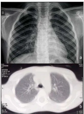
The other sister J.K. was born two years after the fatal outcome in K.K. Previously, the parents declined referral to genetic counseling. She was diagnosed with GD at two years of age on the basis of bone marrow infiltration with Gaucher cells and low leukocyte ß-glucosidase activity. Genetic test-ing (mutation analysis in Biochemical laboratory of Univer-sity of Amsterdam) revealed that J.K. was homozygous for L444P mutation. Initial manifestations of disease included massive hepatomegaly and splenomegaly, hematologic ab-normalities (thrombocytopenia, anemia), growth retardation and oculomotor apraxia as only neurological sign. That pa-tient was assigned with moderate SSI of 16 at the onset of disease. There were no clinical nor radiological signs of in-trinsic pulmonary disease within GD at the time. Enzyme re-placement therapy was started with imiglucerase at the age of four years with dose of 120 IU/kg/month. After two years of ERT dose was increased to 240 IU/kg/month due to dis-crete neurological progression. Other aspects of disease, however, showed a significant improvement: decreased vis-ceromegaly and compensatory growth spurt. At six years of age routine pulmonary function testing revealed moderately reduced forced expiratory volume in the first second, but without any clinical signs of lung disease. Chest radiography revealed fine reticulonodular pattern of involvement in lung interstitium. Six months later high resolution computerized tomography (HRCT) of the lungs showed marked bilateral interstitial markings with ground-glass appearance (Figure 1). Fiberoptic bronchoscopy with bronchoalveolar lavage (BAL) and transbronchial biopsy of lung parenchyma were performed. Numerous Gaucher cells were identified in BAL fluid (Figure 2). Histopathology of transbronchial biopsy
re-Fig. 1 – a) Chest radiography revealing fine reticulonodular pattern of lung interstitium involvement;
b) High resolution computerized tomography of the lungs showing marked bilateral interstitial markings with
ground-glass appearance
Fig. 2 – a) Numerous Gaucher cells identified in fluid recovered by bronchoalveolar lavage (HE, 400); b) Histopathology shows infiltration of lung interstitium
Volumen 68, Broj 12 VOJNOSANITETSKI PREGLED Strana 1073
Djordjević M, et al. Vojnosanit Pregl 2011; 68(12): 1071–1074. vealed infiltration of lung interstitium and alveolar spaces by Gaucher cells. Eighteen months later there were no signifi-cant changes in pulmonary function testing, while a control HRCT three years after baseline evaluation, showed no pro-gression in pulmonary changes. Regular echocardiographic exams showed no signs of pulmonary hypertension. The pa-tient remained without respiratory symptoms during a fol-low-up period.
Discussion
In the case of two siblings with GD intrinsic lung in-volvement several issues previously debated in the literature came to light. Genotype and phenotype correlation in GD, with rare exceptions, seems to be rather vague 8–10. However, several articles pointed out that pulmonary involvement ap-peared significantly more frequent in patients homoallelicfor L444P mutation than in those with other common genotypes 3, 5. The vast majority (90%) of reported pediatric cases with ho-mozygosity for L444P mutation and lung involvement were diagnosed with GD type III 3, 5. Our patient J.K. was a homo-zygote for L444P, and her sister was not genetically tested. A study on variability in phenotype among siblings with GD revealed that in only 4% of families affected members had different genotypes 11. The same study showed substantial discordance between sibs regarding severity of disease, but there was no available data about pulmonary involvement in these patients.
Another aspect of comparison between these two sisters includes possible effects of enzyme replacement therapy. First child died at the age of 3 years and 4 months with frank interstitial lung disease and progressive respiratory failure, while her sister started receiving ERT at 4 years and 2 months of age with no clinical or radiological signs of
pul-monary involvement at the time. Visceromegaly significantly subsided before lung involvement was proven in J.K., while in K.K. the enlargement of abdominal organs most probably contributed substantially to respiratory failure. The presence of chest radiography, HRCT and pulmonary function test ab-normalities in J.K. was noted after two and a half years of ERT and several months after the dosage of imiglucerase was increased to 240 IU/kg/month. In J.K. no echocardio-graphic signs of increased tricuspid incompetence gradient nor signs of pulmonary hypertension on chest X-ray were found. There have been reports that ERT induced pulmonary hypertension in a number of patients 12, 13. However, a recent study on a large group of children with non-neuronopathic GD did not show substantial incidence of pulmonary hyper-tension during ERT 14. Other reports imply that some im-provement of pulmonary function and HRCT findings could be expected in children on high dosage ERT 3, 7, 15.
The presence of Gaucher cells in J.K.'s BAL fluid and transbronchial biopsy histopathology confirmed diagnosis of pulmonary involvement in GD. Several previous studies pointed out the significance of BAL fluid citology in diag-nosing lung complications of GD and other inborn errors of metabolism 16, 17.
Conclusion
Primary lung disease in children homoallelic for L444P mutation in GBA gene has emerged as a significant clinical manifestation of GD with unclear response to enzyme re-placement therapy. Correlation of genotype and phenotype regarding lung disease could be less variable in comparison with other features of GD. Siblings with GD type III reported herein had differencies in progression of lung disease that could be related to ERT.
R E F E R E N C E S
1. Beutler E, Grabowski GA. Gaucher disease. In: Scriver, CR,
Beaudet, AL, Sly, WS, Valle, D, editors. Metabolic and
molecu-lar bases of inherited disease. New York: McGraw-Hill; 2001. p. 3635–68.
2. Lee RE, Yousem SA. The frequencyand type of lung
involve-ment in patients with Gaucher disease.Lab Invest 1988; 58(1): 54A.
3. Goitein O, Elstein D, Abrahamov A, Hadas-Halpern I, Melzer E,
Kerem E, et al.. Lung involvement and enzyme replacement
therapy in Gaucher's disease. Q J Med 2001; 94(8): 407–15.
4. Kerem E, Elstein D, Abrahamov A, Bar Ziv Y, Hadas-Halpern I,
Melzer E, et al. Pulmonary function abnormalities in type I
Gaucher disease. Eur Respir J 1996; 9(2): 340–5.
5. Santamaria F, Parenti G, Guidi G, Filocamo M, Strisciuglio P, Grillo
G, et al.Pulmonary manifestations of Gaucher disease: an in-creased risk for L444P homozygotes. Am J Respir Crit Care Med 1998; 157(3 Pt 1): 985–9.
6. Versteegh C, Avni F, Cuvelier P, Ferster A. Persistence of
pul-monary lesions in a 6-year-old boy with type I Gaucher's dis-ease treated by alglucerase since the age of 20 months. Arch Pediatr 1998; 5(12): 1341–3. (French)
7. Banjar H. Pulmonary involvement of Gaucher's disease in
chil-dren: a common presentation in Saudi Arabia. Ann Trop Pae-diatr 1998; 18(1): 55–9.
8. Mistry PK, Sirrs S, Chan A, Pritzker MR, Duffy TP, Grace ME, et
al. Pulmonary hypertension in type 1 Gaucher's disease: ge-netic and epigege-netic determinants of phenotype and response to therapy. Mol Genet Metab 2002; 77(1–2): 91–8.
9. Grabowski GA. Gaucher disease: gene frequencies and
geno-type/phenotype correlations. Genet Test 1997; 1(1): 5–12.
10.Whitfield PD, Nelson P, Sharp PC, Bindloss CA, Dean C,
Raven-scroft EM, et al. Correlation among genotype, phenotype, and
biochemical markers in Gaucher disease: implications for the prediction of disease severity. Mol Genet Metab 2002; 75(1): 46–55.
11.Amato D, Stachiw T, Clarke JT, Rivard GE. Gaucher disease:
variability in phenotype among siblings. J Inherit Metab Dis 2004; 27(5): 659–69.
12.Harats D, Pauzner R, Elstein D, Many A, Klutstein MW, Kramer
MR, et al. Pulmonary hypertension in two patients with type I Gaucher disease while on alglucerase therapy. Acta Haematol 1997; 98(1): 47–50.
13.Elstein D, Klustein MW, Lahad A, Abrahamov A, Hadas-Halpern I,
Zimran A. Echocardiographic assessment of pulmonary
hy-pertension in Gaucher's disease. Lancet 1998; 351(9115): 1544–6.
14.Rosengarten D, Abrahamov A, Nir A, Farber B, Glaser J, Zimran
Strana 1074 VOJNOSANITETSKI PREGLED Volumen 68, Broj 12
Djordjević M, et al. Vojnosanit Pregl 2011; 68(12): 1071–1074. children with Gaucher disease. Eur J Pediatr 2007; 166(6):
549–51.
15.Lee SY, Mak AW, Huen KF, Lam ST, Chow CB. Gaucher
dis-ease with pulmonary involvement in a 6-year-old girl: report of resolution of radiographic abnormalities on increasing dose of imiglucerase. J Pediatr 2001; 139(6): 862–4.
16.Carson KF, Williams CA, Rosenthal DL, Bhuta S, Kleerup E,
Diaz RP, et al. Bronchoalveolar lavage in a girl with
Gau-cher’s disease. A case report. Acta Cytol 1994; 38(4): 597– 600.
17.Poletti V, Chilosi M, Olivieri D. Diagnostic invasive procedures
in diffuse infiltrative lung diseases. Respiration 2004; 71(2): 107–19.