and Variants in Genes Involved in Immune Response
Beata Jurecka-Lubieniecka1, Rafal Ploski2, Dorota Kula1, Aleksandra Krol1, Tomasz Bednarczuk2, Zofia Kolosza1, Andrzej Tukiendorf1, Sylwia Szpak-Ulczok1, Anita Stanjek-Cichoracka3,
Joanna Polanska4, Barbara Jarzab1*
1Department of Nuclear, Medicine and Endocrine Oncology, Maria Sklodowska-Curie Memorial Cancer, Center and Institute of Oncology, Gliwice Branch, Gliwice, Poland, 2Department of Forensic Medicine, Medical University of Warsaw, Warsaw, Poland,3Department of Isotopic Diagnostic and Radiopharmaceutical, Medical University of Silesia, Katowice, Poland,4Institute of Automatic Control, Silesian Technical University, Gliwice, Poland
Abstract
Background: Graves’ disease (GD) is a complex disease in which genetic predisposition is modified by environmental factors. The aim of the study was to examine the association between genetic variants in genes encoding proteins involved in immune response and the age at diagnosis of GD.
Methods:735 GD patients and 1216 healthy controls from Poland were included into the study. Eight genetic variants in the
HLA-DRB1,TNF,CTLA4,CD40, NFKb, PTPN22, IL4andIL10 genes were genotyped. Patients were stratified by the age at diagnosis of GD and the association with genotype was analysed.
Results:Polymorphism in the HLA-DRB1, TNF and CTLA4 genes were associated with GD. The carriers of the HLA DRB1*03 allele were more frequent in patients with age at GD diagnosis#30 years than in patients with older age at GD diagnosis.
Conclusions:HLADRB1*03 allele is associated with young age at diagnosis of Graves’ disease in polish population.
Citation:Jurecka-Lubieniecka B, Ploski R, Kula D, Krol A, Bednarczuk T, et al. (2013) Association between Age at Diagnosis of Graves’ Disease and Variants in Genes Involved in Immune Response. PLoS ONE 8(3): e59349. doi:10.1371/journal.pone.0059349
Editor:Marco Giorgio Baroni, University of Cagliari, Italy
ReceivedOctober 4, 2012;AcceptedFebruary 13, 2013;PublishedMarch 27, 2013
Copyright:ß2013 Jurecka-Lubieniecka et al. This is an open-access article distributed under the terms of the Creative Commons Attribution License, which permits unrestricted use, distribution, and reproduction in any medium, provided the original author and source are credited.
Funding:This work was supported by Ministry of Science and Higher Education, Grant number N N519 579938. The funders had no role in study design, data collection and analysis, decision to publish, or preparation of the manuscript.
Competing Interests:The authors have declared that no competing interests exist. * E-mail: [email protected]
Introduction
Graves’ disease (GD) is an autoimmune disorder. Genetic predisposition modified by environmental factors is responsible for the pathogenesis if GD. Several genetic variants associated with GD were found, but none of them was introduced into clinical diagnostics [1]. An inherited predisposition to autoimmune thyroid diseases, including GD, currently involves about 20 identified genetic polymorphisms, including both thyroid-specific genes and those regulating the autoimmune response. Analyzed variants showed little impact on the development of GD with OR = 1,2–1,5 [2]. Interaction among genetic variants may increase the risk of GD disease [3]. Environment has a proven impact on the GD risk [4–5]. The impact of genetic factors may be higher than environmental factors in patients with young age at GD diagnosis [6]. GD is more frequent in the children of parents with young age at GD diagnosis [7–8]. Disease phenotype is also different between patients with young age at GD diagnosis than in patients with older age at GD diagnosis [9–12].
The aim of the study was to examine the association between genetic variants in genes encoding proteins involved in immune response and the age at GD diagnosis in polish population. Genetic variants in following genes were analyzed: HLADRB1, CTLA4, PTPN22, CD40, TNFa, NFkB, IL4 and IL10. Proteins
coded by these genes are involved in development of autoimmune diseases [13–15].
Materials and Methods
Subjects
group (n = 872) included anonymous, unrelated adults collected in the Regional Blood Transfusion Center.
Methods
Genomic DNA was extracted from whole blood according to standard protocols by salt protein precipitation. HLA-DRB1 genotyping in Gliwice was performed using sequence-specific oligonucleotides (SSO, Innolipa HLA-DRB1, Innogenetics) and sequence-specific primers (MSSP Class II DRB Only, One Lambda, Dynal All Set SSP DR test, Dynal Biotech) [16] and by Dynal All Set SSP Dr test (Dynal Biotech) in Warsaw [17]. The TNF, CTLA-4, NFKB, CD40, and PTPN22 genetic variants were genotyped by the PCR-RFLP method (Table 2) [16], [18–20]. PCR products were digested by appropriate restriction endonu-clease and digestion products were separated on the 3% agarose gel.
Statistical Analysis
Statistical analysis was performed using the statistical program STATA12.0. The distribution of genotypes and alleles in both groups was compared using the chi-squared test or the Fisher’s Exact Test. Bonferroni correction was applied for multiple comparisons. Hardy-Weinberg equilibrium was analyzed.
Due to the non-random allele association, the linkage disequi-librium ratios D’ were estimated. We performed multivariate logistic regression taking into the model HLA and TNF. To estimate the haplotypes effect on the disease risk we performed
analysis using ‘‘haplologit’’ command (STATA). The EM algo-rithm was used to calculate the haplotype frequency of two polymorphisms (HLADRB1*03, 2308A TNF), localized on chromosome 6.
Results
Allele Frequency Analysis
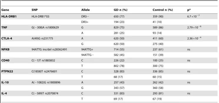
The genotype frequency in control group was in Hardy-Weinberg equilibrium. HLADRB1*03, 2308A TNF and 49G CTLA4 genetic variants had different allele frequency in GD patients than in controls (Table 3). GD patients and healthy controls did not differ when allele frequency of genetic variants in the CD40, NFkB, PTPN22, IL4 and IL10 genes was analyzed (Table 3). The frequency of the DRB1*03 allele in the GD patients was higher than the control group (23% vs. 10%, p = 1,76*1026) (Table 3). Similarly the2308A allele of theTNFgene and the 49G allele of theCTLA4gene occurred significantly more frequently in GD patients than in the control group (25% vs. 14%, p = 7,33*1028 and 50% vs. 40%, p = 6,21*1024 respectively) (Table 3).
Genotype Frequency Analysis
We observed significantly more frequent occurrences of at least one DRB1*03 allele in the GD patients compared to healthy subjects (OR 3,41; 95%CI 2,24–5,26) (Table 4).
Analysis of theTNFgene polymorphism revealed significantly higher incidence of AG and AA genotypes in GD patients
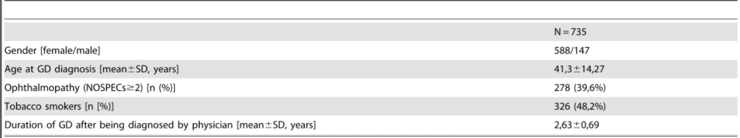
Table 1.Clinical characteristics of patients with Graves’ disease.
N = 735
Gender [female/male] 588/147
Age at GD diagnosis [mean6SD, years] 41,3614,27
Ophthalmopathy (NOSPECs$2) [n (%)] 278 (39,6%)
Tobacco smokers [n [%)] 326 (48,2%)
Duration of GD after being diagnosed by physician [mean6SD, years] 2,6360,69
doi:10.1371/journal.pone.0059349.t001
Table 2.Methods of genotyping of analyzed polymorphism.
Gene SNP Location Method PCR Primer F 59-39Primer R 59-39 RFLP enzyme
HLA-DRB1 HLA-DRB1*03 exon 2 PCR-SSP/PCR-SSO – –
TNF G(-308)A rs1800629 promoter PCR-RFLP F:AGGCAATAGGTTTTGAGGGCCAT R:TCCTCCCTGCTCCGATTCCG
NcoI
CTLA-4 A(49)G rs231775/ rs57563726
Codon 1, exon 3 PCR-RFLP F:CCAAGTCTCCACTTAGTTATCC R:CCTCCATCTTCATGCTCC
Bst71I
NFKB 94ATTG ins/del rs28362491
promoter PCR-RFLP F:TTTAATCTGTGAAGAGATGTGAATG R:CTCTGGCTTCCTAGCAGGG
Van91I
CD40 C(21)T rs1883832 Kozak sequence PCR-RFLP F:CCTCTTCCCCGAAGTCTTCC R:GAAACTCCTGCGCGGTGAAT
StyI
PTPN22 C(1858)T rs2476601 codon 620, exon 14
PCR-RFLP F:TCACCAGCTTCCTCAACCACA R:GATAATGTTGCTTCAACGGAATTT
XcmI
IL10 A(21082)G rs1800896 promoter ARMS-PCR FA:ACTACTAAGACTTCTTTGGGAA FG:CTACTAAGGCTTCTTTGGGAG R:AGAAGTCCTGATGTCACTGC
-IL4 C(2589)T rs2243250 promoter PCR-RFLP F:CACCTAAACTTGGGAGAACATTG R:GGAAAGATAGAGTAATATCA
AvaII
compared to controls, with increased risk for carriers of at least one2308A allele (OR = 2,70; 95%CI 1,99–3,70) (Table 4).
In case of theCTLA4 gene genotypes showed more frequent presence of GG homozygotes among patients compared to controls and increased GD risk in G allele carriers (OR 1,67, 95% CI 1,19–2,39) (Table 4).
Haplotype Frequency Analysis
SinceTNFgene is located within theHLAgenes we examined the haplotypes containing alleles HLADRB1*03 and 2308A of TNF. A strong linkage disequilibrium among genetic variants in the TNF and HLADRB1 genes was observed (D’.0,8). The most common haplotype in GD patients was the one containing all two
risk alleles. It was present in 20% of patients, which was associated with a significantly elevated GD risk (OR = 3,16, p = 1,94*10211) relative to 9% in healthy people.
Relationship between the HLA and the CTLA4 Susceptibility Genes
The interaction of HLADRB1*03 and A(49)G CTLA4 poly-morphisms was tested to check whether they exert any additional influence on GD development. They were found to act independently: the combined OR for the two alleles was 1,01.
Table 3.Allele frequency in patients with Graves’ disease (GD)and control group.
Gene SNP Allele GD n (%) Control n (%) p*
HLA-DRB1 HLA-DRB1*03 DR32 650 (77) 359 (90) 6.761027
DR3+ 194 (23) 41 (10)
TNF G(2308)A rs1800629 G 829 (75) 589 (86) 2.7961028
A 281 (25) 93 (14)
CTLA-4 A(49)G rs231775 A 620 (50) 411 (60) 2.3661024
G 620 (50) 275 (40)
NFKB 94ATTG ins/del rs28362491 94ATTG+ 714 (55) 237 (61) ns
94ATTG2 582 (45) 151 (39)
CD40 C(21)T rs1883832 C 226 (22) 100 (25) ns
T 802 (78) 300 (75)
PTPN22 C(1858)T rs2476601 C 328 (83) 336 (85) ns
T 68 (17) 60 (15)
IL-10 A(21082)G rs1800896 A 257 (43) 262 (42) ns
G 343 (57) 360 (58)
IL-4 C(2589)T rs2070874 C 331 (83) 293 (81) ns
T 69 (17) 67 (19)
*with Bonferroni correction. doi:10.1371/journal.pone.0059349.t003
Table 4.Genotypes of 4 polymorphisms with significant differences in allele frequencies between patients with Graves’ disease (GD) and control group.
Gene SNP Genotype GD n(%) Control n(%) p/OR/95% CI
HLADRB1 HLA-DRB1*03 DRB32/2 238 (56) 163 (81)
DRB32/+ 174 (41) 33 (17)
DRB3+/+ 10 (3) 4 (2)
DRB32/+and+/+ 1,75*1028
vs. DRB32/2 3,41 (2,24–5,26)
TNF G(2308)A GG 299 (54) 259 (76)
GA 231 (42) 71 (21)
AA 25 (4) 11 (3)
GA+AA vs. GG 5,78*102102,70 (1,99–3,70)
CTLA4 A49G AA 159 (26) 126 (37)
AG 302 (49) 156 (46)
GG 159 (25) 58 (17)
AG+GG vs. AA 0,03 1,68 (1,25–2,26)
Genotype-phenotype Correlation
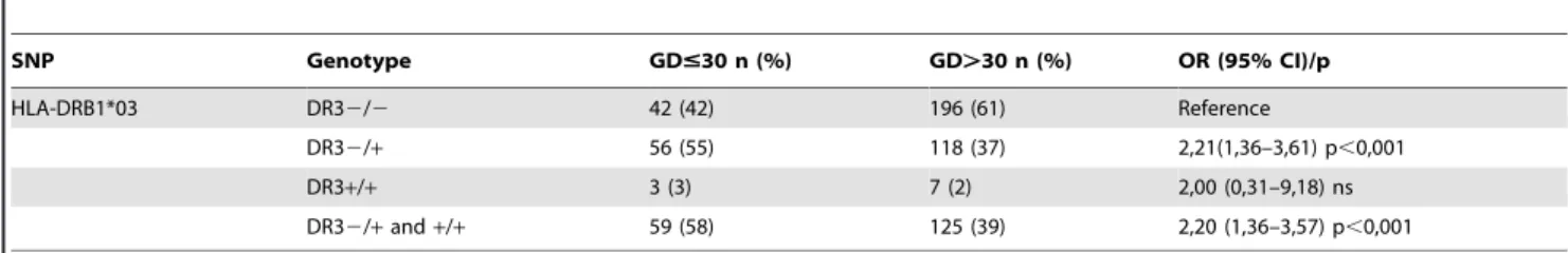
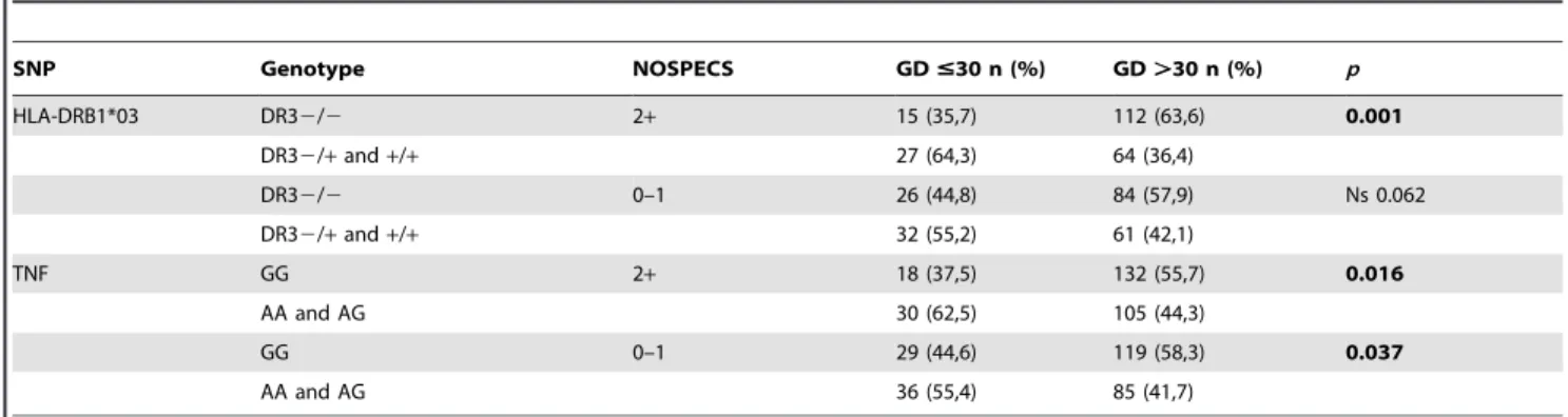
The genotype containing at least one DRB1*03 allele was associated with younger age at GD diagnosis (Table 5). No association was observed between DRB1*03 allele and duration of pharmacotherapy, GD relapse rate, number of radioiodine treatment courses, thyroid receptor (TR) Ab level and gender. The association between the HLADRB1*03 allele and age at GD diagnosis was confirmed in multivariable analysis. Graves’ ophthalmopathy of NOSPECS$2 was diagnosed in 278 (39.6%) patients (Table 1). Carriers of the HLADRB1*03 allele were significantly more common in young patients with Graves’ ophthalmopathy (p = 0.001) (Table 6).
Discussion
We confirmed previous observations, that genetic variants in HLA-DRB1, TNF and CLTA-4 genes are associated with GD. We found an association between the HLA-DRB1-03 polymor-phism and young age at GD diagnosis. A similar association between genetic markers and age at diagnosis was reported in type 1 diabetes [21], rheumatoid arthritis [22] and multiple sclerosis [23]. GD was diagnosed at younger age in patients with family history of this disease [9], [24].
We found a significant association between HLA DRB1*03 and the age at GD diagnosis when study population was stratified by the age of 30 years at GF diagnosis. Patients with age#30 years at GD diagnosis had higher frequency of the HLADRB1*03 allele. Genotypes containing the HLADRB1*03 allele occurred almost twice as much in patients at younger age at GD diagnosis. In our research, young carriers of the DR3 polymorphism were more common than older ones, both in family and sporadic GD. These results suggest the existence of different genetic conditions for the development of GD in young people. It can be assumed that earlier recognition of the disease in case of young patients with positive family history (FH) results from the increased alertness of ill parents. However, a violent course of disease in case of young people with distinct clinical symptoms forces to quick medical consultation regardless to FH [9], [24]. Young carriers of HLADRB1*03 allele are likely to be more susceptible to GD, despite a shorter exposure to environmental factors. HLADRB1*03 is necessary for induction of the immune response. The effect of HLA on the age at GD diagnosis can be explained by the molecular mechanism of initiation of the autoimmune processes involving the identification of an antigen bound to HLA molecule by T cells [25]. It has been suggested that the presence of the HLADRB1 allele with arginine at position 74 of the B1 chain changes the shape of the antigen-binding groove, allowing effective presentation of the TSH receptor peptide in the Caucasian population [26].
This is the first study in population of Poland showing association between HLADRB1*03 and age at GD diagnosis. There are only few studies which showed the association of this
polymorphism with younger age at GD diagnosis. In the Caucasian population, the association of HLADRB1 with GD diagnosis at a younger age has previously been shown in two studies. Lavard et al. affirmed that the presence of the HLADRB1*0301 allele resulted in a higher relative risk of GD among ninety Danish young patients [27]. Farid et al. observed an association between the HLADRB1*03 allele and eighty-three younger GD patients, taking the age of 30 as a cut-off line [28]. The association of otherHLAalleles with the age at GD diagnosis was also demonstrated in other ethnic groups - in the Chinese [29] and Japanese population [30]. The value of our study is a large well characterized population of GD patients from polish population. The carriers of the HLA DRB1*03 allele who are predisposed to earlier onset of GD may require further diagnostics for other autoimmune diseases.
We observed a higher frequency of the HLADRB1*03 allele in young patients with Graves’ ophthalmopathy. The results of genetic tests assessing the effect ofHLADR3on the development of ophthalmopathy in whole, age-irrespective groups of patients are incompatible [31–35]. On the other hand, there are no reports concerning the role of HLA in the development of ophthalmo-pathy in young GD patients, despite the fact that the clinical course of the disease is different in different age groups [36-39]. Objective difficulties in explaining the genetic background of ophthalmopathy in young patients are due to its significantly lower incidence in this age group, while in adults those difficulties are associated with the impact of environmental factors, especially smoking [40]. Other factors interfering with the research include clinical course of ophthalmopathy, and the time of its occurrence in relation to hyperthyroidism [41], which requires long-term observation of the patient.
While analysing the interaction between HLADRB1*03 and CTLA4 genetic variants we found no additive effect on the risk of GD. The impact of HLADRB1*03 [28], [42] and 49G of CTLA4 [43-44] on GD development was proven in many studies. Similarly as in case of our research, other authors reported only a weak association between GD and genetic variants in the CTLA4 gene [45].
We did not observed an HLA-independent effect of 2308A TNF variant on the risk of GD. This is consistent with previous reports [46–47]. Following multivariate logistic regression, the statistically significant effect of HLA has been established, only; the effect of polymorphism TNF was statistically insignificant. This suggests the leading effect of the HLA polymorphism on GD.
We were not able to confirm the association between PTPN22 and GD, what was reported by other research groups. However we observed a higher frequency of the risk T allele in GD patients than in control group, the differences was not significant. This may be caused by a potential dissimilarity among analysed populations [48].
Table 5.HLA-DRB1-03 genotypes in patients with Graves’ disease (GD) stratified by the age at GD diagnosis.
SNP Genotype GD#30 n (%) GD.30 n (%) OR (95% CI)/p
HLA-DRB1*03 DR32/2 42 (42) 196 (61) Reference
DR32/+ 56 (55) 118 (37) 2,21(1,36–3,61) p,0,001
DR3+/+ 3 (3) 7 (2) 2,00 (0,31–9,18) ns
DR32/+and+/+ 59 (58) 125 (39) 2,20 (1,36–3,57) p,0,001
Conclusion
Polymorphism of HLADRB1*03 is associated with early age at diagnosis of Graves’ disease.
Author Contributions
Conceived and designed the experiments: BJL BJ. Performed the experiments: BJL DK RP TB. Analyzed the data: ZK AT JP. Contributed reagents/materials/analysis tools: AK SSU ASC. Wrote the paper: BJL.
References
1. Davies TF (2007) Really significant genes for autoimmune thyroid disease do not exist - so how can we predict disease. Thyroid 17: 1027–29.
2. Zeitlin AA, Simmonds MJ, Gough SCL (2007) Genetic developments in autoimmune thyroid disease: an evolutionary process. Clin Endocrinol 68: 671– 82.
3. Tomer Y (2010) Genetic susceptibility to autoimmune thyroid disease: past, present and future. Thyroid 7: 715–25.
4. Rioux JD, Abbas AK (2005) Paths to understanding the genetic basis of autoimmune disease. Nature 435: 584–9.
5. Prummel MF, Strieder T, Wiersinga WM (2004) The environment and autoimmune thyroid diseases. Europ J Endocrinol 150: 605–18.
6. Tomer Y, Menconi F, Davies TF, Barbesino G, Rocchi R et al (2007) Dissecting genetic heterogeneity in autoimmune thyroid diseases by subset analysis. J Autoimmun 29: 69–77.
7. Hemminki K, Li X, Sundquist J, Sundquist K (2010) The epidemiology of Graves’ disease: evidence of a genetic and an environmental contribution. J Autoimmun 34: J307–13.
8. Segni M, Wood J, Pucarelli I, Toscano V, Toscano R et al (2001) Clustering of autoimmune thyroid diseases in children and adolescent: a study of 66 families. J Pediatr Endocrinol 14 Supl 5: 1271–5; discussion 1297–8.
9. Manji N, Carr-Smith JD, Boelaert K, Allahabadia A, Armitage M et al (2008) Influences of age, gender, smoking, and family history on autoimmune thyroid disease phenotype. J Clin Endocrinol & Metabolism 91: 4873–880. 10. Bossowski AT, Reddy V, Perry LA, Johnston LB, Banerjee K et al (2007)
Clinical and endocrine features and long-term outcome of Graves’ disease in early childhood. J Endocrinol Invest 30: 388–92.
11. Vos XG, Smit N, Endert E, Tijssen JG, Wiersinga WM (2009) Variation in phenotypic appearance of Graves’ disease: effect of genetic anticipation and duration of complaints. Europ J Endocrinol 16: 113–8.
12. Perrild H, Lavard L, Brock-Jacobsen B (1997) Clinical aspects and treatment of juvenile Graves’ disease. Exp Clin Endocrinol Diabetes 105 Suppl 4: 55–7. 13. Tomer Y, Huber A (2009) The etiology of autoimmune thyroid disease: a story
of genes and environment. J Autoimmun 32: 231–9.
14. Khalilzadeh O, Anvari M, Momen-Heravi F, Esteghamati A, Rashidi A et al (2010) Gene polymorphisms of interleukin-4, interleukin-10 and transforming growth factor-beta in Graves’ disease. Clin Exp Med 10(2): 123–8.
15. O’Sullivan B, Thompson A, Thomas R (2007) NF-kappa B as a therapeutic target in autoimmune disease. Expert Opin Ther Targets 11: 111–22. 16. Kula D, Bednarczuk T, Jurecka-Lubieniecka B, Polanska J, Hasse-Lazar K et al
(2006) Interaction of HLA-DRB1 alleles with CTLA-4 in the predisposition to Graves’ disease: the impact of DRB1*07. Thyroid 16: 447–53.
17. Bednarczuk T, Hiromatsu Y, Seki N, Płoski R, Fukutani T et al (2004) Association of tumor necrosis factor and human leukocyte antigen DRB1 alleles with Graves’ ophthalmopathy. Hum Immunol 65: 632–9.
18. Bednarczuk T, Hiromatsu Y, Fukutani T, Jazdzewski K, Miskiewicz P et al (2003) Association of cytotoxic T-lymphocyte-associated antigen-4 (CTLA-4) gene polymorphism and non-genetic factors with Graves’ ophthalmopathy in European and Japanese populations. Eur J Endocrinol 148: 13–8.
19. Kurylowicz A, Hiromatsu Y, Jurecka-Lubieniecka B, Kula D, Kowalska M et al (2007) Association of NFKB1–94ins/del ATTG promoter polymorphism with susceptibility to and phenotype of Graves’ disease. Genes Immun 8: 532–8.
20. Kurylowicz A, Kula D, Ploski R, Skorka A, Jurecka-Lubieniecka B et al (2005) Association of CD40 gene polymorphism (C-1T) with susceptibility and phenotype of Graves’ disease. Thyroid 15: 1119–24.
21. Paterson AD, Kennedy JL, Petronis A (1996) Evidence for genetic anticipation in non-Mendelian diseases. Am J Hum Genet 59: 264–8.
22. Radstake TR, Barrera P, Albers MJ, Swinkels HL, van de Putte LB et al (2001) Genetic anticipation in rheumatoid arthritis in Europe. European Consortium on Rheumatoid Arthritis Families. J Rheumatol 28: 962–7.
23. Cocco E, Sardu C, Lai M, Spinicci G, Contu P et al (2004) Anticipation of age at onset in multiple sclerosis: a Sardinian cohort study. Neurology 62: 1794–8. 24. Brix TH, Petersen HC, Iachine I, Hegedus L (2003) Preliminary evidence of
genetic anticipation in Graves’ disease. Thyroid 13: 447–51.
25. Sawai Y, DeGroot J (2000) Binding of human thyrotropin receptor peptides to a Graves’ disease – predisposing human leukocite antigen class II molecule. J Clin Endocrinol & Metabolism 85: 1176.
26. Ban Y, Davies TF, Greenberg DA, Conception ES, Osman R et al (2004) Arginine at position 74 of the HLA-DR beta1 chain is associated with Graves’ disease. Genes Immun 5: 203–8.
27. Lavard L, Madsen HO, Perrild H, Jacobsen BB, Svejgaard A (1997) HLA class II associations in juvenile Graves’ disease: indication of a strong protective role of the DRB1*0701,DQA1*0201 haplotype. Tissue Antigens 50: 639–41. 28. Farid NR, Stone E, Johnson G (1980) Graves’ disease and HLA: clinical and
epidemiologic association. Clin Endocrinol (Oxf) 13: 535–44.
29. Wong GW, Cheng SH, Dorman JS (1999) The HLA-DQ associations with Graves’ disease in Chinese children. Clin Endocrinol (Oxf) 50: 493–5. 30. Onuma H, Ota M, Sugenoya A, Inoko H (1994) Association of
HLA-DPB1*0501 with early-onset Graves’’ disease in Japanese. Hum Immunol 39: 195–201.
31. Weetman AP, So AK, Warner CA, Foroni L, Fells P et al (1988) Immunogenetics of Graves’ ophtalmopathy. Clin. Endocrinol (Oxf) 28(6): 619–28.
32. Allahabadia A, Heward JM, Nithiyanathan R, Gibson SM, Reuser TTQ et al (2001) MHC class II region, CTLA4 gene, and ophthalmopathy in patients with Graves’ disease. Lancet 358: 984–985.
33. Frecker M, Stenszky V, Balazs C, Kozma L, Kraszits E et al (1986) Genetic factors in Graves’ ophthalmopathy. Clin Endocrinol 25: 479–85.
34. Farid NR, Balazs C (1998) The genetic of thyroid associated ophthalmopathy. Thyroid 8: 407–9.
35. Yin X, Latif R, Bahn R, Davies TF (2012) Genetic profiling in Graves’ disease: further evidence for lack of distinct genetic contribution to Graves’ ophthalmo-pathy. Thyroid 22: 730–6.
36. Krassas GE, Segni M, Wiersinga WM (2005) Childhood Graves’ ophthalmo-pathy: resulf a European questionaire study. Europ J Endocrinol 153: 515–20. 37. Chan W, Wong GWK, Fan DSP, Cheng ACK, Lam DSC et al (2002)
Ophthalmopathy in childhood Graves’ disease. Br J Ophthalmol 86: 740–42. 38. Goldstein MG, Katowitz WR, Moshang T, Katowitz JA (2008) Pediatric
Thyroid-Associated Orbitopathy: The Children’s Hospital of Philadelphia Experience and Literature Review. Thyroid 9: 997–9.
39. Godakos AI, Boboridis K, Krassas GE (2010) Pediatric aspects in Graves’ orbitopathy. Pediatr Endocrinol Rev 7 Suppl 2Ł234+44.
Table 6.Distribution of HLA DRB1*03 and TNF polymorphisms in GD patients stratified by the diagnosis of Graves’ ophthalmopathy.
SNP Genotype NOSPECS GD#30 n (%) GD.30 n (%) p
HLA-DRB1*03 DR32/2 2+ 15 (35,7) 112 (63,6) 0.001
DR32/+and+/+ 27 (64,3) 64 (36,4)
DR32/2 0–1 26 (44,8) 84 (57,9) Ns 0.062
DR32/+and+/+ 32 (55,2) 61 (42,1)
TNF GG 2+ 18 (37,5) 132 (55,7) 0.016
AA and AG 30 (62,5) 105 (44,3)
GG 0–1 29 (44,6) 119 (58,3) 0.037
AA and AG 36 (55,4) 85 (41,7)
40. Villanueva R, Inzerillo AM, Tomer Y, Barbesino G, Meltzer M et al (2000) Limited genetic susceptibility to severe Graves’ ophthalmopathy: no role for CTLA-4 but evidence for an environmental etiology. Thyroid 10: 791–6. 41. Bednarczuk T, Gopinath B, Ploski R, Wall JR (2007) Susceptibility genes in
Graves’ ophthalmopathy: searching for a needle in a haystack? Clin Endocrinol 67: 3–19.
42. Zamani M, Spaepen M, Bex M, Bouillon R,Cassiman JJ (2000) Primary role of the HLA class II DRB1*0301 allele in Graves Disease. Am J Med Genet 95(5): 432–7.
43. Kouki T, Sawai Y, Gardine CA, Fisfalen ME, Alegre ML et al (2000) CTLA-4 gene polymorphism at position 49 in exon 1 reduces the inhibitory function of CTLA-4 and contributes to the pathogenesis of Graves’ disease. J Immunol 165: 6606–11.
44. Ueda H, Howson JM, Esposito L, Heward J, Snook H et al (2003) Association of the T-cell regulatory gene CTLA4 with susceptibility to autoimmune disease. Nature 423: 506–11.
45. Kavvoura FK, Akamizu T, Awata T, Ban Y, Chistiakov DA et al (2007) Cytotoxic T-lymphocyte associated antigen 4 gene polymorphism and autoimmune thyroid disease. J Clin Endocrinol & Metabolism 92(8): 3162–70. 46. Simmonds MJ, Heward JM, Howson JM, Foxall H, Nithiyananthan R et al
(2004) A systematic approach to the assessment of known TNF-alpha polymorphism in Graves’ disease. Genes Immun 5: 267–73.
47. Hunt PJ, Marshall SE, Weetman AP, Bunce M, Bell JI et al (2001) Histocompatibility leukocite antigens and closely linked immunomodulatory genes in autoimmune thyroid disease. Clin Endocrinol (Oxf) 55: 491. 48. Heward JM, Brand OJ, Barrett JC, Carr-Smith JD, Franklyn JA et al (2007)