Carotid intima-media thickness in spondyloarthritis patients
Espessamento da camada média-íntima da carótida em pacientes com
espondiloartrite
Thelma Larocca Skare
I, Guilherme Cortez Verceze
II, André Augusto de Oliveira
II, Sonia Perreto
IIIRheumatology and Echocardiography Units, Hospital Universitário Evangélico de Curitiba, Curitiba, Paraná, Brazil
ABSTRACT
CONTEXT AND OBJECTIVE: Accelerated atherosclerosis has become a major problem in rheumatic inlammatory disease. The aim here was to analyze carotid intima-media thickness (IMT) in spondyloarthri-tis (SpA) patients and correlate this with clinical parameters and inlammatory markers.
DESIGN AND SETTING: Cross-sectional analytical study at Rheumatology Outpatient Clinic, Evangelical University Hospital, Curitiba.
METHODS: IMTs (measured using Doppler ultrasonography) of 36 SpA patients were compared with controls. The IMT in SpA patients was associated with inlammatory markers, like erythrocyte sedimenta-tion rate (ESR), C-reactive protein (CRP) and Bath Ankylosing Spondylitis Disease Activity Index (BASDAI); and with clinical parameters, like axial or peripheral involvement, dactylitis, HLA B27, uveitis occurrence, Bath Ankylosing Spondylitis Functional Index (BASFI) and lipid proile.
RESULTS: The mean IMT in SpA patients was 0.72 ± 0.21 mm; in controls, 0.57 ± 0.13 mm (P = 0.0007). There were no associations with ESR, CRP, BASDAI or clinical data. In univariate analysis, greater IMT was seen in patients with longer disease duration (P = 0.014; Pearson R = 0.40; 95% confidence in-terval, CI = 0.06 to 0.65); higher triglycerides (P = 0.02; Spearman R = 0.37; 95% CI = 0.03 to 0.64); and older age (P = 0.0014; Pearson R 0.51; 95% CI = 0.21 to 0.72).
CONCLUSION: SpA patients have a higher degree of subclinical atherosclerosis than in controls, thus supporting clinical evidence of increased cardiovascular risk in rheumatic patients.
RESUMO
CONTEXTO E OBJETIVO: A aterogênese acelerada tem se tornado um grande problema nas doenças reumáticas inlamatórias. O objetivo foi analisar a espessura da camada íntima-média (ECIM) da carótida em pacientes com espondiloartrite (ES) e relacioná-la com parâmetros clínicos e marcadores inlamatórios.
TIPO DE ESTUDO E LOCAL: Estudo transversal analítico no Ambulatório de Reumatologia do Hospital Universitário Evangélico de Curitiba.
MÉTODOS: A ECIM (medida por Doppler) de 36 pacientes com ES foi comparada com controles. A ECIM de pacientes com ES foi associada com marcadores inlamatórios, como velocidade de hemossedimentação (VHS), proteína C-reativa (PCR), Bath Ankylosing Spondylitis Disease Activity Index (BASDAI), e com parâmetros clínicos, como envolvimento axial e periférico, dactilite, HLA B27, ocorrência de uveíte, Bath Ankylosing Spondylitis Functional Index (BASFI) e peril lipídico.
RESULTADOS: A ECIM média em pacientes com ES foi de 0,72 ± 0,21 mm, enquanto nos controles foi de 0,57 ± 0,13 mm (P = 0,0007). Não se encontrou associação com VHS, PCR, BASDAI e dados clínicos. Em análise univariada, maior ECIM foi encontrado nos indivíduos com maior duração de doença (P = 0,014; R Pearson = 0,40; 95% intervalo de coniança, IC = 0,06 to 0,65), aumento nos triglicerídeos (P = 0,02; R Spearman = 0,37; 95% IC = 0,03 to 0,64) e maior idade (P = 0,0014; R Pearson 0,51; 95% IC = 0,21 to 0,72).
CONCLUSÃO: Pacientes com ES têm maior grau de aterosclerose subclínica do que controles, dando suporte às evidências clínicas de aumento de risco cardiovascular em pacientes com doenças reumáticas.
IMD, PhD. Head of Rheumatology Unit, Hospital
Universitário Evangélico de Curitiba, Paraná, Brazil.
IIStudent, School of Medicine, Faculdade
Evangélica do Paraná (Fepar), Curitiba, Paraná, Brazil.
IIIMD. Cardiologist, Echocardiography Service,
Hospital Universitário Evangélico de Curitiba, Curitiba, Paraná, Brazil.
KEY WORDS:
Spondylarthropathies. Carotid artery diseases. Inlammation. Atherosclerosis. Spondylitis, ankylosing.
PALAVRAS-CHAVE:
Espondiloartropatias. Doenças das artérias carótidas. Inlamação.
INTRODUCTION
Chronic inlammatory rheumatic diseases are considered nowa-days to present a risk of cardiovascular events and increased car-diovascular mortality.1 Circulating mediators such as interleu-kin (IL)-6, tumor necrosis factor-alpha (TNF-alpha), C-reactive protein (CRP) and adhesion molecules secondary to rheumatic inlammatory processes contribute to all stages of atherosclero-sis: from endothelial dysfunction to atheroma formation, plaque instability and thrombus development.2
he association between subclinical atherosclerosis and severity of inlammatory response has been clearly demonstrated in patients with rheumatoid arthritis3,4 and systemic lupus ery-thematosus.4 However, in relation to spondyloarthritis (SpA), studies have produced divergent results.5,6 In this latter group of diseases, inlammatory markers do not accurately relect the underlying pathological events. It is well known that the associa-tion between CRP levels and inlammaassocia-tion is lower in SpA than in rheumatoid arthritis.7
SpA encompasses a group of diseases that include ankylos-ing spondylitis (AS), reactive arthritis (ReA), psoriatic arthri-tis (PsA), inlammatory bowel disease (IBD), related arthriarthri-tis and undiferentiated SpA (uSpA).8 his wide clinical spectrum results from combinations of diferent features such as inlamma-tory spinal involvement, peripheral arthritis, enthesitis, dactyli-tis, uveidactyli-tis, aortic incompetence and the presence of human leu-kocyte antigen (HLA)-B27.9
An analysis10 on mortality rate and causes of death among 398 patients with longstanding AS found that the group of patients that died had higher erythrocyte sedimentation rate (ESR). Gonzalez-Juanatey et al.6 found that carotid artery intima-media thickness (IMT) in AS patients without clinically evident cardiovascular disease was higher than in matched controls, although Chloe et al.11 could not conirm this inding.11
OBJECTIVE
In the present study, we analyzed carotid artery IMT in a cohort of local patients with SpA, comparing them with patients without rheumatic inlammatory disease, in order to investigate the pres-ence of subclinical atherosclerosis. We further analyzed the asso-ciation of clinical parameters and inlammatory activity in SpA with cardiovascular risks.
METHODS
his was a cross-sectional analytical study that was approved by the local Research Ethics Committee, and all participants signed a consent statement. We included 72 patients: 36 SpA patients who fulilled the ESSG (European Spondyloarthropathy Study Group)12 classiication criteria for diagnosing SpA and 36 con-trols. he sample of SpA patients was formed by all the patients
with a SpA diagnosis who were seen at our Outpatient Clinic from January 2011 to July 2011 (number estimated to be 55 patients) and who agreed to participate in the study.
he control group was selected among patients who sought the hospital for cataract and lower-leg varicose vein surgery without any known inlammatory disease. We excluded patients who had already experienced cardiovascular disease, heart failure, cerebro-vascular events, peripheral arterial disease or renal insuiciency (creatinine above 1.2 mg/dl), or who were on anticoagulants.
he patients and controls were interviewed to obtain demo-graphic data and data on associated diseases and tobacco and medication use. Patients were considered to have hypertension if their blood pressure was greater than 150/90 mm on at least two occasions.13 Information relating to diabetes mellitus was accessed through the patient’s history. For all patients and con-trols, cholesterol, triglyceride, low-density lipoprotein (LDL) cholesterol and high-density lipoprotein (HDL) cholesterol levels were determined ater fasting overnight, by means of the enzy-matic colorimetric method.
he SpA patients also gave responses to the Bath Ankylosing Spondylitis Disease Activity Index (BASDAI)14 and Bath Ankylosing Spondylitis Functional Index (BASFI)15 question-naires. heir ESR was determined using the Westergreen method and CRP levels using immunoturbidimetry.
BASDAI is an instrument that measures disease activity in SpA, and this is done through measurement of ive variables: spi-nal pain, peripheral joint pain, pain at enthesopathic sites, morn-ing stifness and fatigue, usmorn-ing a visual analogue scale. BASDAI values range from zero to 10, and values greater than 4 are consid-ered to be suggestive of high levels of disease activity.14 BASFI is a functional index constructed through ten questions about daily activities that also score from zero to ten (where zero is the best possible performance and ten, the worst).15 Both BASDAI and BASFI have been validated for use in the Portuguese language.16
of SpA without fulilling the diagnostic criteria for one deined disease.20,21
A history of uveitis was considered to be present when diag-nosed by an ophthalmologist. Peripheral arthritis was considered to be present when current or past synovitis was diagnosed by a doctor (in the 44-joint count).22 Assessment of axial involve-ment (sacroiliitis and/or spondylitis) was done through imaging the sacroiliac joints, lumbar and cervical spine by radiography and/or computed tomography scans.22 Enthesitis was consid-ered to be present when the patient reported spontaneous pain or tenderness on examination at enthesis sites (according to the Maastricht Ankylosing Spondylitis Score).22 Dactylitis was con-sidered to be present when current or past dactylitis was diag-nosed by a doctor.22
Carotid IMT measurements were made on both sides using color Doppler equipment (ESAOTE, model MEGA CVX,with 7.5 MHz linear transducers). Transversal and longitudinal slices were imaged at the common carotid vessel, 3 cm below the bul-bus. All tests were read by a single cardiologist who was blinded to clinical information. We considered that the patient had no thickening if the measurement on the intima-media complex was less than 0.8 mm. hickening was present if the measurements were between 0.8 mm and 1.5 mm; and atheromatous plaques was present when the measurement was greater than 1.5 mm.23-25 For statistical calculations, we took the carotid IMT value that was greater, between the two sides.
he data obtained were studied using contingency and fre-quency tables. For association studies on nominal data, the chi-square and Fisher tests were used and for association stud-ies on numerical data, the Mann-Whitney and unpaired t tests were used. For correlation studies, the Pearson and Spearman tests were applied. Calculations were done with the aid of the GraphPad Prism sotware, version 4.0. To further study the cor-relation of IMT with variables with P < 0.05, we performed mul-tivariate analysis using the Medcalc sotware, version 12.1.3.0. he signiicance level used was 5%.
RESULTS
Among the 36 SpA patients studied: 1/36 (2.7%) was ReA, 3/36 (8.33%) were PsA; 9/36 (25%) were USpA and 23/36 (63.88%) were AS. In the SpA group, the age range was from 19 to 74 years (mean 43.9 ± 10.97 years), the mean disease duration was 10.59 ± 10.57 years (range: from 1 to 48 years) and 47.2% were males and 52.7% were females. In this sample, we found that 90.62% had axial involvement, 53.12% enthe-seal involvement, 50% peripheral arthritis, 25% uveitis and none dactylitis. For 17 patients, there was data on HLA B27 and 76.4% were positive. Regarding treatment, 52.7% were using non-steroidal anti-inflammatory drugs; 11.1% were on methotrexate; 38.8% were using sulphasalazine, 19.4% were
on prednisone and 25% were on anti-TNF-alpha drugs (19.4% were on etanercept and 5.5% on adalimumab).
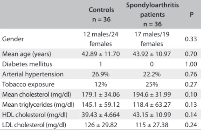
he data relating to sample pairing is shown in Table 1. he carotid IMT in the controls ranged from 0.3 to 0.67 mm (mean 0.57 ± 0.13 mm); in SpA patients, it ranged from 0.44 to 1.40 mm (mean 0.72 ± 0.21 mm), with P = 0.0007 (Figure 1).
In the SpA sample, the ESR ranged from 2 to 100 mm (median 13.5 mm); the CRP values were between 0.3 and 4.64 mg/dl (median 0.34 mg/dl). Analysis on carotid IMT according to the functional index and inlammatory activity parameters did not ind any diference, as seen in Table 2.
From studying the carotid IMT according to disease dura-tion, we observed a positive correladura-tion, with P = 0.014 (Pearson R = 0.409; 95% conidence interval, CI = 0.06 to 0.65) (Figure 2). From analysis on the lipid proile and IMT in SpA patients, we did not ind any correlation with total cholesterol (P = 0.42), HDL cholesterol (P = 0.39) or LDL cholesterol (P = 0.39). However, there were positive correlations with triglycerides (P = 0.02;
Controls n = 36
Spondyloarthritis patients
n = 36
P
Gender 12 males/24
females
17 males/19
females 0.33
Mean age (years) 42.89 ± 11.70 43.92 ± 10.97 0.70
Diabetes mellitus 1 0 1.00
Arterial hypertension 26.9% 22.2% 0.76
Tobacco exposure 12% 25% 0.27
Mean cholesterol (mg/dl) 179.1 ± 34.06 194.6 ± 31.99 0.10 Mean triglycerides (mg/dl) 145.1 ± 59.12 118.4 ± 63.27 0.13 HDL cholesterol (mg/dl) 39.43 ± 4.664 43.15 ± 10.99 0.14 LDL cholesterol (mg/dl) 126 ± 29.82 115 ± 27.38 0.24 Table 1. Gender, age, diabetes mellitus and lipid proile among 36 patients with spondyloarthritis and 36 controls
HDL = high-density lipoprotein; LDL = low-density lipoprotein.
Figure 1. Carotid intima-media thicknesses in 36 patients with spondyloarthritis and 36 controls.
SpA = spondyloarthritis; IMT = intima-media thickness.
control SpA
0.0 0.5 1.0 1.5
Spearman R = 0.37; 95% CI = 0.03 to 0.64) and the patient’s age (P = 0.0014; Pearson R = 0.51; 95% CI = 0.21 to 0.72). From studying carotid IMT in the SpA patients according to SpA sub-types, no diferences could be found (P = 0.29). From analyzing carotid IMT according to clinical manifestations, we found the data shown in Table 3.
Patients on anti-TNF-alpha drugs had IMT between 0.49 and 1.5 mm (mean 0.8 ± 0.33 mm) and those not using these drugs had IMT between 0.50 and 1.2 mm (mean 0.73 ± 0.18 mm); P = 0.49.
DISCUSSION
Arterial wall thickening has a strong prognostic value for cardio-vascular events, and carotid IMT assessment allows easy identii-cation of patients at risk, as shown in a recent systematic review and meta-analysis.26 In particular, it was demonstrated that an absolute diference of 0.1 mm increases the risk of future myocar-dial infarction by 10 to 15% and the stroke risk by 13 to 18%.26,27 he use of carotid IMT as a predictor of cardiovascular events has predominantly been reported in the literature in relation to patients with rheumatic diseases.3
It is well known that in chronic inlammatory rheumatic dis-eases, systemic inlammation can act independently or synergis-tically with traditional risk factors in relation to development of cardiovascular complications, but each rheumatic disease may difer regarding the amount and type of inlammation.27 In rheu-matoid arthritis, the severity of disease has consistently been proven to be the major determinant of myocardial infarction and stroke.28 he same has been proven for PsA,29 but other types of SpA have not been so well studied. In cardiovascular disease, ele-vated CRP and interleukin (IL)-6 levels correlate with adverse events.5 In rheumatoid arthritis, CRP levels are increased, and this correlates well with disease activity but in SpA, CRP is usu-ally not a good guide for inlammation. It is elevated in only 50-60% of the cases of active disease.7 Despite these diferences between SpA and rheumatoid arthritis, we found in the present study that SpA patients were also at risk of developing acceler-ated atherosclerosis.
Our indings conirm those of Gonzalez-Gay et al.,4 who studied 64 SpA patients and found that they had greater carotid IMT and carotid plaque than shown by controls. hese authors also found an association between this inding and disease dura-tion, as we also did. his inding probably relects the additive efect of prolonged inlammation.
Although the atherosclerotic process is associated with inlammation, we could not demonstrate any association for carotid IMT in relation to ESR, CRP or BASDAI. As already mentioned, CRP levels are not good markers for disease inlam-mation in SpA. Peters et al.30 also found that carotid IMT did not show any association with CRP or BASDAI in ankylosing spondylitis patients. hese indings can be explained by the fact
that BASDAI and ESR are measurements that relect current inlammation while the atherosclerotic process is built up over time. Furthermore, the use of anti-TNF-alpha did not alter the IMT in the present analysis. Even though this drug has well-known anti-inlammatory properties,31 its action on the lipid proile is debatable.32-34 An increase in triglyceride levels has been shown in anti-TNF-alpha users with PsA.32 in RA cases, some authors33 have found an increase in HDL cholesterol, while others have not noted any diference in the atheroscle-rotic index (total cholesterol/HDL cholesterol).34 Modiications to the lipid proile may compensate for the anti-inlammatory efect of this drug.
R 95% CI P
BASDAI 0.19* - 0.14 to 0.49 0.26
ESR (mm, 1st hour) 0.046† - 0.35 to 0.31 0.89
CRP (mg/dl) 0.046† - 0.29 to 0.37 0.79
BASFI 0.25* - 0.07 to 0.53 0.13
Table 2. Correlation of inlammatory disease activity markers and functional index with carotid intima-media thickness in 36 patients with spondyloarthritis
BASDAI = Bath Ankylosing Spondylitis Activity Index; BASFI = Bath Ankylosing Spondylitis Functional Index; ESR = erythrocyte sedimentation rate; CRP = C-reactive protein; CI = conidence interval; *Pearson correlation; †Spearman correlation.
Figure 2. Correlation of carotid intima-media thickness with disease duration in 36 patients with spondyloarthritis.
0.0 0.5 1.0 1.5 2.0
0 10 20 30 40 50 60
disease duration
carotid IMT
IMT = intima-media thickness.
With Without P
Axial involvement 0.75 ± 0.22 0.77 ± 0.28 0.91
Peripheral arthritis 0.76 ± 0.24 0.74 ± 0.20 0.82
Uveitis 0.86 ± 0.32 0.71 ± 0.17 0.12
Entheseal involvement 0.74 ± 0.25 0.77 ± 0.19 0.69 Positive HLA B27 (n = 17) 0.82 ± 0.28 0.66 ± 0.17 0.30 Table 3. Mean values of carotid intima-media thickness (in mm) in patients with and without spondyloarthritis, according to clinical indings
No special clinical characteristic was associated with higher carotid IMT in SpA patients in the present study. he same has been noted by others.4 hus, surveillance of atherosclerotic and inlammatory processes should also be done for all SpA patients, in order to avoid their severe consequences.
CONCLUSIONS
Concluding, patients with SpA have higher carotid IMT than shown by controls. Every efort should be made in order to con-trol inlammation and traditional risk factors in this population, to avoid the consequences of accelerated atherogenesis.
REFERENCES
1. Goodson NJ, Symmons DP, Scott DG, et al. Baseline levels of
C-reactive protein and prediction of death from cardiovascular
disease in patients with inlammatory polyarthritis: a ten-year
followup study of primary care-based inception cohort. Arthritis
Rheum. 2005;52(8):2293-9.
2. Huang AL, Vita JA. Efects of systemic inlammation on
endothelium-dependent vasodilation. Trends Cardiovasc Med. 2006;16(1):15-20.
3. Salmon JE, Roman MJ. Subclinical atherosclerosis in rheumatoid
arthritis and systemic lupus erythematosus. Am J Med. 2008;121(10
Suppl 1):S3-8.
4. Gonzalez-Gay MA, Gonzalez-Juanatey C, Piñeiro A, et al.
High-grade C-reactive protein elevation correlates with accelerated
atherogenesis in patients with rheumatoid arthritis. J Rheumatol.
2005;32(7):1219-23.
5. Heeneman S, Daemen MJ. Cardiovascular risks in spondyloarthritides.
Curr Opin Rheumatol. 2007;19(4):358-62.
6. Gonzalez-Juanatey C, Vasquez-Rodriguez TR, Miranda-Filloy JA, et
al. The high prevalence of subclinical atherosclerosis in patients
with ankylosing spondylitis without clinically evident cardiovascular
disease. Medicine (Baltimore). 2009;88(6):358-65.
7. van der Horst-Bruinsma IE, Lems WF, Dijkmans BA. A systematic
comparison of rheumatoid arthritis and ankylosing spondylitis. Clin
Exp Rheumatol. 2009;27(4 Suppl 55):S43-9.
8. Toussirot E, Wendling D. Late-onset ankylosing spondylitis and related
spondylarthropathies: clinical and radiological characteristics and
pharmacological treatment options. Drugs Aging. 2005;22(6):451-69.
9. Toussirot E. Late-onset ankylosing spondylitis and spondylarthritis:
an update on clinical manifestations, diferential diagnosis and
pharmacological therapies. Drugs Aging. 2010;27(7):523-31.
10. Lehtinen K. Mortality and causes of death in 398 patients admitted
to hospital with ankylosing spondylitis. Ann Rheum Dis. 1993;52(3):
174-6.
11. Choe JY, Lee MY, Rheem I, et al. No diferences of carotid intima-media
thickness between young patients with ankylosing spondylitis and
healthy controls. Joint Bone Spine. 2008;75(5):548-53.
12. Dougados M, van der Linden S, Juhlin R, et al. The European
Spondylarthropathy Study Group preliminary criteria for
the classiication of spondylarthropathy. Arthritis Rheum.
1991;34(10):1218-27.
13. Gonzalez-Gay MA, Gonzalez-Juanatey C, Lopez-Diaz MJ, et al.
HLA-DRB1 and persistent chronic inlammation contribute to
cardiovascular events and cardiovascular mortality in patients with
rheumatoid arthritis. Arthritis Rheum. 2007;57(1):125-32.
14. Garrett S, Jenkinson T, Kennedy LG, et al. A new approach to deining
disease status in ankylosing spondylitis: the Bath Ankylosing
Spondylitis Disease Activity Index. J Rheumatol. 1994;21(12):2286-91.
15. Calin A, Garrett S, Whitelock H, et al. A new approach to deining
functional ability in ankylosing spondylitis: the development of
the Bath Ankylosing Spondylitis Functional Index. J Rheumatol.
1994;21(12):2281-5.
16. Torres TM, Ciconelli RM. Instrumentos de avaliação em espondilite
anquilosante [Outcome measures in ankylosing spondylitis]. Rev Bras
Rheumatol. 2006;46(Suppl 1):52-9.
17. van der Linden S, Valkenburg HA, Cats A. Evaluation of diagnostic
criteria for ankylosing spondylitis. A proposal for modiication of the
New York criteria. Arthritis Rheum. 1984;27(4):361-8.
18. Moll JM, Wright V. Psoriatic arthritis. Semin Arthritis Rheum.
1973;3(1):55-78.
19. Kingsley G, Sieper J. Third International Workshop on Reactive
Arthritis. 23-26 September 1995, Berlin, Germany. Report and
abstracts. Ann Rheum Dis. 1996;55(8):564-84.
20. Cruzat V, Cuchacovich R, Espinoza LR. Undiferentiated
spondyloarthritis: recent clinical and therapeutic advances. Curr
Rheumatol Rep. 2010;12(5):311-7.
21. De La Mata J, Maese J, Martinez JA, Rosario P, Loza E. Current
evidence of the management of undiferentiated spondyloarthritis: a
systematic literature review. Semin Arthritis Rheum. 2011;40(5):421-9,
429.e1-3.
22. Sieper J, Rudwaleit M, Baraliakos X, et al. The Assessment of
SpondyloArthritis international Society (ASAS) handbook: a guide to
assess spondyloarthritis. Ann Rheum Dis. 2009;68 Suppl 2:ii1-44.
23. Bots ML, Grobbee DE, Hofman A, Witteman JC. Common carotid
intima-media thickness and risk of acute myocardial infarction: the
role of lumen diameter. Stroke. 2005;36(4):762-7.
24. O’Leary DH, Polak JF, Kronmal RA, et al. Carotid-artery intima and
media thickness as a risk factor for myocardial infarction and stroke
in older adults. Cardiovascular Health Study Collaborative Research
Group. N Engl J Med. 1999;340(1):14-22.
25. Simon A, Gariepy J, Chironi G, Megnien JL, Levenson J. Intima-media
thickness: a new tool for diagnosis and treatment of cardiovascular
risk. J Hypertens. 2002;20(2):159-69.
26. Lorenz MW, Markus HS, Bots ML, Rosvall M, Sitzer M. Prediction of
clinical cardiovascular events with carotid intima-media thickness:
a systematic review and meta-analysis. Circulation. 2007;115(4):
27. Tyrrell PN, Beyene J, Feldman BM, et al. Rheumatic disease and carotid
intima-media thickness: a systematic review and meta-analysis.
Arterioscler Thromb Vasc Biol. 2010;30(5):1014-26.
28. Mathieu S, Joly H, Baron G, et al. Trend towards increased arterial
stifness or intima-media thickness in ankylosing spondylitis patients
without clinically evident cardiovascular disease. Rheumatology
(Oxford). 2008;47(8):1203-7.
29. Gonzalez-Juanatey C, Llorca J, Amigo-Diaz E, et al. High prevalence
of subclinical atherosclerosis in psoriatic arthritis patients without
clinically evident cardiovascular disease or classic atherosclerosis risk
factors. Arthritis Rheum. 2007;57(6):1074-80.
30. Peters MJ, van Eijk IC, Smulders YM, et al. Signs of accelerated
preclinical atherosclerosis in patients with ankylosing spondylitis.
J Rheumatol. 2010;37(1):161-6.
31. Silva LC, Ortigosa LC, Benard G. Anti-TNF-α agents in the treatment
of immune-mediated inlammatory diseases: mechanisms of action
and pitfalls. Immunotherapy. 2010;2(6):817-33.
32. Castro KR, Aikawa NE, Saad CG, et al. Inliximab induces increase in
triglyceride levels in psoriatic arthritis patients. Clin Dev Immunol.
2011;2011:352686.
33. Spanakis E, Sidiropoulos P. Papadakis J, et al. Modest but sustained
increase of serum high density lipoprotein cholesterol levels in
patients with inlammatory arthritides treated with inliximab.
J Rheumatol. 2006;33(12):2440-6.
34. Vis M, Nurmorhamed MT, Wolbink G, et al. Short term efects of
inliximab on the lipid proile in patients with rheumatoid arthritis.
J Rheumatol. 2005;32(2):252-5.
Sources of funding: Guilherme Cortez Verceze received a scholarship grant from the Programa Institucional de Bolsas de Iniciação Cientíica -
Conselho Nacional de Desenvolvimento Cientíico e Tecnológico
(PIBIC-CNPq), Brazil
Conlict of interest: None
Date of irst submission: December 5, 2011
Lastreceived: August 11, 2012
Accepted: August 16, 2012
Address for correspondence:
Thelma Larocca Skare
Rua João Alencar Guimarães, 796
Santa Quitéria – Curitiba (PR) – Brasil
CEP 80310-420
Tel. (+55 41) 3274-1659