INTRODUCTION
The acute-phase response is a pathophysiological defense mechanismassociated with inlammatory states that, despite the already established name, occurs in both acute and chronic inlammation. It is characterized by the increase or decrease in serum concentration of certain proteins in consequence of a stimulus that causes tissue damage.1-3 Currently, the term
inlammatory biomarker is preferred when referring to proteins involved in this response.
Analysis of inlammation biomarkers is used in rheumatologic diseases to monitor disease activity, correlated with other clinical and laboratory data, and to differentiate between active disease and the presence of infection. This article reviews the use of inlammatory activity tests currently available in health care.
HISTORY
In 1930, investigators discovered a protein that reacted with the C-polysaccharide capsule of the S. pneumoniae from the blood of patients during the acute-phase of pneumococcal
he use of inlammatory laboratory
tests in rheumatology
Nilton Salles Rosa Neto1, Jozélio Freire de Carvalho2
Received on 10/14/2008. Approved on 03/03/2009. We declare no conflict of interest. 1. Resident Doctor at Rheumatology at Hospital das Clínicas (FMUSP)
2. Assistant Professor, Discipline of Rheumatology, Medicine School (USP), and Assistant doctor of the Rheumatology Department at Hospital das Clínicas (FMUSP)
Correspondence to: Dr. Jozélio Freire de Carvalho. Disciplina de Reumatologia. Av. Dr. Arnaldo, 455, 3º andar, sala 3190, Cerqueira César, São Paulo, SP - Brasil. Tel./Fax: 55 11 3061-7490. E-mail: [email protected]
ABSTRACT
Inlammation is the hallmark of rheumatic diseases. Tissue injury response promotes several modiications, which
result in elimination of the offending agent, limitation of tissue damage, and restoration of affected structures. Such
modiications depend on the increase or decrease of the serum concentration of certain proteins known as inlamma -tory biomarkers. Labora-tory analysis of these markers assists in monitoring disease activity and treatment response.
Rheumatologists have available methods that evaluate inlammatory reaction such as C-reactive protein, erythrocyte
sedimentation rate, and protein electrophoresis, among others. In this paper, we review some of those biomarkers and their use in rheumatic diseases.
Keywords: acute-phase proteins, C-reactive protein, erythrocyte sedimentation rate, rheumatic diseases, inlammatory response.
pneumonia. This protein was named C-reactive protein (CRP). Since then, studies on changes of plasma proteins in serum of patients acutely ill due to infections are performed. The proteins found in this situation were called acute-phase proteins; and the inlammatory reaction, or the organism’s response to tissue injury, called acute-phase reaction.4
Subsequently, the presence of these proteins was veriied after other events, such as trauma, ischemia, neoplasm, and hypersensitivity reaction. Their concentration were also altered in chronic inlammatory states.1,2
ACUTE-PHASE RESPONSE
because of the availability of the method, or the cost of its results. Tables 1 and 2 present some of the acute-phase proteins, divided according to their original biological function.
Behavioral changes and physiological, biochemical, and nutritional alterations add up to complete the acute-phase response.2,3
Neuroendocrine Changes
One of the major characteristic of this phase is the presence of fever, a response that promotes an optimal enzyme function and promotes the stabilization of cell membranes. Indisposition and drowsiness is present, which reduce the body’s energy consumption.
Modulating the inlammatory response, there is increased secretion of corticotropin releasing hormone (CRH), adrenocorticotropic hormone (ACTH) and cortisol, antidiuretic hormone (ADH) and catecholamines; and decrease of insulin-like growth factor (IGF-1).
Hematopoietic Changes
As a result of inlammation, leukocytosis and thrombocytosis can be found and, in prolonged cases, also anemia of chronic disease.
Metabolic Changes
These include muscle loss and negative nitrogen balance, resulting in chronic cases; there is growth limitation in children and cachexia in adults. There is a decrease of gluconeogenesis and acceleration of osteoporosis. Increased liver lipogenesis and lipolysis in adipose tissue are found, as well as a decrease in muscular and adipose lipoprotein lipases activity, with the objective, in some cases of infection, to increase the concentration of lipoproteins, promoting a larger connection to lipopolysaccharides (LPS), resulting in less toxicity to the body.
Hepatic Changes
The liver participates in the production and liberation of innumerous proteins related to the inlammatory response. Physiologically, there is an increase in metallothionein, nitric oxide synthase, heme oxygenase, superoxide dismutase, tissue inhibitor of metalloproteinase-1, and decrease in phosphoenolpyruvate carboxykinase activity.
Changes in Other Components of the Plasma
As a control of reactions in progress, there is consumption of zinc, iron and copper, and increased serum retinol and antioxidants, such as glutathione.
Table 1
Positive inlammatory biomarkers.
Coagulation and fibrinolytic system
Fibrinogen
Plasminogen
Tissue plasminogen activator
Urokinase
Protein S
Vitronectin
Plasminogen-activator inhibitor 1
Complement system
C3; C4; C9
Factor B
C1 inhibitor (C1 INH)
C4b-binding protein (C4b)
Mannose-binding lectin (MBL)
Transport proteins
Ceruloplasmin
Haptoglobin
Hemopexin
Participants of inflammatory responses
Secreted phospholipase A2 (sPLA2-IIA)
Lipopolysaccharide-binding protein
Interleukin-1-receptor antagonist (IL-1 RA)
Granulocyte colony-stimulating factor (G-CSF)
Antiproteases
α1-Protease inhibitor
α1-Antichymotripsin
Pancreatic secretory trypsin inhibitor
Inter-α-trypsin inhibitors
Others
C-reactive protein
Serum amyloid A
α1-acid glycoprotein (AGP)
Fibronectin
Ferritin
Angiotensinogen
Retinol binding protein
Adapted from Kushner and Gabay2 with permission of the authors. Copyright
CLASSIFICATION
The biomarkers of inlammation are divided into four groups:1
Host defense proteins - they participate in the recognition and elimination of pathogens: C-reactive protein, mannose binding lectin, lipopolysaccharide-binding protein, complement protein, ibrinogen;
Serum protease inhibitors - they act to limit tissue damage, neutralizing proteolytic enzymes and oxygen metabolites: α1- antiproteinas, α1-anti-chymotrypsin, α2-antiplasmin, C1 inhibitor;
Transport protein with antioxidant activity - responsible for containing the inlammatory reaction and restoring the original structure injured: ceruloplasmin, hemopexin, haptoglobin;
Others - serum amyloid A (SAA) protein, IL-1 receptor antagonist, α1-acid glycoprotein, group IIA secretory phospholipase A2 (sPLA2-IIA).
There is significant difference in terms of kinetics, magnitude and response duration among the inlammatory biomarkers. The CRP and the SAA can be detected after four hours of injury, and they have peak concentration after 24-72 hours, which can reach a thousand times the normal value.3
Fibrinogen has peak concentration 7 to 10 days after injury, and rises 2 to 3 times the normal (Figure 1).
Only some of these markers are available for rheumatologist’s routine practice, and this paper addresses the origin and biological functions of the proteins involved, and the methods to determine their activities. In the following text the uses of these markers in different rheumatic diseases are found in greater detail.
C-REACTIVE PROTEIN
This is the most investigated biomarker, which promotes the interaction between humeral and cellular immunity. It
is produced by the liver and classiied as a pentraxin – a pentamer with a binding site for phosphatidylcholine (calcium ion dependent) and other sites in the opposite side that bind to the complement C1q system component and to the Fc immunoglobulin (Fcγ portion).1,3
C-reactive protein (CRP) binds to pathogens and damaged/apoptotic cells (phosphatidylcholine), causing their elimination by activating the complement system and the phagocytes (C1q and Fcγ). Because of the binding and cellular attraction function, it can be considered as an opsonin.5,6 It
also acts regulating the extension and the intensity of the inlammatory reaction.
Although its function is similar to that of an antibody and it participates in the innate immunity, there is no description of CRP disabled states, which in principle is incompatible with life.
Complement activation occurs through the classic pathway, by the positioning of C3 and C4 fragments on CRP and ligand, formation of C3 convertase by cleaving C3 into C3a, an anaphilatoxin that induces the realize of histamine from basophiles and mast cells, and C3b, that acts as an opsonin, attracting phagocytes (macrophages) to the inflammation site. The activation does not convert C5; therefore, there is no ampliication of the pro-inlammatory effects or formation of membrane attack complex (MAC) directly by the CRP. CRP and the complement classic pathway system act together, promoting clearance of apoptotic cells without causing cell
Adapted from Gitlin and Colten48 with permission of the authors.
Figure 1. Characteristic pattern of inlammatory biomarkers in tissue damage.
Plasma concentr
ation (% c
hange)
30,100 30,000 700 600 500 400 300 200 100 0
0 7
Inflammatory stimulus
14 21
Days C-reactive protein Haptoglobin Albumin Serum amyloid A Fibrinogen
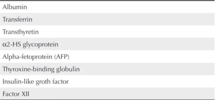
Table 2
Negative inlammatory biomarkers
Albumin
Transferrin
Transthyretin
α2-HS glycoprotein
Alpha-fetoprotein (AFP)
Thyroxine-binding globulin
Insulin-like groth factor
Factor XII
Adapted from Kushner and Gabay2 with permission of the authors. Copyright
lysis, minimizing the liberation of mediators that would increase the inlammatory reaction.5 It is known that, in rheumatoid
arthritis (RA), the complement system is activated by the CRP, especially in those with increased disease activity, although the participation of the complement activation in the maintenance of the inlammatory reaction and joint destruction is not clear.7
The interaction between CRP and Fc portion of immunoglobulins is made in phagocytes through FcγRI (CD64) and FcγRIIa (CD32) receptors, leading to the induction of phagocytosis and secretion of proinlammatory cytokines, such as interleukin (IL) -1 and tumor necrosis factor (TNF)-α. In neutrophils, the interaction promotes down-regulation of the inlammation. There is inhibition of the chemotactic response and cleavage of L-selectin, reducing the margination of leukocytes, and endocytosis of IL-6 receptors.4,8
Therefore, it is established that CRP has pro- and anti-inlammatory function.
The determination of CRP is more sensitive, evaluating a quick response by a direct measurement. It reflects the extension of the inflammatory process or clinical activity, especially in bacterial infections (and not viral), hypersensitivity reactions, ischemia and tissue necrosis. Slightly elevated values of CRP can be found in obesity, smoking, diabetes, uremia, hypertension, physical inactivity, use of oral contraceptives, sleep disorders, among other situations. It is also a marker of artheriosclerosis, used as a predictor of myocardial infarction, sudden death or stroke, and should have a role in the pathogenesis of atherogenesis.4,8-10
The methodology most used is the imunonephelometric measurement, which allows the release of quantitative results, facilitating the clinical interpretation and allowing laboratorial follow up of each case.
CRP is also important as a marker of endothelial activation and inducer of vascular injury related to inflammation, especially in atheroma plaques. It can be used as a predictor of coronaropathy (angina and myocardial infarction), by accelerating the process of atherosclerosis. The designation of sensitive or oversensitive PCR concerns the methods that can detect lower values (lower then the 97,5% percentile) than the limit of usual methods (lower than the 90%); i.e., more sensitive tests, which have identiied inlammatory changes in apparently healthy patients or with known risk factors, and allow to access the cardiovascular risk.11 In
patients with RA and Systemic Lupus Erythematosus(SLE), the persistent inlammation, demonstrated by the sequential dosages of CRP, implies early cardiovascular morbidity and mortality.12
FIBRINOGEN
Fibrinogen is a protein abundantly found in plasma that has a fundamental role in hemostasis. It has a probable role in tissue repair and healing in the inlammatory reactions.
Its molecule is composed of two subunits linked by a disulide bridge. The cleavage by thrombin results in two ibrinopeptides, and the resulting molecule is polymerized remaining stable through the factor XIII and inter-platelet bridges (binding of ibrinogen to glycoprotein IIb/IIIa) to form ibrin.
It interacts with endothelium due to receptors similar to glycoprotein IIb/IIIa, and interferes in the adhesion, motility and organization of the cytoskeleton. Once formed, ibrin stimulates adhesion, dispersion and proliferation of endothelial cells.1
Erythrocyte sedimentation rate
The erythrocyte sedimentation rate (ESR) reflects the increase of acute-phase proteins plasmatic concentration, especially of fibrinogen. Therefore, it can determine a slow response by an indirect measurement. The sedimentation depends on hemoglobin aggregation. Due to the negative charges, they tend to repel, but the presence of other positively charged molecules can neutralize the repulsion and allow the formation of rouleaux (erythrocyte aggregation around its own axis), which, being heavier, tends to deposit on the bottom. The more macromolecules, the greater the aggregation and deposit of red blood cells, and the greater the distance between the aggregation and the top of the column, which means higher value of ESR in the time of analysis. In the plasmatic proteins, fibrinogen has the best aggregation effect, followed by globulins and albumin.13
There are several factors that can interfere with the interpretation of the ESR value. Among the analytical interferences, there is the dilution error, slope of the tube, evaluation delay after collection, and room temperature. The use of medicine and oral contraceptives can also interfere. There are also the physiological differences, such as lower ESR in women and higher in the elderly and pregnant women.
Non inflammatory pathological states can also alter ESR, such as: low red blood count, macrocytosis and hypercholesterolemia tend to increase the velocity, and hypoibrinogenemia, hypogammaglobulinemia, polycythemia, microcytosis, hemolytic anemia and hemoglobinopathies tend to decrease the velocity.13
SERUM AMYLOID A PROTEIN
The SAA protein is also a pentraxin, like CRP, and has three isoforms, but only two of them are acute-phase proteins (acute SAA) and the other one is a constitutive serum amyloid A protein.14 The major productive site is the liver.
It participates in the cholesterol metabolism by binding to a high density lipoprotein (HDL-3). During the inlammation, it takes the position of the apolipoprotein A-1, forming a link, which can be pro-atherogenic.14 It also has a defense role in
the chemotaxis of neutrophils, monocytes and lymphocytes-T; catalysis of the secretory phospholipase A2 activity, which facilitates the action of CRP; and it has a tissue repair role, inducing the formation of metalloproteinase of matrix 2 and 3, collagenases and stromelysin. Like the CRP, it also acts in the opsonization of apoptotic cells, binding to the phosphatidylethanolamine, activating the complement system classic pathway. The binding sites of the pentraxins include lipids and microbial polysaccharide matrix component, and nuclear antigens exposed during cell death. In order to have phagocytosis, it is necessary to have interaction with FcγR.6
The main cytokinesinvolved in the inducement of acute SAA are IL-1, TNF- α and IL-6.
It is the most sensitive marker of acute inlammation, and is related to the clinical activity of RA. The chronic stimulation of its production can have a relevant role in the progression of RA, especially by the inducement of enzymes that destroy the extracellular matrix.14 The control of the acute-phase
response and inlammation resolution, not only regarding the role of SAA function, require inhibition of transcription and transduction factors, formation of antagonist or false receptors, release of anti-inlammatory cytosines and glucocorticoid by the body, in order to maintain the homeostasis.15
In chronic inlammation, the increase in SAA production associated with the decrease in degradation creates tissue deposit and can evolve to systemic AA subtype amyloidosis.1,16
The methodology used to measure SAA protein is imunonephelometric.
ACID α1-GLYCOPROTEIN
The α1 acid glycoprotein (AGP) is composed of a high percentage of carbohydrates and of sialic acid residues, presenting great negative charge and solubility in water. It is synthesized by the liver, granulocytes and monocytes. During the acute-phase inlammatory state, it suffers a change in the glycolisation pattern, which modiies its biological function. It has both pro- and anti-inlammatory activity. Among its functions, there is the inhibition
of the quimiotactic response and production of superoxide by neutrophils, the inhibition of platelet aggregation and induction of the release of cytosines by monocytes (IL-1β, IL-6, IL-12, TNF-α, IL-1Ra and receptor of soluble TNF-α).1,17,18
The used methodology to access the AGP is imunonephe-lometric assay.
PROTEIN ELECTROPHORESIS
The human plasma is composed of a series of proteins possible to be identiied, and protein electrophoresis is a simple technique to separate them from serum. The test consists of placing the serum in a particular environment, and applying electric charge to ensure displacement of proteins from positive to negative pole, according to their molecular weight and physical properties. More speciic separations can be performed with immunologically active agents that result in immunoluorescence and immunoixation. In normal conditions, ive bands are found: albumin, alpha-1, alpha-2, beta (with the possible subdivision in beta-1 and beta-2), and gamaglobulins.19
The albumin band is relatively homogeneous; however, the others are composed of a different protein mixture.
Albumin
It is the most abundant protein in plasma, approaching 60% of the total protein concentration. It is synthesized exclusively by the liver. Its functions include transport of various substances and maintenance of the plasma oncotic pressure. Levels of albumin are reduced in hepatic illnesses, malnutrition, nephrotic syndrome, chronic infections, hormonal therapy, pregnancy and burns, and they can be increased in dehydrated patients.
Inlammatory illnesses (acute and chronic) are the major causes of albumin concentration decline in plasma. Among the factors that predispose the decline are hemodilution; increased vascular permeability, leading to extravascular loss; increase in local cellular consumption; and lower cell synthesis, due to the inhibition by cytokines.
α1-Globulin
Levels α1-globulins are increased in the acute and chronic inlammatory illnesses, neoplasm, after traumas or surgeries, and during pregnancy. In hepatocarcinomas, its increase can happen by the increase of alfafetoproteína. However, hepatic illnesses can provoke overall reduction of this band.
α2-Globulin
It includes haptoglobin, α2-macroglobulin and ceruloplasmin, and is increased in adrenal insufficiency, corticotherapy, advanced diabetes mellitus, and nefrotic syndrome; and diminished in malnutrition, megaloblastic anemia, protein-losing enteropathy, serious liver diseases and Wilson’s disease.
β-Globulin
It has two peaks: beta-1, essentially composed by transferrin; and beta-2, composed by β-lipoproteins (for example, low density lipoprotein – LDL). C3 and other components of the complement system, β2-microglobulin, and antithrombin III are also found in this band. There is an increase of these globulins in the acute inlammatory process, nefrotic syndrome, hypothyroidism, iron deiciency anemia, malignant hypertension, obstructive jaundice, pregnancy, and some cases of diabetes mellitus. Diminished rates are found in cases of malnutrition.
γ-Globulin
Consisting of immunoglobulins, predominantly IgG, the immunoglobulin A, D, E, M and PCR are in the beta-gamma junction area. The absence or the reduction of the gamma band indicates congenital or acquired immunodeiciency. The increase suggests elevation of gamaglobulins, associated with chronic inlammatory illnesses, immune or not, hepatic illnesses, or even neoplasm, all of policlonal character.
The monoclonal gammopathies produce a specific pattern (Figure 2). The monoclonal bands occur due to the proliferation of one plasmocyte clone, which liberates a certain immunoglobulin that, when found in great amount in the plasma, becomes responsible for the peak found in the assay. Lower quantities of polyclonal immunoglobulins are gradually produced as neoplastic plasmocytes replace the normal ones. The most common diagnostic use of protein electrophoresis is for the recognition of these paraproteins, especially the M component of multiple mieloma. Monoclonal peaks can also be found in Waldenström’s macroglobulinemia, primary amyloidosis, and monoclonal gammopathy of undetermined
signiicance, plasma cell leukemia, solitary plasmacytoma, and heavy chain disease.
The methodology used is automated agarose gel electrophoresis. Hemolysis and hyperlipemia can interfere. In rheumatology, it is used mainly in the evaluation of polyclonal gammopathies. In the differential diagnosis, different infections are found: viral (hepatitis, HIV, mononucleosis and varicela), bacterial (osteomyelitis, endocarditis and bacteremia), tuberculosis; rheumatic illnesses: LES, mixed connective tissue disease, temporal arthritis, RA, sarcoidosis; hepatic illnesses: cirrhosis, abusive use of ethanol, autoimmune hepatitis; malignant neoplams: ovary, lung, hepatocellular, renal and stomach; hematologic illnesses: linfoma, leukemia, thalassemia, falciform anemia; and other inflammatory diseases, such as Crohn’s disease, and ulcerative proctocolitis, cystic ibrosis, bronquiectasis, among others.19
Figure 3 shows the comparison between the result of a test in a patient with inlammatory status and another patient in normal conditions.
Figure 2. Electrophoretic comparison between monoclonal polyclonal and gammopathies.
Polyclonal Gammopathy Monoclonal Gammopathy
Figure 3. Electrophoretic comparison of α1 and α2 – difference between inlammatory and non-imlammatory states.
Ferritin
Ferritin is a protein found in every cell, especially in those involved in the synthesis of ferric compounds, the metabolism and in the iron reserve. Its concentration increases in response to infections, traumas and acute inlammatory process. The increase occurs in the irst 24 to 48 hours, with a peak on the third day, and is maintained for several weeks.20,21
The widely used methodologies are quimioluminescense and immunonephelometric. The presence of hemolysis and hyperlipemia in the serum act as interfering factors.
Classically used in the monitoring of Still’s Disease, where it correlates with activity and remission, ferritin can be used for evaluation of macrophage activation syndrome (hemophagocytic), in which values greater than 10,000 μg/L can be found.20Currently, it has been used as a predictor of
premature childbirth, severity of acute respiratory stress syndrome, cranial encephalic trauma, and as a predictor of cardiovascular disease, such as CRP.
Ferritin levels may be elevated in cases of acute and chronic leukemias, neuroblastoma, malignant melanoma, germinal tumors, acute hepatic necrosis, and hemocromatose. However, in these conditions, its levels are rarely found above 3,000 μg/L.21-25
CLINICAL APPLICATIONS
Rheumatoid Arthritis
CRP levels can be used to establish diagnosis, especially to differentiate from osteoarthritis, in which, however, the levels are also higher than in the normal population.
As explained previously, the ESR relects the activity during several weeks (slow increase and decrease) and is inluenced by different factors, such as gender and anemia, while the CRP relects short term changes, and it does not suffer the same inluence from the factors previously stated, being more sensitive to changes in disease activity.26,27
High initial levels of CRP correspond to a poor prognostic and progressive erosive disease. CRP has a good correlation with therapeutic and radiological progression, better than the ESR.26However, there can be great discrepancies between the
results of ESR and CRP in up to 28% of the patients, being of worse clinical status those with high CRP/ high ESR. Next in classiication are those with high CRP/low ESR, low CRP/ high ESR, and low CRP/low ESR, in that order, when the evaluation of affected joints, strength HAQ (Health Assessment Questionnaire), pain, and overall severity are taken into consideration.28
In some cases, as in early RA, the ESR may be better for the patient initial follow-up, but with the warning that the test must be analyzed within 2 hours after collection, preferably still in the doctor’s ofice,28 which is incompatible with the
routine of the rheumatologist.
It cannot be forgotten that the disease progresses independently from the CRP values normalization. Before the onset of symptoms, high levels of CRP are associated with higher levels of rheumatoid factor anti-peptide cyclic citrullinated factor (anti-CCP). However, the presence of anti-CCP was not statistically correlated to the initial values of ESR and PCR.1,29
Recently, it was demonstrated that high levels of the enzyme matrix metalloproteinase (MMP)-3 are better than PCR to predict progressive erosive disease in early RA, since (MMP)-3 is more articulation speciic, in addition to having less systemic inluence; however, the sensitivity and the speciicity have not yet been examined systematically. Transversal studies have shown conlicting results, and the cost-beneit is bad.30,31
The scores and indices of disease activity include ESR or PCR in their calculations, and studies show that DAS28 (disease activity index) with ESR is as useful as DAS28 with CRP in the clinical follow-up.27 Studies comparing acute-phase
tests and the use of sonogram in RA evaluation showed that ESR and CRP had greater association to the Power Doppler measurements (that evaluates vascularization and relects inflammation) than the evaluations by Modo-B (sinovial morphology).32
We must remember that, besides CRP, SAA can be used to monitor these patients and that both play an important role in the pathophysiology of joint damage.33
Juvenile Idiopathic Arthritis
High levels of CRP are associated with polyarticular or systemic onset, but not particularly. There is good correlation with remission of symptoms (except those with amyloidosis within ive years of onset).1
Adult Still’s Disease
rates are in general less than 20%. General inlammatory diseases have rates that oscillate between 20 and 50%. However, they are not part of the criteria for the disease deinition.20,22,34,35
Ankylosing Spondylitis and Psoriatic Arthritis
In these diseases, the correlation between CRP and ESR and clinical activity index is debatable, as well as the indices of clinical activity. Discrepancies between their values and the symptoms presented by patients were veriied, especially in ankylosing spondilitis, in which about 50 to 70% of patients with active disease have elevated PCR.36-38There was no correlation
found between the severity of illness and ESR.38The highest levels
of CRP, in this group of illnesses, are seen in patients with AS that present peripheral arthritis or uveitis, questioning its utility in patients without these manifestations.1,37,38
According to Bath Ankylosing Spondylitis Disease Activity Index (BASDAI), which monitors the activity of AS, it was veriied a greater association with the levels of CRP than with ESR, haptoglobin or β2-microglobulin.37,39
The determination of SAA may be more sensitive in monitoring the disease activity than ESR and CRP, with good correlation with BASDAI.36,40
In patients in treatment with inliximabe, some studies showed good correlation of CRP and ESR with a clinical response just two weeks after the initial use, in contrast to others that veriied low sensitivity and speciicity to distinguish respondents from non-respondents.41,42The use of both is considered more useful
than one or the other separately and, although the correlation isn’t ideal, they should not be ignored.37,42
Rheumatic Fever
Determination of AGP concentration has great importance in acute rheumatic fever, because it assists in the diagnosis, allows evolutional monitoring of disease activity (presents a later increase than ESR and CRP in the disease natural history), and its normalization implies clinical remission. The electrophoretic band of α2-globulins can also be used to access disease activity.
The increase of ESR demonstrates disease activity, but the levels are not correlated to the severity of the manifestations.43
Gout
The levels of CRP correlate the number of joints affected with joint temperature and the value of ESR. The levels return to normal with treatment or with the end of inlammation.1
Giant Cell Arthritis and Rheumatic Polymyalgia
Bothconditionsare characterized by great increases of CRP and ESR levels. It has good correlation with clinical response to prednisone. Patients with conirmed diagnosis and with low ESR levels can respond to lower doses of prednisone and enter into remission in less time. The values of ESR and CRP are not determinant for the disease diagnosis, but they present an excellent negative predictive value.44-47
Systemic Lupus Erythematosus
The values of CRP are normal or discreetly increased, even in patients with disease clinically active and high levels of ESR.1
In a similar way, they also present lower levels of SAA and lesser incidence of amyloidosis. Levels of IL-6 have better correlation with clinical activity than CRP.
In patients with lupus, high levels of CRP are more associated with infections, although it can also be increased in patients with joint manifestations (synovitis), or, more importantly, serositis. Discreet increase of the CRP in lupus can also be associated with the presence of atherosclerosis, since this protein plays an important role in atherogenesis, as previously commented in this article.2,4
CONCLUSION
The acute-phase inlammatory response includes changes in humoral and cellular components derived from stimuli of cytokines released after tissue injury. The analysis of markers involved in these reactions allows monitoring of the response evolution, and is very useful in the follow-up of rheumatic patients. Even simple tests, such as ESR, have a distinctive position in the rheumatologic patient care setting. However, it does not prescind the clinical data and results of other complementary exams.
The peculiar characteristics of each test and its role in rheumatic diseases are information that should be embedded in the clinical reasoning
It is expected that in the future, other tests, already applied in clinical research, may also be used for the daily activity of the rheumatologist in the medical care of their patients.
REFERENCES
2. Kushner I, Gabay C. Acute-phase Proteins and Other Systemic Responses to Inlammation. N Eng J Med 1999;340:448-54. 3. James K. Cellular and Humoral Mediators of Inflammation:
Nonspeciic Humoral Response to Inlammation. In: McClatchey KD. Clinical Laboratory Medicine. 2 ed. Philadelphia: Lippincott Williams & Wilkins, 2002.
4. de Carvalho JF, Hanaoka B, Szyper-Kravitz M, Shoenfeld Y. C-Reactive Protein and Its Implications in Systemic Lupus Erythematosus. Acta Reum Port 2007;32: 317-22.
5. Gershov D, Kim S, Brot N, Elkon KB. C-Reactive Protein Binds to Apoptotic Cells, Protects the Cells from assembly of the Terminal Complement Components, and Sustains an Antiinlammatory Innate Immune Response: Implications for Systemic Autoimmunity. J Exp Med 2000;192:1353-63.
6. Mold C, Baca R, Du Clos TW. Serum Amyloid P Component and C-Reactive Protein Opsonize Apoptotic Cells for Phagocytosis through Fcγ Receptors. J Autoimmun 2002;19:147-54.
7. Atkinson JP. C-Reactive Protein: A Rheumatologist´s Friend Revisited. Arthritis Rheum 2001;44:995-6.
8. Sheldon J. Laboratory Testing in Autoimmune Rheumatic Diseases. Best Pract Res Clin Rheumatol 2004;18:249-69.
9. Kushner I. C-Reactive Protein Elevation can be Caused by Conditions Other than Inlammation and May Relect Aging. Cleve Clin J Med 2001;68:535.
10. Kavanaugh A. The Role of Laboratory in the Evaluation of Rheumatic Diseases. Clin Cornerstone 1999;2:11-25.
11. Rifai N, Ridker PM. High-Sensitivity C-Reactive Protein: A Novel and Promising Marker of Coronary Heart Disease. Clin Chem 2001;47:403-11.
12. Salmon JE, Roman MJ. Subclinical Atherosclerosis in Rheumatoid Arthritis and Systemic Lupus Erythematosus. Am J Med 2008;121:53-8.
13. Collares GB, Vidigal PG. Recomendações para o uso da velocidade de hemossedimentação. Rev Med Minas Gerais2004;14:52-7. 14. Uhlar CM, Whitehead AS. Serum Amyloid A, the Major Vertebrate
Acute-Phase Reactant. Eur J Biochem 1999;265:501-23.
15. Jense LE, Whitehead AS. Regulation of Serum Amyloid A Protein Expression during Acute-Phase Response. Biochem J 1998;334:489-503.
16. Marhaug G, Dowton SB. Serum Amyloid A: an Acute Phase Apolipoprotein and Precursor of AA Amyloid. Baillieres Clin Rheumatol 1994;8:553-73.
17. Hochepied T, Berger FG, Baumann H, Libert C. α1-Acid Glycoprotein: an Acute-Phase Protein with Inflammatory and Immunomodulating Properties. Cytokine Growth Factor Rev 2003;14:25-34.
18. Fournier T, Medjoubi-N N, Pourquet D. Alpha-1-acid Glycoprotein. Biochim Biophys Acta 2000;1482:157-71.
19. O’Connel TX, Horite TJ, Kasravi B. Understanding and Interpreting Serum Protein Electrophoresis. Am Fam Phys2005;71:105-12. 20. Meijvis SC, Endeman H, Geers AB, ter Borg EJ. Extremily High
Serum Ferritin Levels as Diagnostic Tool in Adult-onset Still’s Disease. Neth J Med 2007;65:212-4.
21. Yamanishi H, Kimura S, Hata N, Iyama S, Kanakura Y, Iwatani Y. Evaluation of a model of latent pathologic factor in relation to serum ferritin elevation. Clin Biochem 2007;40:359-64.
22. Schwarz-Eywill M, Heilig B, Bauer H, Breitbart A, Pezzutto A. Evaluation of Serum Ferritin as a Marker for Adult Still´s Disease Activity. Ann Rheum Dis1992;51:683-5.
23. Lee MH. Means Jr RT. Extremely Elevated Serum Ferritin Levels in a University Hospital: Associated Diseases and Clinical Signiicance. Am J Med1995;98:566-71.
24. Koorts AM, Viljoen M. Ferritin and Ferritin Isoforms I: Structure-Function Relationships, Synthesis, Degradation and Secretion. Arch Physiol Biochem 2007;113:30-54.
25. Koorts AM, Viljoen M. Ferritin and Ferritin Isoforms II: Protection against Uncontrolled Cellular Proliferation, Oxidative Damage and Inlammatory Processes. Arch Physiol Biochem 2007;113:55-64. 26. Emery P, Gabay C, Kraan M, Gomez-Reino J. Evidence-Based
Review of Biologic Markers as Indicators of Disease Progression and Remission in Rheumatoid Arthritis. Rheumatol Int 2007;27:793-806.
27. Matsui T, Kuga Y, Kaneko A, Nishino J, Eto Y, Chiba N et al. Disease Activity Score 28 (DAS28) using C-Reactive Protein Underestimates Disease Activity and Overestimates EULAR Response Criteria Compared with DAS28 Using Erythrocyte Sedimentation Rate in a Large Observational Cohort of Rheumatoid Arthritis Patients in Japan. Ann Rheum Dis 2007;66:1221-26.
28. Paulus HE, Brahn E. Is Erythrocyte Sedimentation Rate the Preferable Measure of the Acute Phase Response in Rheumatoid Arthritis? J Rheumatol 2004;31:838-40.
29. Shovman O, Gilburd B, Zandman-Goddard G, Sherer Y, Orbach H, Gerli R et al. The Diagnostic Utility of Anti-Cyclic Citullinated Peptide Antibodies, Matrix Metalloproteinases-3, Rheumatoid Factor, Erythrocyte Sedimentation Rate, and C-Reactive Protein in Patients with Erosive and Non-Erosive Rheumatoid Arthritis. Clin Dev Immunol 2005;12:197-202.
30. Young-Min S, Cawston T, Marshall N, Coady D, Christgau S, Saxne T et al. Biomarkers Predict Radiographic Progression in Early Rheumatoid Arthritis and Perform Well Compared with Traditional Markers. Arthritis Rheum 2007;56:3236-47.
31. Ateş A, Türkçapar N, Olmez U, Tiryaki O, Düzgün N, Uğuz E et al.
Serum Pro-Matrix Metalloproteinase-3 as an Indicator of Disease Activity and Severity in Rheumatoid Arthritis: Comparison with Traditional Markers. Rheumatol Int 2007;27:715-22.
32. Hameed B, Pilcher J, Heron C, Kiely PD. The Relation between Composite Ultrasound Measures and the DAS28 Score, its Components and Acute Phase Markers in Adult RA. Rheumatology 2008;47:476-80.
33. Badolato R, Oppenheim JJ. Role of Cytokines, Acute-Phase Proteins, and Chemokines in the Progression of Rheumatoid Arthritis. Semin Arthritis Rheum 1996;26:526-38.
34. Vignes S, Le Moël G, Fautrel B, Wechsler B, Godeau P, Piette JC. Percentage of Glycosilated Serum Ferritin Remains Low Throughout the Course of Adult-Onset Still’s Disease. Ann Rheum Dis 2000;59:347-50.
35. Hamidou MA, Denis M, Barbarot S, Boutoille D, Belizna C, Le Moël G. Usefulness of Glycosilated Ferritin in Atypical Presentations of Adult-onset Still’s Disease (letter). Ann Rheum Dis 2004;63:605. 36. Jung SY, Park MC, Park YB, Lee SK. Serum Amyloid A as a
37. Ozgocmen S, Godekmerdan A, Ozkurt-Zengin F. Acute-Phase Response, Clinical Measures and Disease Activity in Ankylosing Spondylitis. Joint Bone Spine 2007;74:249-53.
38. Kaya T, Bal S, Gunaydin R. Relationship between the Severity of Enthesitis and Clinical and Laboratory Parameters in Patients with Ankylosing Spondylitis. Rheumatol Int 2007;27:323-7.
39. Yildirim K, Erdal A, Karatay S, Melikoğlu MA, Uğur M, Senel K. Relationship Between Some Acute Phase Reactants and the Bath Ankylosing Spondylitis Disease Activity Index in Patients with Ankylosing Spondylitis. South Med J 2004;97:350-3.
40. Lange U, Boss B, Teichmann J, Klör HU, Neeck G. Serum Amyloid A: an Indicator of Inflammation in Ankylosing Spondylitis. Rheumatol Int 2000;19:119-22.
41. Visvanathan S, Marini JC, Smolen JS, Clair EW, Pritchard C, Shergy W et al. Inlammatory Biomarkers, Disease Activity and Spinal
Disease Measures in Patients with Ankylosing Spondylitis after Treatment with Inliximab. Ann Rheum Dis 2008;67:511-17. 42. Romero-Sánchez C, Robinson WH, Tomooka BH, Londoño J,
Valle-Oñate R, Huang F et al. Identiication of Acute Phase Reactants and
Cytokines Useful for Monitoring Inliximab Therapy in Ankylosing Spondylitis. Clin Rheumatol 2008;27:1429-35.
43. Roodpeyma S, Kamali Z, Zare R. Rheumatic Fever: The Relationship Between Clinical Manifestations and Laboratory Tests. J Pediatr Child Health 2005;41:97-100.
44. Ellis ME, Ralston S. The ESR in the Diagnosis and Management of the Polymyalgia Rheumatica/Giant-cell Arteritis Syndrome. Ann Rheum Dis 1983;42:168-70.
45. Helfgot S, Kieval RI. Polymyalgia Rheumatica in Patients with a Normal Erythrocyte Sedimentation Rate. Arthritis Rheum 1996;39:304-7.
46. Caliani L, Paira SO. Polymyalgia Rheumatica in Patients with a Normal Erythrocyte Sedimentation Rate: Comment on the Article by Helgot and Kieval. Arthritis Rheum 1997;40:1725-6.
47. Cimmino MA, Accardo S, Scudeletti M, Indiveri F. The Diagnosis of Polymyalgia Rheumatica in Patients with a Low Erythrocyte Sedimentation Rate: Comment on the Article by Helfgot and Kieval (letter). Arthritis Rheum 1997;40:1726-7.