DNA damage in the kidney tissue cells of the fish
Rhamdia quelen
after
trophic contamination with aluminum sulfate
Tatiane Klingelfus, Paula Moiana da Costa, Marcos Scherer and Marta Margarete Cestari
Departamento de Genética, Universidade Federal do Paraná, Curitiba, PR, Brazil.
Abstract
Even though aluminum is the third most common element present in the earth’s crust, information regarding its toxic-ity remains scarce. It is known that in certain cases, aluminum is neurotoxic, but its effect in other tissues is unknown. The aim of this work was to analyze the genotoxic potential of aluminum sulfate in kidney tissue of the fishRhamdia quelen after trophic contamination for 60 days. Sixty four fish were subdivided into the following groups: negative control, 5 mg, 50 mg and 500 mg of aluminum sulfate per kg of fish. Samples of the posterior kidney were taken and prepared to obtain mitotic metaphase, as well as the comet assay. The three types of chromosomal abnormalities (CA) found were categorized as chromatid breaks, decondensation of telomeric region, and early separation of sister chromatids. The tests for CA showed that the 5 mg/kg and 50 mg/kg doses of aluminum sulfate had genotoxic poten-tial. Under these treatments, early separation of the sister chromatids was observed more frequently and decon-densation of the telomeric region tended to increase in frequency. We suggest that structural changes in the proteins involved in DNA compaction may have led to the decondensation of the telomeric region, making the DNA suscepti-ble to breaks. Moreover, early separation of the sister chromatids may have occurred due to changes in the mobility of chromosomes or proteins that keep the sister chromatids together. The comet assay confirmed the genotoxicity of aluminum sulfate in the kidney tissue ofRhamdia quelen at the three doses of exposure.
Keywords: fish, metal contamination, chromosomal abnormalities, comet assay. Received: November 11, 2014; Accepted: June 22, 2015.
Introduction
Aluminum is the third most common element in the earth’s crust (Nayak, 2002) and can be found in small quan-tities in several types of food (Koivistoinen, 1980). Small amounts of aluminum are released from cooking utensils and are dissolved in the food, particularly when the food is acidic. Furthermore, aluminum compounds are often used in water purification (Lione, 1983), as catalysts in the chemical and paper industries, in the dyeing of textiles, and in other applications (Ganrot, 1986). Despite the extensive use of aluminum, the information related to its toxicity is scarce (Lankoffet al., 2006).
The toxicity of metals, including aluminum, is an ex-tremely complex matter (Guthrie and Perry, 1980; Hamond and Beliles, 1980) that is related to at least three types of in-fluences: blocking of functional groups that are essential to the performance of a biomolecule, displacing other metals found in the system, and changing the conformation of ac-tive sites and the quaternary structures of proteins. In at
least some ways or under some environmental conditions, many metals are capable of inducing tumors or interacting with genetic materials (Costaet al., 1984; Kazantsis and
Lilly, 1986; Norseth, 1988; Wooet al., 1988).
The interaction of a xenobiotic with DNA can dam-age the chromosomes, cause single- or double-stranded breaks, form DNA adducts, or interfere with the mecha-nisms involved in repairing these damages. Some of those substances are called aneugenics because they cause changes in the distribution of chromosomes during cell di-vision, leading to numerous chromosomal changes. Some others, called clastogenics, induce breaks and changes in the chromosome structure. For both of these types, it is pos-sible to assess the effects of a certain compound through genotoxicity tests (Rabello-Gayet al., 1991).
The formation of chromosomal abnormalities (CA) is a complex cellular process that is not fully understood, nei-ther at the molecular genetic level nor at the ultrastructural level (Palitti, 1998), despite the fact that CAs are micro-scopically visible and represent part of a wide range of DNA alterations caused by different mechanisms (Obeet al., 2002). In fish, the hematopoietic tissue found in the
kid-ney is customarily used to obtain mitotic chromosomes (Bertolloet al., 1978) and also to assess chromosomal
ab-normalities when evaluating genotoxicity (Aleet al., 2004;
DOI: http://dx.doi.org/10.1590/S1415-475738420140327
Send correspondence to Tatiane Klingelfus. Laboratório de Cito-genética Animal e Mutagênese Ambiental, Departamento de Gené-tica, Setor de Ciências Biológicas, Universidade Federal do Para-ná, Jardim das Américas, Caixa Postal 19071, 81531-990 Curitiba, PR, Brazil. E-mail: [email protected].
Cestariet al., 2004; Ferraroet al., 2004; Ramsdorfet al.,
2009a).
The comet assay is a fast, sensitive and relatively in-expensive testing tool for the genotoxic potential of chemi-cal substances (Provostet al., 1993; Singh and Stephens,
1997; Belpaeme et al., 1998). In fish, the blood, liver,
branchial and renal tissues have been evaluated for geno-toxicity using the comet assay (Balpaeme et al., 1998;
Ferraroet al., 2004; Ramsdorfet al., 2009, 2012; Ghisiet al., 2011; Benincáet al., 2012; Vicariet al., 2012).
The use of fish as bioindicators allows for the early detection of pollutants in the environment (Frenzilliet al.,
2004; Domingoset al., 2009; Katsumitiet al., 2009;
Benin-cáet al., 2012). Fish present several advantages in
ecotoxi-cological studies because they comprise the most diverse group of vertebrates and have a high ecological relevance when exposed to toxic substances. Moreover, fish may present similar results to other vertebrates, humans in-cluded (Al-Sabti and Metcalfe, 1995), and are also useful as human food sources (De Camargo and Pouey, 2005; FAO, 2010). The fish speciesRhamdia quelen(Jundiá) has been
used by several researchers as an efficient model for testing the genotoxicity of several classes of xenobiotics (Ghisiet al., 2011; Pamplonaet al., 2011). Furthermore, this species
is extremely useful and of economic interest in pisciculture in Brazil (Marchioro and Baldisserotto, 1999; Piaia and Baldisseroto, 2000).
The main objective of this study was to evaluate the genotoxic potential of aluminum sulfate in the kidney tis-sue ofRhamdia quelenthrough subchronic trophic
contam-ination. This method of exposure is considered a realistic model for assessing the effects of xenobiotic contamination of predatory and omnivorous species (Cestariet al., 2004).
Material and Methods
Animal maintenance and tissue sampling
Rhamdia quelenspecimens were obtained from
fish-eries without any record of contamination and were divided into four groups of 16 fish each (n = 64). The fish were ac-climated for approximately 30 days in 250-liter tanks and were later divided into pairs that were kept in 18-liter aquariums for ease of individual feeding. The fish were kept at a temperature of 28°-29° with constant aeration and controlled luminosity (12 h light and 12 h dark). At first, the fish were conditioned for 20 days with single, individual doses of food prepared in blisters using commercial crumb feed and unflavored gelatin (Dr. Oetker). To induce con-tamination, 5 mg, 50 mg and 500 mg of aluminum sulfate were added to the feed according to the weights of the fish. The fish were fed every three days for 60 days, receiving a total of 20 doses of the does corresponding to each treat-ment group. The negative control group received only the feed with gelatin in the same way as the groups receiving
contaminated feed and the experiments for each group were conducted at the same time.
For tissue sampling, each specimen was anesthetized with 150 mg of benzocaine per L of water (Gontijoet al.,
2003), and weighed before and after the bioassays. Further-more, the fish were measured and sexed. Then, an incision was made from the urogenital pore to the pectoral fin and two 3 mm3piece of the posterior kidney were obtained
us-ing tweezers. One was immediately placed in a Petri dish containing 5 mL of culture medium (RPMI solution with 20% of fetal bovine serum and 0.1% of antibiotic-anti-mycotic) for assessing mitotic metaphases. The second was placed into a microtube filled with 1 mL of fetal bovine se-rum, which had been kept in ice and in the absence of light, for applying the comet assay technique.
Tests of chromosomal abnormalities
Metaphases were obtained according to the indirect method described by Fenocchioet al.(1991), with some
changes as explained here. An approximately 3 mm3piece
of the posterior kidney was taken and placed in a Petri dish with 5 mL of culture medium (10.40 g/L RPMI medium 1640, 0.1% of penicillin/streptomycin and the antimycotic Cultilab, and 20% fetal bovine serum, at pH 7.4). The tissue portion was teased apart with tweezers and a glass syringe without a needle to obtain a cell suspension, which was then transferred to tissue culture flasks. The samples were incu-bated at 29 °C for six hours. Next, 34 mL of colchicine (0.025%) was added to the culture and the samples were in-cubated for 45 min at 29 °C. The samples were made hypotonic by adding KCl (0.075 M) for 45 min before fix-ing with a 3:1 methanol/acetic acid solution. Two to three drops of the sample were dropped onto a histological slide that was pre-heated at 54 °C. The slides were air dried and then stained with Giemsa (5% diluted in phosphate buffer, pH 6.8), for 10 min. For each sample, 50 metaphases were analyzed and the chromosomes were visually inspected for possible chromosomal abnormalities.
The comet assay technique was previously described by Speit and Hartmann (1995) and modified by Ramsdorf
et al. (2009a). The kidney tissue was homogenized at
1500 rpm (homogenizer Potter type) in fetal bovine serum to obtain a cell suspension. Fetal bovine serum is the most appropriate solution for maintaining cell viability for up to 48 h (Ramsdorfet al., 2009b). Moreover, this type of tissue
homogenization has been successfully used in several stud-ies (Ramsdorfet al., 2009, 2012; Ghisiet al., 2011;
4 °C. Subsequently, the slides were immersed in an alkaline buffer [NaOH (10 N) and EDTA (200 mM), pH 13], for 25 min to induce DNA denaturation. The samples were then submitted to electrophoresis at 300 mA per V/cm for 25 min. The reaction was neutralized using Tris-HCl (0.4 M, pH 7.5, 4 °C) and the samples were fixed in abso-lute ethanol for 10 min. The slides were stained with 2mg/mL of ethidium bromide. One hundred nucleoids of each slide were visually categorized using a Leica epi-fluorescence microscope according to damage rating from 0 (no apparent damage) to 4 (maximum damage) (Collinset al., 1997). The scores were calculated by multiplying the number of nucleoids found in each category and adding the resultant values.
Statistical analysis
The Kruskal-Wallis non-parametrical test was used with the Student-Newman-Keulspost-hoctest. Values of
p < 0.05 were considered to be significant.
Ethical issues
The experiments conducted in this study were ap-proved by the Ethics Committee for Animal Experimenta-tion (CEEA) of the Federal University of Paraná Protocol #23075.053046/2010-33.
Results
From the 64 specimens ofRhamdia quelenused in the
bioassay, only 44 (11 specimens from each group) pre-sented mitotic metaphases of sufficient quality for analysis. They showed structural type chromosomal abnormalities (CA) that were categorized as chromatid breaks, DNA decondensation, and early separation of sister chromatids (Figure 1). Abnormal chromosome numbers were not found in any of the groups.
A total of 1,966 metaphases were analyzed, which in-cluded 561 in the negative control group, 531 in the 5 mg/kg group, 470 in the 50 mg/kg group and 404 in the
500 mg/kg group. The total frequency of CAs was signifi-cantly higher in the groups contaminated with 5 mg/kg (p = 0.0154) and 50 mg/kg (p = 0.0245) of aluminum sul-fate compared to the negative control group. The frequen-cies of CAs were similar in the 500 mg/kg group and the negative control (p > 0.05) (Figure 2).
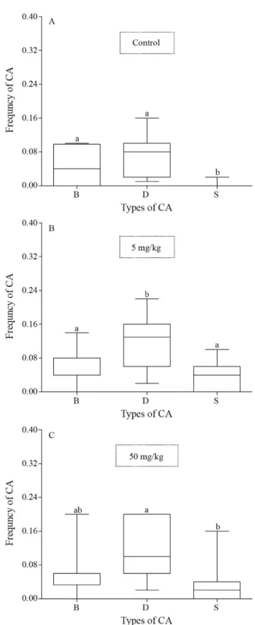
With regard to the types of CAs found in each treat-ment, the negative control group showed a higher fre-quency of chromatid breaks and decondensation relative to the early separation of sister chromatids (p = 0.0055, p < 0.0001, respectively) (Figure 3A). In the group contam-inated with 5 mg/kg of aluminum sulfate, the frequency of decondensation was significantly higher than the frequency of either chromatid breaks or separations (p = 0.0206, p = 0.0023, respectively) (Figure 3B). The group contami-nated with 50 mg/kg showed differences between the fre-quencies of decondensation and chromatid separation (p = 0.0014) (Figure 3C). There was no difference among the types of CA in the group contaminated with 500 mg/kg (p > 0.05).
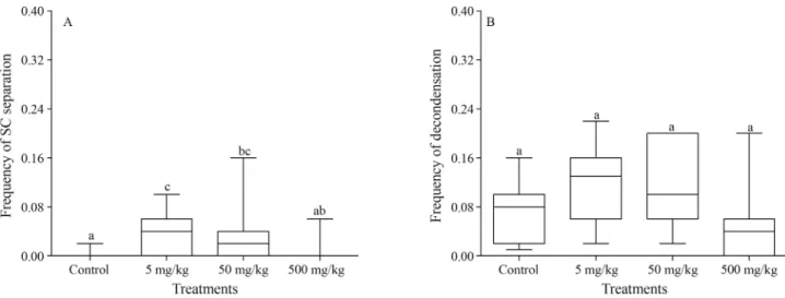
The analysis of each type of CA showed that, relative to the control group, only the early separation of sister chromatids was significantly higher in the groups treated with 5 mg/kg and 50 mg/kg aluminum sulfate (p = 0.0099, p = 0.0329, respectively) (Figure 4A). However, the groups contaminated with 5 mg/kg and 50 mg/kg tended to present differences in the decondensation frequency (p = 0.0570) (Figure 4B).
The comet assay showed significantly higher damage scores in the groups treated with 5 mg/kg, 50 mg/kg and 500 mg/kg compared to the negative control group (p = 0.001, p < 0.0001, p < 0.0001, respectively). This find-ing confirms how sensible and easy it is to obtain the results through this genotoxic technique when compared to the CA
Figure 2- Comparison of the total frequency of chromosomal
abnormali-ties (CA) between the treatments. The box and whisker plots show the me-dian and the first and third quartiles. Different letters indicate statistically significant differences (p < 0.05).
Figure 3- Comparison of the frequency of the types of chromosomal
technique. Nonetheless, no dose-response relationship was observed because the comet assay showed no increase in the amount of DNA damage with increasing doses (Figu-re 5).
Discussion
There is prior evidence that aluminum leads to chro-mosomal abnormalities, micronuclei and the exchange of sister chromatids in human lymphocytes (Royet al., 1990;
Miglioreet al., 1999; Banasiket al., 2005), and
contamina-tion by aluminum and fluoride has been shown to cause telomeric damage in cultured human lymphocytes. Dam-age in the telomeric region caused by toxic compounds leads to chromosomal instability, resulting in cell death as a consequence (Patelet al., 2009).
In fish, different CA types have been described fol-lowing exposure to metal contaminants, such as lead and tributyltin (Cestariet al., 2004; Ferraroet al., 2004., Aleet al., 2004; Ramsdorfet al., 2009a), leading us to suggest that the decondensation of the telomeric regions of the chromosomes may have occurred due to structural modifi-cations to the proteins involved in DNA compaction, thus making the DNA more susceptible to breaks. This may have also caused the chromatid breaks. Telomeric decon-densation and chromatid breaks have been described for contaminations with lead in trophic bioassays conducted for 60 days in the fish species Hoplias malabaricus
(Erythrinidae) (Ferraroet al., 2004; Cestariet al., 2004).
The centromere is a region of the chromosome with com-pact heterochromatin, in which the histones are responsible for the compaction process (Hagele, 1977; Michailovaet al., 1997). In the species Chironomus riparius (Diptera,
Chironomidae), decondensation of the centromeric region observed in response to to aluminum contamination was at-tributed to the inhibition of histone synthesis (Michailova
et al., 2003).
The early separation of sister chromatids observed in this work may have occurred due to changes to the proteins that keep the sister chromatids in cohesion, or that facilitate the mobility of the chromosomes. Using yeast, Michaeliset al.(1997) showed that cohesins are responsible for the
co-hesion of sister chromatids during the cell cycle. In cells containing mutant forms of the proteins SCC1, SCC2, SMC1 and SMC3, the sister chromatids undergo an early separation. The SCC1 protein connects to the chromosome in the final stage of the G1 phase and remains connected to the sister chromatids until metaphase. Moreover, other re-ports have shown that aluminum affects chromosomal mo-bility during the cell division process (Lukiw and Mclachlan, 1995).
Figure 4- Effect of the aluminium sulfate concentrations on sister chromatid separation and chromatin decondensation. (A) Comparison of the frequency of the early separation of the sister chromatids across the treatments. (B) Comparison of the frequency of the decondensation across the treatments. The box and whisker plots show the median and the first and third quartiles. Different letters indicate statistically significant differences (p < 0.05).
Figure 5- Comparison of the DNA damage scores (comet assay) in the
Several studies discussed by Durante et al. (2013)
have shown that chromosomal abnormalities, such as chro-matid breaks, chromosomal breaks or rearrangements, are caused by failures in the repair system when there is a dou-ble-stranded break in the DNA. Nonetheless, the exact mechanisms are not yet clear.
The comet assay showed the genotoxicity of alumi-num, even though the damage detected may have been sub-jected to DNA repair mechanisms. Aluminum toxicity has been studied in the mononuclear leukocytes of people who used aluminum utensils on a daily basis to cook or store food. In that study, high levels of DNA damage were re-lated to the generation of oxidative stress (Celik et al.,
2012). In another study, the comet assay was used to check the genotoxic potential of aluminum in the fishProchilodus lineatus contaminated by acute, subchronic exposure in
water. An increase in the DNA damage in erythrocytes was observed after 6 h and 96 h of exposure, whereas there were no differences relative to the negative control after 24 h and 15 days. These results indicated that the DNA underwent a damage repair process (Galindoet al., 2010).
In the present work, the comet assay was used to con-firm the genotoxic effects observed in the CA test. Ramsdorfet al.(2009a) used the comet assay to verify the
DNA damage seen inHoplias malabaricussubjected to
in-organic lead contamination by intraperitoneal injection. Benincáet al.(2012) also demonstrated DNA damage in
this tissue by monitoring the Santa Marta and Camacho Lakes (Santa Catarina Coast-Southern of Brazil). It is known that the comet assay can detect DNA damage, in-cluding both single- and double-stranded breaks, in indi-vidual cells (Singh et al., 1988; Fairbairn et al., 1995;
Sazakiet al., 1999). This property makes it a very useful
as-say for several tissues. In line with observations from other genotoxicity studies (Ghisiet al., 2011; Pamplonaet al.,
2011), we could confirm this in the present study, showing thatRhamdia quelenappears to be a good test organism for
genotoxicity assays.
In conclusion, we demonstrated that aluminum sul-fate was genotoxic in the kidney tissue cells ofRhamdia quelen. The test for chromosomal abnormalities showed clastogenic effects, whereas the comet assay confirmed the genotoxic potential of aluminum sulfate. Even at very low doses, the comet assay was highly effective at showing DNA damage, and the CA test was shown to be an efficient biomarker in sub-chronic bioassays. In our future works, we will evaluate the damage to other tissues ofRhamdia quelenandHoplias intermediussubjected to trophic alumi-num sulfate contamination. Also, additional studies must be conducted to investigate the mechanisms that cause the chromosomal abnormalities.
Acknowledgments
The authors thank Fundação Araucária (14/2009 -Protocol 18910).
References
Ale E, Fenocchio AS, Pastori MC, Ribeiro CO, Cestari MM and Zacharzewski C (2004) Evaluation of the effects of (NO3)Pb2onOreochromis niloticus(Pisces, Cichlidae) by means of cytogenetic techniques. Cytology 69:453-45. Al-Sabti K and Metcalfe CD (1995) Fish micronuclei for
assess-ing genotoxicity in water. Mutat Res Genet Toxicol 343:121-135.
Banasik A, Lankoff A, Piskulak A, Adamowska K, Lisowska H and Wojcik A (2005) Aluminum-induced micronuclei and apoptosis in human peripheral blood lymphocytes treated during different phases of the cell cycle. Environ Toxicol 20:402-406.
Belpaeme K, Cooreman K and Kirsch-Volders M (1998) Devel-opment and validation of thein vivoalkaline comet assay for detecting genomic damage in marine flatfish. Mutat Res 415:167-184.
Benincá C, Ramsdorf WA, Vicari T, Oliveira-Ribeiro CA, Al-meida MIM, Silva de Assis HC and Cestari MM (2012) Chronic genetic damages in Geophagus brasiliensis ex-posed to anthropic impact in Estuarine Lakes at Santa Cata-rina Coast-Southern of Brazil. Environ Monit Assess 184:2045-2056.
Bertollo LAC, Takahashi CS and Moreira-Filho O (1978) Cyto-taxonomic considerations on Hoplias lacerdae (Pisces, Erythrinidae). Rev Bras Genet 1:103-120.
Celik H, Celik N, Kocyigit A and Dikilitas M (2012) The relation-ship between plasma aluminum content, lymphocyte DNA damage, and oxidative status in persons using aluminum containers and utensils daily. Clinical Biochemistry 45:1629-1633.
Cestari MM, Lemos PMM, Oliveira Ribeiro CA, Costa JRMA, Peletier E, Ferraro MVM, Mantovani MS and Fenocchio AS (2004) Genetic damage induced by trophic doses of lead evaluated by means of the comet assay and chromosomal aberrations in the neotropical fish Hoplias malabaricus (Characiformes, Erythrinidae). Genet Mol Biol 27:270-274. Collins AR, Dobson VL, Dusinska M, Kennedy G and Stetina R
(1997) The comet assay: What can it really tell us? Mutat Res 376:183-193.
Costa M, Kraker AJ and Patierno SR (1984) Toxicity and carcino-genicity of essential and non-essential metals, In: Foreman DT (ed) Progress in Clinical Biochemistry. Vol 1. Springer-Verlag, Berlin, pp 1-45.
De Camargo SGO and Pouey JLOF (2005) Aqüicultura: Um mercado em expansão. Rev Bras Agrociênc 11:393-396. Domingos FXV, Assis HCS, Silva MD, Damian RC, Aleida AIM,
Cestari MM, Randil MAF and Ribeiro CAO (2009) Anthro-pic impact evaluation of two Brazilian estuaries trough bio-markers in fish. J Braz Soc Ecotoxicol 4:21-30.
Durante M, Bedford JS, Chen DJ, Conrad J, Cornforth MN, Natarajan AT, van Gent DC and Obe G (2013) From DNA damage to chromosome aberrations: Joining the break. Mutat Res 756:5-13.
Fairbairn DW, Olive PL and ONeil KL (1995) The comet assay: A comprehensive review. Mutat Res 339:37-59.
Fenocchio AS, Venere PC, Cesar ACG, Dias AL and Bertollo LAC (1991) Short term culture from solid tissues of fishes. Caryologia 44:161-166.
Ferraro MVM, Fenocchio AS, Mantovani MS, Ribeiro CO and Cestari MM (2004) Mutagenic effects of tributyltin and in-organic lead (Pb II) on the fishH. malabaricusas evaluated using the comet assay and the piscine micronucleus and chromosome aberration tests. Genet Mol Biol 27:103-107. Frenzilli G, Scarcelli V, Del Barga I, Nigro M, Förlin L,
Bolog-nesi C and Sturve J (2004) DNA damage in eelpout (Zoarces viviparus) from Göteborg harbour. Mutat Res 552:187-195. Galindo BA, Troilo G, Cólus IMS, Martinez CBR and Sofia SH
(2010) Genotoxic effects of aluminum on the Neotropical FishProchilodus lineatus. Water Air Soil Pollut 212:419-428.
Ganrot R (1986) Metabolism and possible health effects of alumi-num. Environ Health Perspect 65:363-441.
Ghisi NC, Ramsdorf WA, Ferraro MVM, Almeida MIM, Ribeiro CA and Cestari MM (2011) Evaluation of genotoxicity in Rhamdia quelen (Pisces, Siluriformes) after sub-chronic contamination with Fipronil. Environ Monit Assess 180:589-599.
Gontijo ÁMMC, Barreto RE, Speit G, Reyes VAV, Volpato GL and Salvadori DMF (2003) Anesthesia of fish with benzo-caine does not interfere with comet assay results. Mutat Res 534:165-172.
Guthrie FE and Perry JJ (1980) Introduction to Environmental Toxicology. Elsevier, New York, 484 p.
Hagele K (1977) Differential staining of polytene chromosome
bands in Chironomus by Giemsa banding methods.
Chromosoma 59:207-216.
Hamond PB and Beliles RP (1980) Metals. In: Doull J, Klaasen CD and Amdour M (eds) Casarett and Doull’s Toxicology, The Basic Science of Poisons. Macmillan, New York, pp 406-467.
Katsumiti A, Domingos FX, Azevedo M, da Silva MD, Damian RC, Almeida MI, de Assis HC, Cestari MM, Randi MA, Ribeiro CA,et al.(2009) An assessment of acute biomarker responses in the demersal catfishCathorops spixiiafter the Vicuña oil spill in a harbour estuarine area in Southern Brazil. Environ Monit Assess 152:209-229.
Kazantzis G and Lilly LJ (1986) Mutagenic and carcinogenic ef-fects of metals. In: Friberg L, Nordberg F and Vouk V (eds) Handbook on the Toxicology of Metals. 2ndedition. Vol 2. Elsevier, Amsterdam, pp 319-390.
Koivistoinen P (1980) Mineral element composition of Finnish foods: N, K, Ca, Mg, P, S, Fe, Cu, Mn, Zn, Mo, Co, Ni, Cr, F, Se, Si, Rb, Al, B, Br, Hg, As, Cd, Pb, and ash, Acta Agri-cultura e Scandinavica, Supplementum 22, Stockholm, pp 171.
Lankoff A, Banasik A, Duma A, Ochniak E, Lisowska H, Kuszewski T, Gózdz S and Wojcik A (2006) A comet assay study reveals that aluminum induces DNA damage and in-hibits the repair of radiation-induced lesions in human pe-ripheral blood lymphocytes. Toxicol Lett 161:27-36. Lione A (1983) The prophylactic reduction of aluminum intake.
Food Chem Toxicol 21:103-109.
Lukiw WJ and Mclachlan DRC (1995) Neurotoxicity of alumi-num. In: Chang L and Dyer R (eds) Handbook of Neuro-toxicity II: Effects and Mechanisms. Marcel Dekker, New York, pp 105-142.
Marchioro MI and Baldisserotto B (1999) Survival of fingerlings of the Jundiá (Rhamdia quelenQuoy&Gaimard, 1824) to changes on water salinity. Ciênc Rural 29:315-318. Michaelis C, Ciosk R and Nasmyth K (1997) Cohesins:
Chromo-somal proteins that prevent premature separation of sister chromatids. Cell 91:35-45.
Michailova P, Ilkova J and White KN (2003) Functional and structural rearrangements of salivary gland polytene chro-mosomes ofChironomus riparius(Diptera, Chironomidae) in response to freshly neutralized aluminum. Environ Pollut 123:193-207.
Michailova P, Ramella L, Sella G and Bovero S (1997) C band variation in polytene chromosomes ofChironomus riparius (Diptera, Chironomidae) from a polluted Piedmont station (Italy). Cytobios 90:139-151.
Migliore L, Cocchi L, Nesti C and Sabbioni E (1999) Micronuclei assay and FISH analysis in human lymphocytes treated with six metal salts. Environ Mol Mutagen 34:279-284. Nayak P (2002) Aluminum: Impacts and disease. Environ Res
89:101-115.
Norseth T (1988) Metal carcinonesis. Ann N Y Acad Sci 534:377-386.
Obe G, Pfeiffer P, Savage JR, Johannes C, Goedecke W, Jeppesen P, Natarajan AT, Martínez-López W, Folle GA and Drets ME (2002) Chromosomal aberrations: Formation, identifi-cation and distribution. Mutat Res 504:17-36.
Palitti F (1998) Mechanisms of the origin of chromosomal aberra-tions. Mutat Res 404:133-137.
Pamplona JH, da Silva TA, Ramos LP, Ramsdorf WA, Cestari MM, Oliveira Ribeiro CA, Zampronio AR and Silva de Assis HC (2011) Subchronic effects of dipyrone on the fish
species Rhamdia quelen. Ecotoxicol Environ Safety
74:342-349.
Patel TN, Chakraborty S, Sahoo S, Mehta G, Chavda D, Patel C and Patel P (2009) Genotoxic potential of aluminum and flu-oride on human peripheral blood lymphocytes. Res Environ Life Sci 2:147-152.
Piaia R and Baldisseroto B (2000) Stocking density and growth of Rhamdia quelen(Quoy&Gaimard, 1824) fingerlings. Ciênc Rural 30:509-513.
Provost GS, Kretz PI, Hammer RT, Matthews CD, Rogers BJ, Lundberg KS, Dycaico MJ and Short JM (1993) Transgenic systems forin vivomutation analyses. Mutat Res 288:133-149.
Rabello-Gay MN, Rodrigues MAR and Monteleone-Neto R (1991) Mutagênese teratogênese e carcinogênese: Métodos e critérios de avaliação. Rev Bras Genet 14:246.
Ramsdorf WA, Ferraro MVM, Oliveira-Ribeiro CA, Costa JRM and Cestari MM (2009a) Genotoxic evaluation of different doses of inorganic lead (PbII) inHoplias malabaricus. Envi-ron Monit Assess 158:77-85.
Ramsdorf WA, Guimaraes F, Ferraro MVM, Gabardo J, Trindade E and Cestari MM (2009b) Establishment of experimental conditions for preserving samples of fish blood for analysis with both comet assay and flow cytometry. Mutation Res 673:78-81.
Roy AK, Talukder G and Sharma A (1990) Effects of aluminum-sulfate on human leukocyte chromosomes in vitro. Mutat Res 244:179-183.
Sazaki YF, Fujikawa K, Ishida K, Kawamura N, Nishikawa Y, Ohta S, Satoh M, Madarame H, Ueno S, Susa N,et al.(1999) The alkaline single cell gel electrophoresis assay with mou-se multiple organs: Results with 30 aromatic amines. Mutat Res 440:1-18.
Singh NP, Mccoy MT, Tice RR and Schneider EL (1988) A sim-ple technique for quantitation of low levels of DNA damage in individual cells. Exp Cell Res 175:184-191.
Singh NP and Stephens RE (1997) Microgelelectroforesis: Sen-sitivy, mechanisms, and DNA electrostretching. Mutat Res 383:167-175.
Speit G and Hartmann A (1995) The contribution of excision re-pair to the DNA effects seen in the alkaline single cell gel test (comet assay). Mutagenesis 10:555-559.
Vicari T, Ferraro MVM, Ramsdorf WA, Mella M, Oliveira-Ribeiro CA and Cestari MM (2012) Genotoxic evaluation of different doses of methylmercury (CH3Hg+) in Hoplias malabaricus. Ecotoxicol Environ Safety 82:47-55. Woo YT, Lai DJ, Arcos JC and Argus MF (1988) Chemical
In-duction of Cancer, Structural Bases and Biological Mecha-nisms. Academic Press, San Diego, 869 p.
Associate Editor: Daisy Maria Fávero Salvadori