Neopolystoma fentoni
n. sp. (Monogenea: Polystomatidae)
a Parasite of the Conjunctival Sac of Freshwater Turtles in
Costa Rica
Thomas R Platt
Department of Biology, Saint Mary’s College, Notre Dame, IN 46556, USA
Neopolystoma fentoni n. sp. is described from the conjunctival sac of Kinosternon leucostomum (Duméril, Bibron, and Duméril 1851) and Rhinoclemmys pulcherrima (Gray 1855) from the Guanacaste Conservation Area in Costa Rica. The new species differs from all other species of Neopolystoma, except N. elizabethae Platt 2000 in possessing a circle of eight genital spines that are recurved and possess a crescent-shaped base. N. fentoni n. sp. differs from N. elizabethae in lacking cecal diverticula and in a number of morphometric criteria.
Key words: Monogenea - freshwater turtle parasite - Neopolystoma - conjunctival sac - Costa Rica
The monogene family Polystomatidae contains three genera parasitic exclusively in turtles. The majority of species of polystomatids described from turtles inhabit either the oral cavity or the urinary bladder as adults (du Preez & Lim 2000). A small number of species have been reported from the conjunctival sacs of freshwater turtles (Platt 2000). Examination of specimens of two species of turtles, Kinosternon leucostomum (Duméril, Bibron, and Duméril 1851) and Rhinoclemmys pulcherrima (Gray 1855), from the Guanacaste Conservation Area, Costa Rica, yielded specimens of Neopolystoma from the conjunctival sac that are herein described as a new species.
MATERIALS AND METHODS
Turtles were collected by hand from permanent pools in dry stream beds in the vicinity of the op-erations center of the Guanacaste Conservation Area during June 1998. Turtles were returned to the laboratory and examined within 48 h of cap-ture. The animals were decapitated and necropsies were conducted following standard procedures. The lower eyelids were resected and worms were removed from the conjunctival sac with fine for-ceps and a camel hair brush.
Worms were killed, without flattening, in hot (steaming) 5% formalin. Specimens were placed in a vial with enough saline to cover them. Hot formalin was added to the vial, the vial; was quickly
capped and gently shaken. The worms were stored in the same medium, stained in Mayer’s haematoxylin (Pritchard & Kruse 1982), follow-ing standard procedures, and mounted in Canada balsam for examination as whole mounts. Defini-tions of prevalence and mean intensity follow the usage of Bush et al. (1997).
Specimens were observed with a Nikon Optiphot light microscope using bright field and phase contrast illumination. Drawings were made with the aid of a camera lucida and measurements were made using an ocular micrometer. All mea-surements are in micrometers with the mean fol-lowed by the range in parentheses, and n refers to the number of specimens measured.
DESCRIPTION
Neopolystoma fentoni n. sp. Morphometrics: based on ten specimens
(Figs 1-3; Table I)
With the characteristics of the genus. Body pyriform, maximum width near midbody (Fig. 1). Tegument aspinose. Haptor slightly wider than long; six type 2 haptoral suckers (Pichelin 1995). Marginal hooks 16, 12.5 (11.8-12.7; n = 4) with blade oriented toward haptoral margin; hook pairs VI-VIII between anterior pair of suckers; hook pairs III-V in base of each sucker; hook pairs I-II between posterior-most pair of suckers. Marginal hooks rarely present, however, hoods typically present. Oral sucker large, wider than long. Phar-ynx large, wider than long; PW/OSW = 0.81:1. Genital bulb (Fig. 2) round to slightly oval; genital spines 8, recurved from base to tip with crescent-shaped handle at right angle to blade; eight genital spines 11 long (n = 8). Seminal vesicle small, oval. Testis located near midbody (excluding haptor).
Financial support: Saint Mary’s College research grant Fax: 219-284-4716. E-mail: [email protected] Received 7 January 2000
Neopolystoma fentoni n. sp. in ventral view. Fig. 1: whole mount, ventral (holotype), bar = 500. Fig. 2: genital bulb and genital spines, ventral (paratype), bar = 50. Fig. 3: genital spines, ventral (paratype), bar = 20. Fig. 4: marginal spine, ventral (paratype), bar = 20.
Genital system exhibits situs inversus. Ovary oval, anterior to testis. Genito-intestinal canal present. Uterus conspicuous, elliptical; ova elliptical. Va-ginae lateral, uncapped, in second 1/4 of body. Vaginae from anterior end/total length as a per-cent = 39.97 (37.5-44.3). Vitellarium follicular from level of genital pore to anterior margin of haptor, filling intercecal space not occupied by genital structures; not extending into penduncle.
Taxonomic summary
Type host: Kinosternon leucostomum (Duméril, Bibron, and Duméril 1851): Kinosternidae Other known host: Rhinoclemmys pulcherrima (Gray 1855): Bataguridae
Site of infection: conjunctival sac
Type locality: Quebrada Costa Rica (10o 49.666' N, 85o 38.216 W), Guanacaste Conservation Area, Santa Rosa, Guanacaste, Costa Rica
Other known localities: Quebrada el Duende (10o 50.236’N, 85o 36.724’W) and San Gerardo (10o 52.55’N, 85o 23.27’W), Guanacaste Conservation Area, Santa Rosa, Guanacaste, Costa Rica. Type material: holotype and four paratypes: United States National Parasitological Collection, holotype (collection no. 89943); four paratypes (nos 89944-6): four paratypes Helminthological Collection of the Oswaldo Cruz Institute (collection nos 34292, 34293a-b, 34294).
my father-in-law, Mr William S Fenton, as a trib-ute to his friendship and counsel over the last quar-ter century.
REMARKS
The family Polystomatidae contains three gen-era that exclusively parasitize turtles. The genus Neopolystoma Price, 1939 is differentiated from Polystomoides Ward, 1917 and Polystomoidella Price, 1939, by the absence of anchors between the posterior-most pair of suckers on the haptor. The new species clearly belongs to the genus Neopolystoma. Specimens collected from K. leucostomum and R. pulcherrima overlapped mor-phometrically in all attributes and are treated as a single species, N. fentoni, which is considered new to science. Prevalence and intensity of N. fentoni were low in both host species (Table II).
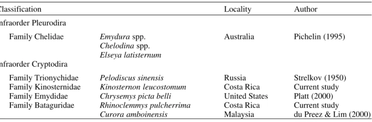
Seven species of Neopolystoma have been de-scribed from the conjunctival sac of freshwater turtles (Table III). N. fentoni n. sp. differs from all other species in the genus, except N. palpabrae Strelkov, 1950, and N. elizabethae Platt, 2000, in having genital spines with a crescent-shaped base. N. palpabrae was described by Strelkov (1950) from the soft-shelled turtle, Pelodiscus sinensis
(Wiegmann 1835), from Lake Chanka, north of Vladivostok, in the former Soviet Union. The geni-tal spines in N. palpabrae are identical in shape to those of the new species; however, they numbered 16 and are arranged in a circle of alternating large and small spines. Strelkov (1950) did not give sepa-rate measurements for the large and small spines for N. palpabrae, however, the range of spine length was given as 10-16 µm, while the lengths of the spines of N. fentoni and N. elizabethae are 11 and 10, respectively.
TABLE I
Morphometric comparison of Neopolystoma fentoni n. sp. and Neopolystoma elizabethae Platt, 2000 (mean followed by the range in parentheses)
Character N. fentoni (n = 10) N. elizabethae (n = 4) Body L 1985 (1500-2450) 3125 (2550-3675)
W 568 (426-760) 823 (640-990) Haptor L 571 (449-690) 865 (790-970) W 683 (550-850) 975 (880-1070) Marginal hooks (n = 4;8) L 12.5 (11.8-12.7) 12.3 (11.8-12.7) Haptor sucker (n = 60;24) D 265 (210-326) 372 (344-408) Oral sucker L 230 (150-303) 271 (251-292) W 370 (240-496) 473 (449-540) Pharynx L 216 (156-257) 255 (216-268) W 278 (185-367) 305 (268-320) Ph/OS 0.82:1 (0.75-0.93:1) 0.65:1 (0.61-0.71) Genital bulb L 55 (43-70) 58 (50-63)
W 60 (30-83) 57 (45-63) Seminal vesicle L 79 (50-100) 151 (140-175)
W 50 (35-65) 63 (50-68)
Testis L 225 (98-367) 208 (178-262)
W 181 (78-251) 155 (140-192)
Ovary L 103 (80-245) 301 (218-350)
W 105 (55-169) 122 (100-146) Ova (n = 7;3) L 286 (245-332) 348 (332-367) W 136 (122-146) 120 (117-122) Vaginae to anterior end 788 (510-920) 1071 (880-1250) VAE/TL (%) 40.0 (37.5-44.3) 34.3 (33.2-35.4) Uterus (empty; n = 3) L 99 (50-133) 330 (188-402)
W 35 (33-38) 151 (140-192) Ph/OS: pharynx width-oral sucker width ratio; VAE/TL (%): vaginae to anterior end as a percent of total length
TABLE II
Prevalence and mean intensity of Neopolystoma fentoni n. sp. in freshwater turtles from the Guanacaste
Conservation Area, Santa Rosa, Costa Rica Host % prevalence X intensity
(range)
Kinosternon leucostomum 12.5 (2/16) 2.0 (1-3)
TABLE III
Classification of extant turtle species (Order Testudines) known to harbor Neopolystoma spp. in the conjunctival sac (turtle classification based on various sources)
Classification Locality Author
Infraorder Pleurodira
Family Chelidae Emydura spp. Australia Pichelin (1995)
Chelodina spp.
Elseya latisternum
Infraorder Cryptodira
Family Trionychidae Pelodiscus sinensis Russia Strelkov (1950) Family Kinosternidae Kinosternon leucostomum Costa Rica Current study Family Emydidae Chrysemys picta belli United States Platt (2000) Family Bataguridae Rhinoclemmys pulcherrima Costa Rica Current study
Curora amboinensis Malaysia du Preez & Lim (2000)
Pichelin (1995) described four species of Neopolystoma from the conjuntival sacs of fresh-water turtles in Australia. Three species had 20 or more genital spines which would clearly separate them from the American forms. Only N. cribbi Pichelin, 1995 possesses a smaller number of geni-tal spines (range 6-10), however, they possess typi-cal fan-shaped roots (Pichelin 1995) and lack the crescent-shaped base present in N. fentoni and N. elizabethae. du Preez and Lim (2000) described N. liewi from the conjunctival sac of the batagurid turtle, Curora amboinensis, from Malaysia. This species also possesses a small number of genital spines (8-11); however, they also have typical “fan-shaped” roots and lack the recurved base present in N. fentoni and N. elizabethae.
N. fentoni appears most closely related to N. elizabethae as the genital and marginal spines are nearly identical in shape and size; however, the new species differs from the North American form in lacking intestinal diverticula present in the pos-terior half of the cecae. The two forms also differ in number of morphometric criteria (Table I). N. fentoni is significantly smaller, the ratio of pha-ryngeal and oral suck width is greater, the diam-eter of the haptoral suckers is larger, the testis is larger, the seminal vesicle is smaller, and the eggs are, on average, 60 shorter. These differences are sufficient to warrant recognizing a new species.
The conjunctival sac has been overlooked as a site for the presence of polystomatids. The discov-ery of N. fentoni is only the seventh species re-corded from this location since the first report of N. palpabrae from Pelodiscus (=Amyda) sinensis (Wiegmann, 1835) by Strelkov (1950). Pichelin (1995) described four species of Neopolystoma from the side-necked turtles (Chelidae) in Austra-lia and Platt (2000) described N. elizabethae from the western painted turtle, Chrysemys picta belli
(Gray, 1831) from northern Michigan in the United States.
The range of turtle taxa harboring Neo-polystoma spp. in the conjunctival sac is shown in Table III. In addition to the fact that species in both major lineages of extant turtles harbor these worms, the five host families represent nearly 70% of gen-era and species of living turtles (Iverson 1992). Nor are these parasites rare in the individual host popu-lations examined. Strelkov (1950) reported a preva-lence of 35% for N. palpabrae, while Platt (2000) reported 80% prevalence for N. elizabethae in a very small sample (n = 5). While published data are not available for the Australian polystomatids, the author found over 20% prevalence in a sample of 70 chelids in Australia (Platt, unpublished ob-servations). Any parasitologist examining turtles should examine the conjunctival sac for polystomes.
coevolution-ary, biogeographic and speciation hypotheses if sufficient material is made available. South America and Asia are two areas of high turtle di-versity, yet recent checklists of monogenes from these areas identify only four and seven species of polystomes reported in the literature, respectively (Kohn & Cohen 1998, Lim 1998). Judicious ex-amination of turtles for helminths from these ar-eas, preserving specimens for both morphological and molecular analysis, has the potential to yield significant information about the history of life.
ACKNOWLEDGMENTS
To Dr Roger Blanco, Research Director at the con-servation area, for the use of the facilities and his gener-ous hospitality. To Dr Daniel R Brooks who arranged the trip and provided valuable assistance in the field. To Drs Anindo Choudhury and Derek Zelmer who provided companionship and assistance in collecting the turtles. To Ms Akmaral Omarova, Saint Mary’s College, for assistance in translating the paper by Strelkov.
REFERENCES
Bush AO, Lafferty KD, Lotz, JM, Shostak AW 1997. Parasitology meets ecology on its own terms: Margolis et al. revisited. J Parasitol 83: 575-583. du Preez, LH, Lim LHS 2000. Neopolystoma liewi sp.n.
(Monogenea: Polystomatidae) from the eye of the Malayan box turtle (Curora amboinensis). Folia Parasitol 47: 11-16.
Iverson JB 1992. A Revised Checklist with Distribution Maps of the Turtles of the World, published by the author, Richmond, IN, 363 pp.
Kohn A, Cohen SC 1998. South American Monogenea - List of species, hosts and geographical distribu-tion. Int J Parasitol28: 1517-1554.
Lim LHS 1998. Diversity of monogeneans in Southeast Asia. Int J Parasitol28: 1495-1515.
Littlewood DTJ, Rohde K, Clough KA 1997. Parasite speciation within or between host species? - Phylo-genetic evidence from site-specific polystome mono-geneans. Int J Parasitol27: 1289-1297.
Pichelin S 1995. The taxonomy and biology of the Polystomatidae (Monogenea) in Australian freshwa-ter turtles (Chelidae, Pleurodira). J Nat Hist29:
1345-1381.
Platt TR 1992. A phylogenetic and biogeographic analy-sis of the genera of Spirorchinae (Digenea: Spirorchidae) parasitic in freshwater turtles. J Parasitol 78: 616-629.
Platt TR 2000. Helminth parasites of the western painted turtle, Chyrsemys picta belli (Gray), including
Neopolystoma elizabethae n. sp. (Monogenea: Polystomatidae), a parasite of the conjunctival sac.
J Parasitol 86: in press.
Pritchard MH, Kruse GOW 1982. The Collection and Preservation of Animal Parasites, Technical Bulle-tin No. 1, University of Nebraska Press, Lincoln, NE, 141 pp.
Rohde K, Pearson JC 1980. Two polystomes (Monogenea) from Australian river tortoises (Pleurodira, Chelidae), Polystomoides australensis
sp. nov. from Emydura krefftii and Neopolystoma chelodinae (MacCallum, 1919) from Chelodina longicollis. Zool Anzgr 204: 191-208.