DNA Damage and Repair Biomarkers in Cervical
Cancer Patients Treated with Neoadjuvant
Chemotherapy: An Exploratory Analysis
Patrizia Vici1☯, Simonetta Buglioni2☯, Domenico Sergi1, Laura Pizzuti11, Luigi Di Lauro1,
Barbara Antoniani2, Francesca Sperati3, Irene Terrenato3, Mariantonia Carosi2, Teresa Gamucci4, Rosanna Dattilo5, Monica Bartucci6, Cristina Vincenzoni7, Luciano Mariani7,8, Enrico Vizza7, Giuseppe Sanguineti9, Angiolo Gadducci10, Ilio Vitale11,12, Maddalena Barba1,12, Ruggero De Maria12*, Marcella Mottolese2‡, Marcello Maugeri-Saccà1,12‡*
1Division of Medical Oncology B,“Regina Elena”National Cancer Institute, Rome, Italy,2Department of Pathology,“Regina Elena”National Cancer Institute, Rome, Italy,3Biostatistics-Scientific Direction,“Regina Elena”National Cancer Institute, Rome, Italy,4Medical Oncology Unit, ASL Frosinone, Frosinone, Italy, 5Department of Hematology, Oncology and Molecular Medicine, Istituto Superiore di Sanità, Rome, Italy, 6Drug Discovery Program, Rutgers Cancer Institute of New Jersey, New Brunswick, New Jersey, United States of America,7Department of Surgery, Gynecologic Oncology Unit,“Regina Elena”National Cancer Institute, Rome, Italy,8HPV-UNIT,“Regina Elena”National Cancer Institute, Rome, Italy,9Department of Radiotherapy,“Regina Elena”National Cancer Institute, Rome, Italy,10Department of Experimental and Clinical Medicine, Division of Gynecology and Obstetrics, University of Pisa, Pisa, Italy,11Department of Biology, University of Rome "Tor Vergata", Rome, Italy,12Scientific Direction,“Regina Elena”National Cancer Institute, Rome, Italy
☯These authors contributed equally to this work.
‡These authors shared senior authorship on this work.
*[email protected](MM-S);[email protected](RDM)
Abstract
Cervical cancer cells commonly harbour a defective G1/S checkpoint owing to the
interac-tion of viral oncoproteins with p53 and retinoblastoma protein. The activainterac-tion of the G2/M
checkpoint may thus become essential for protecting cancer cells from genotoxic insults, such as chemotherapy. In 52 cervical cancer patients treated with neoadjuvant chemother-apy, we investigated whether the levels of phosphorylated Wee1 (pWee1), a key G2/M
checkpoint kinase, andγ-H2AX, a marker of DNA double-strand breaks, discriminated between patients with a pathological complete response (pCR) and those with residual dis-ease. We also tested the association between pWee1 and phosphorylated Chk1 (pChk1), a kinase acting upstream Wee1 in the G2/M checkpoint pathway. pWee1,γ-H2AX and pChk1
were retrospectively assessed in diagnostic biopsies by immunohistochemistry. The degrees of pWee1 and pChk1 expression were defined using three different classification methods, i.e., staining intensity, Allred score, and a multiplicative score.γ-H2AX was ana-lyzed both as continuous and categorical variable. Irrespective of the classification used, elevated levels of pWee1 andγ-H2AX were significantly associated with a lower rate of pCR. In univariate and multivariate analyses, pWee1 andγ-H2AX were both associated with reduced pCR. Internal validation conducted through a re-sampling without replacement procedure confirmed the robustness of the multivariate model. Finally, we found a
OPEN ACCESS
Citation:Vici P, Buglioni S, Sergi D, Pizzuti L, Di Lauro L, Antoniani B, et al. (2016) DNA Damage and Repair Biomarkers in Cervical Cancer Patients Treated with Neoadjuvant Chemotherapy: An Exploratory Analysis. PLoS ONE 11(3): e0149872. doi:10.1371/ journal.pone.0149872
Editor:Yanchang Wang, Florida State University, UNITED STATES
Received:December 3, 2015
Accepted:February 7, 2016
Published:March 1, 2016
Copyright:© 2016 Vici et al. This is an open access article distributed under the terms of theCreative Commons Attribution License, which permits unrestricted use, distribution, and reproduction in any medium, provided the original author and source are credited.
Data Availability Statement:All relevant data are within the paper.
Funding:The authors have no support or funding to report.
Competing Interests:The authors have declared that no competing interests exist.
significant association between pWee1 and pChk1. The message conveyed by the present analysis is that biomarkers of DNA damage and repair may predict the efficacy of neoadju-vant chemotherapy in cervical cancer. Further studies are warranted to prospectively vali-date these encouraging findings.
Introduction
Eukaryotic cells are constantly exposed to endogenous and exogenous sources of DNA damage. The transmission of undamaged DNA to the offspring is ensured by a complex molecular net-work, the DNA damage response (DDR), which operates through the coordinated activity of cell cycle checkpoints, DNA repair mechanisms and apoptotic pathways [1,2]. The presence of genetic lesions triggers checkpoint-mediated arrest of the cell cycle [2]. This event enables DNA repair effectors and apoptotic pathways to repair the lesion or eliminate irremediably damaged cells, respectively.
Cancer cells aberrantly use DNA repair mechanisms to survive stressful conditions, such as exposure to chemotherapy [2]. A common trait to a variety of tumors is the defective nature of the G1/S-phase checkpoint, stemming from mutational or functional inactivation of p53 or ret-inoblastoma protein (pRb) [3]. When this occurs, cancer cells become extremely dependent on the G2/M checkpoint for cell cycle arrest and DNA repair [3]. The ataxia telangiectasia and Rad3-related protein (ATR)-Checkpoint kinase 1 (Chk1)-Wee1-like protein kinase (Wee1) cascade represents the core of the G2/M checkpoint, whose activation leads to the inhibition of the cyclin-dependent kinase 1 and culminates into checkpoint-mediated cell cycle arrest [3]. In such a manner, cancer cells have the time to correct chemotherapy-induced DNA lesions, avoiding entry into a lethal mitosis known as mitotic catastrophe [4]. G2/M checkpoint depen-dency in a p53-defective molecular background is a concept currently exploited for the clinical development of synthetic lethality-based therapeutics. When G1/S-phase checkpoint-defective cells are exposed to chemotherapeutics, the concomitant pharmacological inhibition of G2/M checkpoint kinases is deleterious for cell fitness [3].
We reasoned that G2/M checkpoint“addiction”for compensating p53 or pRb defects upon exposure to genotoxic agents can be exploited in the search for predictive biomarkers foreseeing chemotherapy sensitivity/resistance. In this exploratory analysis we focused on cervical cancer, the prototype of p53- and pRb-defective tumors. Indeed, human papillomavirus E6 and E7 onco-proteins promote ubiquitin-mediated degradation of p53 and pRb, respectively [5]. We thus ret-rospectively investigated the association between the levels of DNA damage and repair
biomarkers, assessed in bioptic samples collected from untreated patients at the time of diagnosis, and pathological complete response (pCR) after neoadjuvant chemotherapy, i.e., chemotherapy delivered in the timeframe between diagnostic biopsy and the surgical resection. All the patients were homogenously treated with paclitaxel, ifosfamide and cisplatin (TIP regimen). We focused on phosphorylated Wee1 (pWee1) as a proxy of G2/M checkpoint activation, and phosphory-lated H2A Histone Family Member X (γ-H2AX) as a marker of DNA double-strand breaks. Phosphorylated Chk1 (pChk1) was tested in a fraction of samples for a signaling study.
Materials and Methods
Study Participants and Procedures
Fifty-two histologically confirmed cervical cancer patients (stage Ib2-IIIa) who received neoad-juvant chemotherapy were included in this retrospective analysis. All patients were treated
hosphorylated Chk1;γ-H2AX, phosphorylated H2A
with the TIP regimen (paclitaxel 175 mg/m2on day 1 + ifosfamide 2500 mg/m2on days 1 and 2 + cisplatin 50 mg/m2on day 2 every 21 days for three or four cycles) followed by radical sur-gery. Patients were considered eligible if they completed the planned treatment, data on clinical features and treatment outcomes were available, and the amount of biological materials in their biopsies was sufficient for molecular analyses. pCR was defined as no residual disease in surgi-cal samples. The immunohistochemisurgi-cal assessment of pWee1,γ-H2AX, and pChk1 was per-formed in formalin-fixed paraffin-embedded (FFPE) tissues, obtained from the biological specimens collected through bioptic procedures in untreated patients, using the following anti-bodies: anti-phospho-H2AX (Ser139) (clone JBW301) mouse monoclonal antibody (MAb) (Upstate, NY, USA) at the dilution of 1:500, anti-phospho-Wee1 (Ser642) (clone D47G5) rab-bit MAb (Cell Signaling, Danvers, MA, USA) at the dilution of 1:100, and anti-phospho-Chk1 (Ser345) (clone 133D3) rabbit MAb (Cell Signaling, Danvers, MA, USA) at the dilution of 1:100. Immunohistochemical staining was performed in an automated autostainer (BOND-III, Leica, Milan, Italy) by a biotin-free polymeric horseradish peroxidase (HRP)-linker antibody conjugate system (Leica, Milan, Italy). For each tumor, three different, 3μm paraffin sections were analyzed and examined by light microscopy. Immunoreaction of tumor cells was counted in four high-power fields (400x magnification) per section. pWee1 and pChk1 were considered positive when10% of the neoplastic cells showed a distinct nuclear immunoreactivity. pWee1 and pChk1 were graded on a four-grade scale based on staining intensity (0: negative, 1+: weak, 2+: moderate, 3+: strong). Tumors were classified as negative (0 = pWee1negand pChk1neg) or positive (1–3 = pWee1posand pChk1pos).The Allred scores were obtained as pre-viously described [6], considering staining intensity and percentage of tumor-expressing cells, and reported according to a scale of 0 to 8. Tumors were classified as low expressing if the Allred score was2 (pWee1allred low, pChk1allred low), or as high expressing if the Allred score was>2 (pWee1allred high, pChk1allred high). The multiplicative scores were obtained by multi-plying staining intensity x the percentage of tumor-expressing cells, and were expressed on a scale of 0 to 300. Tumors were classified as low expressing (pWee1multi lowand pChk1multi low) or high expressing (pWee1multi highand pChk1multi high) using the median score of all tumors as a cut-off point.γ-H2AX expression was considered as the percentage of tumor-expressing cells and analyzed both as continuous (γ-H2AXcont) and as categorical variable, whose modality was defined using the median score of all tumors (γ-H2AXlowandγ-H2AXhigh). Tumor samples were evaluated independently by two investigators (SB and MC) who were blinded to treat-ment outcomes, and discordant cases were reviewed (MM). This retrospective study was con-ducted in accordance with the Declaration of Helsinki and was approved by the Ethic Committee of“Regina Elena”National Cancer Institute of Rome, the coordinating centre. Written informed consents were secured before chemotherapy.
Statistical analysis
end, one hundred datasets were generated by randomly removing approximately 20% of the original sample. For each simulation, we repeated the multivariate logistic regression model and the Cohen's Kappa coefficient, the Positive Predictive Value (PPV), the Negative Predictive Value (NPV), Sensibility and Specificity were calculated. We considered statistically significant p values less than 0.05. Statistical analyses were carried out using SPSS software (SPSS version 21, SPSS Inc., Chicago, IL, USA).
Results
Baseline characteristics of the 52 patients included in this study are summarized inTable 1. Median time between diagnostic biopsy and radical surgery was 4.99 months [IQ Range: 4.01–
5.83]. All the pre-chemotherapy samples, consisting in diagnostic biopsies, were examined for pWee1 andγ-H2AX, whereas pChk1 data were available for 37 samples. Median percentages of nuclear-expressing cells for pWee1, pChk1 andγ-H2AX were 40% (min/max 10/80), 30% (min/max 10/80) and 30% (min/max 0/80), respectively. Representative immunohistochemical staining patterns are illustrated inFig 1. As shown inTable 2, we found a statistically significant association between elevated nuclear pWee1 expression and reduced pCR rate. The association tested significant for all the scoring methods investigated (pWee1posvs pWee1neg, p = 0.016; pWee1allred highvs pWee1allred low, p = 0.016; pWee1multi highvs pWee1multi low, p = 0.034) (Table 2). Likewise, elevated nuclear levels ofγ-H2AX were associated with reduced pCR rate, both when considered as categorical and continuous variable (p = 0.037 and p = 0.026 in
Table 2andFig 2, respectively). When considering the combination of the two markers, only 1 patient out of 16 with double positive tumors experienced a pCR, 9 out of 16 patients with dou-ble negative tumors achieve a pCR, and an intermediate outcome was seen in patients whose tumors expressed only one biomarker (p = 0.009) (Table 3). Six out of 8 deaths were observed in double positive tumors (p = 0.013) (Table 3). In the univariate logistic regression model, pWee1 andγ-H2AX were directly associated with pCR (pWee1posvs pWee1neg: Odds Ratio (OR) 5.31, 95% Confidence Interval (CI): 1.42–19.87, p = 0.013;γ-H2AXhighvsγ-H2AXlow: OR 4.20, 95%CI:1.13–15.59, p = 0.032, respectively) (Table 4); the multivariate model con-firmed the predictive role of pWee1 andγ-H2AX (Table 4). The internal validation performed through a re-sampling procedure confirmed the robustness of the multivariate model. Concor-dance, Positive Predictive Value, Negative Predictive Value, Sensitivity and Specificity are
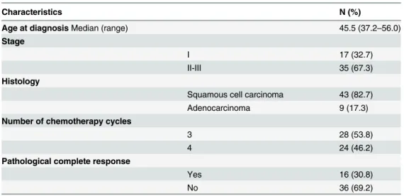
Table 1. Baseline characteristics and treatment outcome of cervical cancer patients treated with neoadjuvant chemotherapy (N = 52).
Characteristics N (%)
Age at diagnosisMedian (range) 45.5 (37.2–56.0)
Stage
I 17 (32.7)
II-III 35 (67.3)
Histology
Squamous cell carcinoma 43 (82.7)
Adenocarcinoma 9 (17.3)
Number of chemotherapy cycles
3 28 (53.8)
4 24 (46.2)
Pathological complete response
Yes 16 (30.8)
No 36 (69.2)
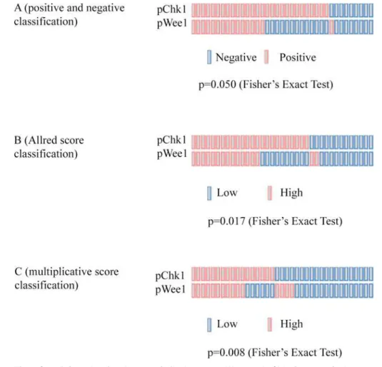
shown inTable 5. Finally, when investigating co-expression patterns, we did not observe any association between pWee1 andγ-H2AX (data available upon request), whereas a statistically significant association was reported between pWee1 and pChk1 (Fig 3).
Discussion
In the present study we retrospectively explored the predictive significance of pWee1 and γ-H2AX expression, evaluated in diagnostic biopsies related 52 cervical cancer patients who received neoadjuvant chemotherapy. We also investigated the association between pWee1 and pChk1 in order to provide clues on whether Wee1 activation in cervical cancer is mediated by Chk1. To our knowledge, this is the first study reporting on DNA damage and repair biomark-ers in cervical cancer that exploited the concept of the defective nature of the G1/S-phase checkpoint. Overall, we observed a statistically significant association between elevated
Fig 1. Representative examples of immunohistochemical expression of DNA damage and repair biomarkers in cervical cancer patients.Three consecutive sections for each tumor are showed. (A-C) A triple positive tumor with nuclearγ-H2AX(A), pWee1(B) and pChk1 (C) immunoreactivity.(D-F) A tumor that
did not expressγ-H2AX (D), and that co-expressed pWee1(E) and pChk1 (F). (G-I) A tumor expressing
nuclearγ-H2AX (G) that lacked both pWee1(H) and pChk1 (I) expression.
doi:10.1371/journal.pone.0149872.g001
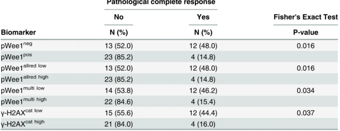
Table 2. Association between biomarkers of DNA damage and repair (pWee1 andγ-H2AX) and
patho-logical complete response in cervical cancer patients treated with neoadjuvant chemotherapy (N = 52).
Pathological complete response
No Yes Fisher's Exact Test
Biomarker N (%) N (%) P-value
pWee1neg 13 (52.0) 12 (48.0) 0.016
pWee1pos 23 (85.2) 4 (14.8)
pWee1allred low 13 (52.0) 12 (48.0) 0.016
pWee1allred high 23 (85.2) 4 (14.8)
pWee1multi low 14 (53.8) 12 (46.2) 0.034
pWee1multi high 22 (84.6) 4 (15.4)
γ-H2AXcat low 15 (55.6) 12 (44.4) 0.037
γ-H2AXcat high 21 (84.0) 4 (16.0)
pWee1, phosphorylated Wee1-like protein kinase;γ-H2AX, phosphorylated H2A Histone Family Member X
expression of pWee1 andγ-H2AX and reduced rate of pCR. Thus, we provided first hints that the elevated expression of DDR biomarkers in diagnostic samples might be associated with suboptimal efficacy of chemotherapy, evaluated through pCR in surgically resected tumors. We also observed a positive association between pWee1 and pChk1 expression that suggests effective G2/M checkpoint activation. We are aware that our results are hypothesis-generating
Fig 2. Box plot of the distribution ofγ-H2AX values by pathologic complete response.In the figure: the
upper horizontal line of the box is the 75th percentile; the lower horizontal line of the box is the 25th percentile; the horizontal bar within box is the median value; the upper horizontal bar outside the box is the maximum value; the lower horizontal bar outside the box is the minimum values.
doi:10.1371/journal.pone.0149872.g002
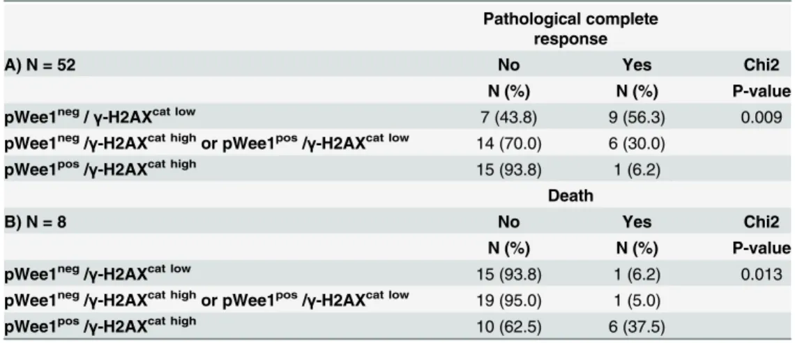
Table 3. Association between the co-expression of pWee1 andγ-H2AX and A) Pathological complete response (N = 52), B) Death (N = 8).
Pathological complete response
A) N = 52 No Yes Chi2
N (%) N (%) P-value
pWee1neg/γ-H2AXcat low 7 (43.8) 9 (56.3) 0.009
pWee1neg/
γ-H2AXcat highor pWee1pos/γ-H2AXcat low 14 (70.0) 6 (30.0)
pWee1pos/
γ-H2AXcat high 15 (93.8) 1 (6.2)
Death
B) N = 8 No Yes Chi2
N (%) N (%) P-value
pWee1neg/
γ-H2AXcat low 15 (93.8) 1 (6.2) 0.013
pWee1neg/
γ-H2AXcat highor pWee1pos/γ-H2AXcat low 19 (95.0) 1 (5.0)
pWee1pos/γ-H2AXcat high 10 (62.5) 6 (37.5)
pWee1, phosphorylated Wee1-like protein kinase;γ-H2AX, phosphorylated H2A Histone Family Member X
in nature given the retrospective design of the study. Nevertheless, beyond the straightforward analytical approach, our study has some important strengths.
First, the neoadjuvant setting offers multiple advantages for the identification and develop-ment of cancer biomarkers: i) the analysis of potential markers in a molecular background not
“polluted”by the exposure of previous anticancer treatments, ii) the identification of predictive markers to select patients who will more likely benefit from chemotherapy, iii) the identifica-tion of biomarkers that also hold prognostic significance, even though evidence on the associa-tion between pCR and long-term survival outcomes in cervical cancer is not as robust as it is in breast cancer [7,8].
Second, thus far, in cervical cancer the search for predictive biomarkers linked to the increased ability of cancer cells to protect their genome when challenged with chemotherapy or radiotherapy has been exclusively focused on nucleotide excision repair (NER) proteins, and in particular on the excision repair cross-complementation group1 (ERCC1) protein [9–13]. NER is deputed to correct bulky helix-distorting lesions, such as those inflicted on the DNA by plati-num-based therapy. However, within the context of the DDR, NER is one of the many distal effectors assigned to maintain genome integrity. A number of molecular networks safeguard the genome, albeit their engagement depends on the type of lesion. DNA repair pathways also include base excision repair (BER), mismatch repair (MMR), direct repair, and the double-strand break (DSB) recombinational repair. This latter encompasses the error-free homologous recombination repair (HRR) and the error-prone non-homologous end-joining (NHEJ) [1]. Therefore, the level of biologic complexity of the DDR might be underestimated when exclu-sively considering one, or few, components collocated in a specific repair network. Moreover, concerns were raised on the reliability, and biological significance, of ERCC1 detection
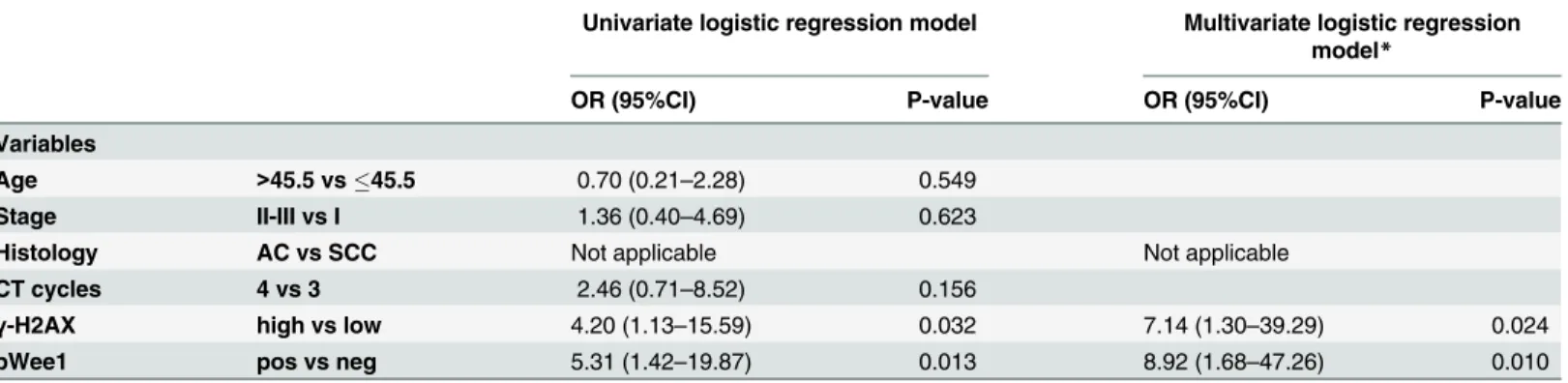
Table 4. Uni and multivariate logistic regression models of patient- and disease-related features and pathological complete response.
Univariate logistic regression model Multivariate logistic regression model*
OR (95%CI) P-value OR (95%CI) P-value
Variables
Age >45.5 vs45.5 0.70 (0.21–2.28) 0.549
Stage II-III vs I 1.36 (0.40–4.69) 0.623
Histology AC vs SCC Not applicable Not applicable
CT cycles 4 vs 3 2.46 (0.71–8.52) 0.156
γ-H2AX high vs low 4.20 (1.13–15.59) 0.032 7.14 (1.30–39.29) 0.024
pWee1 pos vs neg 5.31 (1.42–19.87) 0.013 8.92 (1.68–47.26) 0.010
*Adjusted for age, stage, number of chemotherapy cycles.
AC, Adenocarcinoma; SCC: Squamous cell carcinoma; CT, Chemotherapy.
doi:10.1371/journal.pone.0149872.t004
Table 5. Replication stability of the multivariate analysis after internal validation with a re-sampling procedure.One hundred less-powered simula-tion datasets were generated, each approximately 80% of the original size.
Mean Median Minimum Maximum Standard Deviation
Cohen's kappa coefficient 0.575 0.581 0.408 0.715 0.06
Positive predictive value (PPV) 0.741 0.742 0.600 0.888 0.05
Negative predictive value (NPV) 0.858 0.857 0.800 0.933 0.02
Sensitivity 0.660 0.667 0.440 0.800 0.06
Specificity 0.898 0.899 0.833 0.966 0.03
methods [14,15]. Conversely, our study focused on master DDR components, whose activa-tion is known to be particularly efficient in cervical cancer.
Next, we hypothesized that endogenous levels of DNA damage, mirrored byγ-H2AX, should have been paralleled by increased expression of pWee1 and pChk1.Even though the ATR-Chk1-Wee1 axis is primarily activated by stretched of single-stranded DNAs, these abnormal structures may generate DSBs upon replication fork collapse [16]. Moreover, we rea-soned that the activation of the G2/M checkpoint should be particularly proficient in the pres-ence of high basal levels of endogenous DNA damages, representing an adaptive mechanism through which cancer cells counteract oncogene-induced replication stress [16]. Indeed, it is known that ATR and Chk1 suppress the apoptotic response following DNA replication stress [17], and that tumors characterized by elevated levels of replicative stress, such as Myc-driven cancers, are extremely vulnerable to the pharmacological targeting of G2/M checkpoint kinases [18–23]. We did not observe any association between pWee1 andγ-H2AX, but rather these end-points were independently associated with pCR. We can speculate that two independent repair avenues, particularly efficient in cervical cancer, were captured in this study. A suitable candidate is the Ataxia-telangiectasia mutated (ATM)-Checkpoint kinase 2 (Chk2) pathway, which is mainly activated by DSBs [16]. An extensive cooperation exists between the ATM-Chk2 pathway and ATR-Chk1-Wee1 signaling, and ATM also phosphorylates H2AX [16].
Fig 3. OncoPrints showing the association between pWee1 and pChk1 in 37 cervical cancer samples. (A) Association according to staining intensity-based classification (positive vs negative). (B) Association according to Allred score classification (high vs low). (C) Association according to a multiplicative score classification (high vs low).
Another aspect emerging from this study relates to the association between pWee1 and pChk1 expression. Wee1 is placed downstream Chk1 [3], and Wee1 phosphorylation at Ser642 increases its stability in the nucleus and promotes cell cycle arrest at the G2/M transition [24,
25]. However, to our knowledge formal proof that this regulatory mechanism, namely Chk1-driven phosphorylation of Wee1 at Ser642, operates in mammalian cells is still lacking. Current evidence mostly stems from studies usingXenopusextracts andSchizosaccharomyces pombeas model systems [26,27]. Even though our study was not designed to generate mecha-nistic insights into the dynamics governing Wee1 activation, its results provide a suggestion for future preclinical investigations.
A final point that deserves consideration refers to the protective role of G2/M checkpoint activation in the context of cancer stem cells [28]. Activation of the axis has been associated with therapeutic resistance in different cancer stem cell models, including brain, lung and colon cancers [29–31]. Multiplying the efforts for establishing a collection of patient-derived cervical cancer stem cells for comprehensive molecular characterization is a strategy that should be pursued to further dissect the relationship existing between G2/M checkpoint activa-tion and chemoresistant features. The relevance of this approach is even more evident when considering the need for more accurate cellular and animal models in light of the number of Chk1 and Wee1 inhibitors that entered clinical development [3,32]. For instance, a phase I trial with the first-in-class Wee1 inhibitor AZD1775 (MK1775) in association with cisplatin and radiation therapy is ongoing (ClinicalTrials.govIdentifier: NCT01958658), and a phase I/ II trial in combination with topotecan/cisplatin results as completed (ClinicalTrials.gov Identi-fier:NCT01076400). Moreover, a phase I-II trial of AZD1775 in combination with chemother-apy has been initiated in patients with TP53-mutated epithelial ovarian, fallopian tube, or primary peritoneal cancer [33].
Conclusions
To sum up, pWee1 andγ-H2AX expression in pre-chemotherapy samples showed ability to foresee pCR in cervical cancer patients treated with neoadjuvant paclitaxel, ifosfamide and cis-platin. Based on the extremely promising results herein presented prospective validation or, alternatively, ancillary molecular analyses in the context of prospective trials is warranted to better characterize the predictive ability of these biomarkers.
Acknowledgments
We thank Tania Merlino for technical assistance.
Author Contributions
Conceived and designed the experiments: PV RDM MM-S. Performed the experiments: SB BA MC MM. Analyzed the data: FS IT MB. Contributed reagents/materials/analysis tools: DS LP LDL TG CV LM EV GS AG. Wrote the paper: PV SB DS BA FS IT MC RDM RD IV MB MM LP LDL TG CV LM EV GS AG MM-S. Made substantial contributions to analyses and biologi-cal interpretation of data: RD IV MB.
References
1. Hoeijmakers JH. Genome maintenance mechanisms for preventing cancer. Nature. 2001; 411:366–
374. PMID:11357144
2. Medema RH, Macurek L. Checkpoint control and cancer. Oncogene. 2012; 31:2601–2613. doi:10.
3. Maugeri-SaccàM, Bartucci M, De Maria R. Checkpoint kinase 1 inhibitors for potentiating systemic
anti-cancer therapy. Cancer Treat Rev. 2013; 39:525–533. doi:10.1016/j.ctrv.2012.10.007PMID:
23207059
4. Vitale I, Galluzzi L, Castedo M, Kroemer G. Mitotic catastrophe: a mechanism for avoiding genomic instability. Nat Rev Mol Cell Biol. 2011; 12:385–392. doi:10.1038/nrm3115PMID:21527953
5. Vici P, Mariani L, Pizzuti L, Sergi D, Di Lauro L, Vizza E, et al. Emerging biological treatments for uterine cervical carcinoma. J Cancer. 2014; 5:86–97. doi:10.7150/jca.7963PMID:24494026
6. Leake R, Barnes D, Pinder S, Ellis I, Anderson L, Anderson T, et al. Immunohistochemical detection of steroid receptors in breast cancer: a working protocol. UK Receptor Group, UK NEQAS, The Scottish Breast Cancer Pathology Group, and The Receptor and Biomarker Study Group of the EORTC. J Clin Pathol. 2000; 53:634–635. PMID:11002770
7. Buda A, Lissoni AA, Floriani I,Biagioli E, Gerardi C, Bonazzi C, et al. Long-Term Clinical Benefits of Neoadjuvant Chemotherapy in Women With Locally Advanced Cervical Cancer: Validity of Pathologi-cal Response as Surrogate Endpoint of Survival. Int J Gynecol Cancer. 2015;; 25:1468–1475 doi:10.
1097/IGC.0000000000000515PMID:26222484
8. Cortazar P, Zhang L, Untch M, Mehta K, Costantino JP, Wolmark N, et al. Pathological complete response and long-term clinical benefit in breast cancer: the CTNeoBC pooled analysis. Lancet. 2014; 384:164–172. doi:10.1016/S0140-6736(13)62422-8PMID:24529560
9. Bajpai D, Banerjee A, Pathak S,Thakur B, Jain SK, Singh N. Single nucleotide polymorphisms in the DNA repair genes in HPV-positive cervical cancer. Eur J Cancer Prev. 2015Mar 25. [Epub ahead of print]
10. Muallem MZ, Marnitz S, Richter R,Köhler C, Sehouli J, Arsenic R. ERCC1 expression as a predictive marker of cervical cancer treated with cisplatin-based chemoradiation. Anticancer Res. 2014; 34:401–
406. PMID:24403494
11. Bai ZL, Wang YY, Zhe H, He JL, Hai P. ERCC1 mRNA levels can predict the response to cisplatin-based concurrent chemoradiotherapy of locally advanced cervical squamous cell carcinoma. Radiat Oncol. 2012; 7:221. doi:10.1186/1748-717X-7-221PMID:23259415
12. Park JS, Jeon EK, Chun SH, Won HS, Lee A, Hur SY, et al. ERCC1 (excision repair cross-complemen-tation group 1) expression as a predictor for response of neoadjuvant chemotherapy for FIGO stage 2B uterine cervix cancer. Gynecol Oncol. 2011; 120:275–279. doi:10.1016/j.ygyno.2010.10.034PMID:
21093896
13. Chung HH, Kim MK, Kim JW, Park NH, Song YS, Kang SB, Lee HP. XRCC1 R399Q polymorphism is associated with response to platinum-based neoadjuvant chemotherapy in bulky cervical cancer. Gynecol Oncol. 2006; 103:1031–1037. PMID:16875718
14. Friboulet L, Olaussen KA, Pignon JP, Shepherd FA, Tsao MS, Graziano S, et al. ERCC1 isoform expression and DNA repair in non-small-cell lung cancer. N Engl J Med. 2013; 368:1101–1110. doi:10.
1056/NEJMoa1214271PMID:23514287
15. Olaussen KA, Dunant A, Fouret P,Brambilla E, André F, Haddad V, et al. DNA repair by ERCC1 in non-small-cell lung cancer and cisplatin-based adjuvant chemotherapy. N Engl J Med. 2006; 355:983–991. PMID:16957145
16. Larsson LG. Oncogene- and tumor suppressor gene-mediated suppression of cellular senescence. Semin Cancer Biol. 2011; 21:367–376. doi:10.1016/j.semcancer.2011.10.005PMID:22037160
17. Myers K, Gagou ME, Zuazua-Villar P,Rodriguez R, Meuth M. ATR and Chk1 suppress a caspase-3-dependent apoptotic response following DNA replication stress. PLoS Genet. 2009; 5:e1000324. doi:
10.1371/journal.pgen.1000324PMID:19119425
18. Cole KA, Huggins J, Laquaglia M, Hulderman CE, Russell MR, Bosse K, et al. RNAi screen of the pro-tein kinome identifies checkpoint kinase 1 (CHK1) as a therapeutic target in neuroblastoma. Proc Natl Acad Sci U S A. 2011; 108:3336–3341. doi:10.1073/pnas.1012351108PMID:21289283
19. Murga M, Campaner S, Lopez-Contreras AJ, Toledo LI, Soria R, Montaña MF, et al. Exploiting
onco-gene-induced replicative stress for the selective killing of Myc-driven tumors. Nat Struct Mol Biol. 2011; 18:1331–1335. doi:10.1038/nsmb.2189PMID:22120667
20. Höglund A, Nilsson LM, Muralidharan SV,Hasvold LA, Merta P, Rudelius M, et al. Therapeutic implica-tions for the induced levels of Chk1 in Myc-expressing cancer cells. Clin Cancer Res. 2011; 17:7067–
7079. doi:10.1158/1078-0432.CCR-11-1198PMID:21933891
21. Ferrao PT, Bukczynska EP, Johnstone RW, McArthur GA. Efficacy of CHK inhibitors as single agents in MYC-driven lymphoma cells. Oncogene. 2012; 31:1661–1672. doi:10.1038/onc.2011.358PMID:
22. Brooks K, Oakes V, Edwards B, Ranall M, Leo P, Pavey S, et al. A potent Chk1 inhibitor is selectively cytotoxic in melanomas with high levels of replicative stress. Oncogene. 2013; 32:788–796. doi:10. 1038/onc.2012.72PMID:22391562
23. Saini P, Li Y, Dobbelstein M. Wee1 is required to sustain ATR/Chk1 signaling upon replicative stress. Oncotarget. 2015; 6:13072–13087. PMID:25965828
24. Rothblum-Oviatt CJ, Ryan CE, Piwnica-Worms H. 14-3-3 binding regulates catalytic activity of human Wee1 kinase. Cell Growth Differ. 2001; 12:581–589. PMID:11751453
25. Wang Y, Jacobs C, Hook KE,Duan H, Booher RN, Sun Y. Binding of 14-3-3beta to the carboxyl termi-nus of Wee1 increases Wee1 stability, kinase activity, and G2-M cell population. Cell Growth Differ. 2000; 11:211–219. PMID:10775038
26. Lee J, Kumagai A, Dunphy WG. Positive regulation of Wee1 by Chk1 and 14-3-3 proteins. Mol Biol Cell. 2001; 12:551–563. PMID:11251070
27. O'Connell MJ, Raleigh JM, Verkade HM, Nurse P. Chk1 is a wee1 kinase in the G2 DNA damage checkpoint inhibiting cdc2 by Y15 phosphorylation. EMBO J. 1997; 16:545–554. PMID:9034337
28. Maugeri-SaccàM, Bartucci M, De Maria R. DNA damage repair pathways in cancer stem cells. Mol
Cancer Ther. 2012; 11:1627–1636. doi:10.1158/1535-7163.MCT-11-1040PMID:22844074
29. Bao S, Wu Q, McLendon RE, Hao Y, Shi Q, Hjelmeland AB, et al. Glioma stem cells promote radioresis-tance by preferential activation of the DNA damage response. Nature. 2006; 444:756–760. PMID:
17051156
30. Bartucci M, Svensson S, Romania P, Dattilo R, Patrizii M, Signore M, et al. Therapeutic targeting of Chk1 in NSCLC stem cells during chemotherapy. Cell Death Differ. 2012; 19:768–778. doi:10.1038/ cdd.2011.170PMID:22117197
31. Gallmeier E, Hermann PC, Mueller MT, Machado JG, Ziesch A, De Toni EN, et al. Inhibition of ataxia telangiectasia- and Rad3-related function abrogates the in vitro and in vivo tumorigenicity of human colon cancer cells through depletion of the CD133(+) tumor-initiating cell fraction. Stem Cells. 2011; 29:418–429. doi:10.1002/stem.595PMID:21308861
32. Do K, Doroshow JH, Kummar S. Wee1 kinase as a target for cancer therapy. Cell Cycle.2013; 12:3159–3164. doi:10.4161/cc.26062PMID:24013427