Mo rpho lo gical change s o f caro tid
bo die s in acute re spirato ry distre ss
syndro me : a mo rpho me tric study in
hum ans
Departamento de Patologia, Faculdade de Medicina, Universidade de São Paulo, São Paulo, SP, Brasil E.N.G. Vinhaes,
M. Dolhnikoff and P.H.N. Saldiva
Abstract
Carotid bodies are chemoreceptors sensitive to a fall of partial oxygen pressure in blood (hypoxia). The morphological alterations of these organs in patients with chronic obstructive pulmonary disease (COPD) and in people living at high altitude are well known. However, it is not known whether the histological profile of human carotid bodies is changed in acute clinical conditions such as acute respiratory distress syndrome (ARDS). The objective of the present study was to perform a quantitative analysis of the histology of carotid bodies collected from patients who died of ARDS. A morphometric study of carotid bodies collected during routine autopsies was carried out on three groups: patients that died of non-respiratory diseases (controls, N = 8), patients that presented COPD and died of its complications or associ-ated diseases (N = 7), and patients that died of ARDS (N = 7). Morphometric measurements of the volume fraction of clusters of chief cells were performed in five fields on each slide at 40X magni-fication. The numerical proportion of the four main histological cell types (light, dark, progenitor and sustentacular cells) was determined analyzing 10 fields on each slide at 400X magnification. The propor-tion of dark cells was 0.22 in ARDS patients, 0.12 in controls (P<0.001), and 0.08 in the COPD group. The proportion of light cells was 0.33 (ARDS), 0.44 (controls) (P<0.001), and 0.36 (COPD). These findings suggest that chronic and acute hypoxia have different effects on the histology of glomic tissue.
Co rre spo nde nce
M. Dolhnikoff,
Departamento de Patologia Faculdade de Medicina, USP Av. Dr. Arnaldo, 455 01246-903 São Paulo, SP Brasil
Fax: + 55-11-3062-8098 E-mail: maridol@ usp.br
Research supported by LIM 05-HC-FMUSP, CNPq and FAPESP.
Received May 25, 2001 Accepted May 22, 2002
Ke y words
·Carotid body ·Hypoxia
·Acute respiratory distress
syndrome
·Morphometry
Intro ductio n
Carotid bodies are nodular structures found in the angle of the bifurcation of the common carotid arteries. They are consid-ered to be chemoreceptors sensitive to changes in the partial blood oxygen pressure. De Castro (1) showed that the innervation of the carotid body, originating from the
pres-sure and the fall in arterial blood pH, con-tributing to the genesis of the hyperventila-tion observed under these condihyperventila-tions. Other factors such as temperature, osmolarity and arterial pressure, at least in animals, can stimulate the carotid body (6). Morphologi-cally, the carotid body shows two different types of cells. First, forming clusters, there are the chief cells, with three subtypes (light, dark and progenitor) observed by staining with hematoxylin and eosin (Figure 1A,B). These cells are considered to be the chemore-ceptor cells of the organ. At the level of electron microscopy, the chief cells contain electron-dense granules in their cytoplasm. Many substances have been demonstrated in these granules, mainly biogenic amines (do-pamine, noradrenaline) and some peptides, but the physiological significance of these findings remains unknown. The second type, surrounding the clusters of chief cells, is represented by the sustentacular cells that envelope the nonmyelinated nervous fila-ments and enclose them to the surface of the chief cells (Figure 1C).
There is considerable evidence that ca-rotid bodies can have their structure modi-fied under several conditions, such as nor-mocapnic hypoxia (7-9), chronic obstructive pulmonary disease (COPD) (10-12), aging (13), and arterial hypertension (11,14). In these situations, the changes in the histology of carotid bodies have been presumed to provide a plausible structural basis for the functional abnormalities of respiratory con-trol. For instance, chronic high altitude hy-poxia has been associated with light cell hyperplasia (15-17); an increase of susten-tacular cells has been clearly demonstrated in COPD (18-20). Thus, it is reasonable to assume that the cellular profile of carotid bodies can provide useful information about the physiopathology of the respiratory con-trol. However, the relationship between the morphological and functional alterations of the structure in these situations remains ob-scure.
Figure 1. Photomicrographs of glomic cells. A, Light (L) and dark (d) cells in a cluster of chief cells, in a patient that died of acute myocardial infarction. B, Light (L) and progenitor (p) cells in a patient that died of ARDS and systemic lupus erythematosus. C, Light (L) and sustentacular (s) cells in a patient that died of COPD and rupture of aortic aneurysm (HE; bar = 10 µm).
A AA AA
B BB BB
If chronic hypoxia modifies the histo-logical profile of the carotid bodies, one can speculate that acute episodes of hypoxia may promote histopathological changes as well. It has been well established that patients that survive acute respiratory distress syndrome (ARDS) may have abnormalities of respira-tory control as demonstrated during weaning from mechanical ventilation (21). However, it is not known whether the histology of carotid bodies is modified in this situation.
The present study was designed to per-form a quantitative analysis of the histology of carotid bodies collected at autopsy from patients that died of ARDS. The data were compared with those obtained for two differ-ent groups: patidiffer-ents with non-respiratory and non-chronic cardiovascular diseases (con-trols), and patients with COPD. Patients with COPD were included in the study because the changes found in the glomic tissue in this clinical situation have been well documented (11,12,19).
Patie nts and Me thods
Carotid bodies were collected during rou-tine autopsies performed at our Medical School. Clinical information was obtained from hospital charts and the diagnosis was confirmed at autopsy. Patients were divided into three groups: deaths caused by non-respiratory diseases (group I, N= 8), patients with COPD who died of its complications or associated diseases (group II, N = 7), and patients who died of ARDS (group III, N= 7). For group III, no restrictions were made about time of mechanical ventilation and inspired fraction of oxygen.In all of these patients the duration of ARDS was less than one week. Clinical information about these patients is presented in Table 1.
At autopsy, patients from group I showed no significant changes in lung histology or signs of chronic cardiovascular diseases. Patients with ARDS showed marked histo-logical changes in lung parenchyma,
includ-Table 1. Age, sex and main diagnosis of the patients studied.
Case Group Age (years) Sex M ain diagnosis
1 I 70 M Acute myocardial infarction
2 I 41 M Acute myocardial infarction
3 I 59 M Gastric tumor
4 I 67 M Gastric tumor
5 I 60 F Acute myocardial infarction
6 I 75 M Pancreatic tumor
7 I 50 M Acute myocardial infarction
8 I 33 M AIDS + cryptococcosis
9 II 75 M COPD + cor pulmonale
10 II 82 M COPD + cerebral infarction
11 II 59 M COPD + cor pulmonale
12 II 81 M COPD + bronchopneumonia
13 II 59 M COPD + bronchopneumonia
14 II 58 M COPD + rupture of aortic aneurysm
15 II 74 M COPD + acute myocardial infarction
16 III 17 M ARDS + septicemia
17 III 23 M ARDS + systemic lupus erythematosus
18 III 22 M ARDS + leptospirosis
19 III 54 M ARDS + septic shock
20 III 74 M ARDS + septic shock
21 III 40 M ARDS + leptospirosis
22 III 18 F ARDS + cerebral hemorrhage
Data w ere obtained from patient charts. The main diagnosis w as confirmed at au-topsy. Groups: I = control, II = chronic obstructive pulmonary disease (COPD), III = acute respiratory distress syndrome (ARDS). M = male, F = female.
ing alveolar infiltration of inflammatory cells, intra-alveolar exudate with hyaline mem-branes, alveolar hemorrhage and septal thick-ening by loose connective tissue.
In order to collect the carotid bodies, the common carotid artery on each side of the neck was dissected up to above its bifurca-tion and the whole piece (3 cm above and 5 cm below the bifurcation) was placed in buffered formalin for 48 h. After this, the carotid body was gently dissected with the aid of a 25X magnifying glass (Carl Zeiss, Germany) and routinely processed for paraf-fin embedding and histological sectioning. Five-micrometer thick sections were stained with hematoxylin and eosin. The two carotid bodies (left and right) of each patient were studied. One section per carotid body was analyzed.
(Instru-mentação Técnica Científica, São Paulo, SP, Brazil) containing 50 lines and 100 points. We determined the volume fraction of clus-ters of chief cells (Vc) at 40X magnification. Briefly, five non-coincident fields on each slide were examined across a surface cut of the carotid body. For each field, 100 points were counted and Vc was determined by the following relationship: Vc = pC/ptot (1), where pC represents the number of points hitting chief cell clusters and ptot is the total number of points considered for each micro-scopic field (Figure 2A).
In addition to Vc, the number of profiles of the four main histological subtypes of glomic tissue - light, dark, progenitor and sustentacular cells (4) - was determined in 10 non-coincident microscopic fields on each slide at 400X magnification using the unbi-ased procedure described by Gundersen et al. (22), whereby the cell profiles are counted in each field respecting the inclusion and exclusion limits of the integrating eyepiece (Figure 2B).
The proportion P of the main histological cell types - Plight, Pdark, Pprogenitor and Psustent,
respectively, within the glomus was com-puted by dividing the numerical density of each cell profile by the total number of pro-files counted.
Groups were compared by analysis of variance (ANOVA), with the level of signif-icance set at 5%. When a significant differ-ence was detected, multiple comparisons among the groups were performed by the Student-Newman-Keuls test. Logarithmic transformations were applied when neces-sary in order to test the sensitivity of the results to heteroscedasticity. The statistical calculations were done with the aid of the SPSS v6.0 software (23).
Re sults
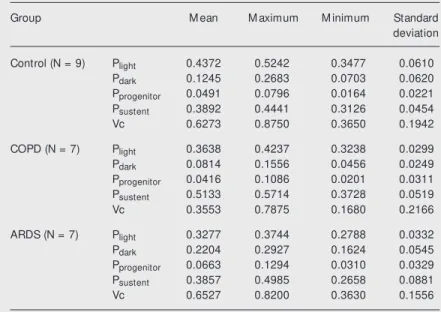
The results of the morphometric meas-urements obtained for the different groups are shown in Table 2. Table 3 shows the
Figure 2. M orphometric studies of glomic tissue. A, Schematic representation of the morphometric measurement of the volume fraction of clusters of chief cells (Vc) using the standard point-counting procedure (40X). Vc w as obtained using the Vc = pC/ptot ratio, w here pC represents the number of points hitting chief cell clusters and ptot is the total number of points considered for each microscopic field. B, Schematic representation of clusters of chief cells in one microscopic field (1000X). The upper and right limits of the integrating eyepiece (dashed lines) w ere considered the exclusion limits. The proportion P of the main histological cell types (Plight, Pdark, Pprogenitor and Psustent) w as obtained by
dividing the numerical density of each cell profile by the total number of profiles counted.
A B
Table 2. M orphometric parameters of carotid bodies in control, COPD and ARDS groups.
Group M ean M aximum M inimum Standard
deviation
Control (N = 9) Plight 0.4372 0.5242 0.3477 0.0610
Pdark 0.1245 0.2683 0.0703 0.0620
Pprogenitor 0.0491 0.0796 0.0164 0.0221
Psustent 0.3892 0.4441 0.3126 0.0454
Vc 0.6273 0.8750 0.3650 0.1942
COPD (N = 7) Plight 0.3638 0.4237 0.3238 0.0299
Pdark 0.0814 0.1556 0.0456 0.0249
Pprogenitor 0.0416 0.1086 0.0201 0.0311
Psustent 0.5133 0.5714 0.3728 0.0519
Vc 0.3553 0.7875 0.1680 0.2166
ARDS (N = 7) Plight 0.3277 0.3744 0.2788 0.0332
Pdark 0.2204 0.2927 0.1624 0.0545
Pprogenitor 0.0663 0.1294 0.0310 0.0329
Psustent 0.3857 0.4985 0.2658 0.0881
Vc 0.6527 0.8200 0.3630 0.1556
Data are reported as means for each group. COPD = chronic obstructive pulmonary disease; ARDS = acute respiratory distress syndrome; Plight = proportion of light cells;
Pdark = proportion of dark cells; Pprogenitor = proportion of progenitor cells; Psustent =
ANOVA output. Patients with COPD had an increase in sustentacular cells and reduced glomic tissue when compared to controls. Patients with ARDS had a significantly higher number of dark cells and a reduction of light cells compared to COPD and controls. These findings suggest that chronic and acute hy-poxia have different effects on the histology of glomic tissue.
D iscussio n
In this study our objective was to deter-mine whether ARDS would change the cel-lular profile of the carotid body, since this is an acute clinical condition characterized by severe hypoxia with a high mortality rate despite an increase in the inspired fraction of oxygen (24-26). We had a “positive con-trol”, since we observed a significant in-crease in the number of sustentacular cells in COPD, as already shown by others (18,19). Although the studies of carotid bodies in COPD patients previously performed did not use unbiased morphometric methods, proliferation of type II cells (sustentacular cells) in hypoxic patients has been reported (10-12). We also observed that the amount of glomic tissue in COPD was reduced in comparison to that of the connective tissue that limits the lobules within the carotid body, suggesting that the parenchyma of COPD patients shows reduced chemosensi-tivity.
ARDS patients had a different histologi-cal profile, with marked reduction of light cells and an increase in dark cells when compared to controls. It is interesting to note that the patients in the ARDS group were younger than in the other groups. The differ-ence in age could be a factor that affects the histological profile of carotid bodies since there is a predominance of dark cells in the chief cell population of children and young adults. However, even the three oldest pa-tients in the ARDS group had an increased dark cell population when compared to
con-trols (mean values of Pdark = 0.125) and the
COPD group. Table 4 shows individual val-ues of Pdark in ARDS patients.
What could be the mechanisms respon-sible for the increase of dark cells in ARDS? Since the increase in dark cells occurred at the same time as a decrease in light cells, it is tempting to consider that these two cell types are variants of the same cell, as suggested by Verna (27). The increase in the dark cell population observed in ARDS could reflect a higher functional activity of carotid bodies in response to hypoxia. In an elegant study, Biscoe and Stehbens (28) investigated in detail the ultrastructural aspects of glomic cells, suggesting that dark cells have a highly complex secretory structure, presenting a large number of electron-dense granules, most probably containing biogenic amines. Furthermore, the number of electron-dense granules seemed to be increased in chief cells after exposure to acute hypoxia in rab-bits (29). Jago et al. (30) suggested that the increase in electron-dense granules was more pronounced in dark cells in a patient with arterial hypertension, leading to a darker cytoplasm at the ultrastructural level. In the human carotid body, dopamine is the
bio-Table 3. ANOVA output of the multiple comparisons among the groups.
Pdark P<0.001 ARDS different from all others
Plight P<0.001 Control different from ARDS and COPD
Psustent P = 0.002 COPD different from all others
Pprogenitor P = 0.282 No differences among groups
Vc P = 0.015 COPD different from all others
Data w ere obtained by comparison of the groups using analysis of variance (ANOVA), w ith the level of significance set at 5% . When a significant difference w as observed, multiple comparisons among the groups w ere performed by the Student-New man-Keuls test. For abbreviations see legend to Table 2.
Table 4. Age and proportion of dark cells (Pdark) of patients w ith acute respiratory
distress syndrome.
Age (years) 17 18 22 23 40 54 74
Pdark 0.286 0.279 0.163 0.180 0.179 0.192 0.239
Patient 1 7 3 2 6 4 5
genic amine present at the highest concen-tration in the electron-dense granules (31), but, interestingly enough, dopamine is con-sidered to decrease the respiratory response to hypoxia (32). The functional significance of these observations remains to be clarified. Since dark cells can represent a higher functional status of chief cells, an increased number of dark cells in ARDS could be seen as an adaptive response to low arterial oxy-gen tension. Consistent with the foregoing view is the report that dark cell hyperplasia was observed in Dutch rabbits kept on Monte Bianco at an altitude of 3370 m for 3 months. However, the number of dark chief cells returned to normal values (11%) when the rabbits were left at the same altitude for a period of 6 months (33). Furthermore, the carotid bodies from patients with asthma also present an increased number of dark chief cells (34) and this response is considered the first morphological alteration of the ca-rotid body cells after exposure to hypoxia (4).
The morphological changes observed in carotid bodies in ARDS indicate that func-tional alterations of respiratory chemosensi-tivity may be present in these patients. In this scenario, our results indicate that studies focusing on respiratory control should be done in patients surviving ARDS in order to determine to what extent the difficulties of weaning from the ventilator are related to abnormal respiratory drive.
Our results demonstrate that patients with ARDS present an increase of the numerical fraction of dark cells in their carotid bodies. These findings are consistent with an initial response of these structures to acute hypoxia and indicate the necessity for functional stud-ies of respiratory control in patients surviv-ing ARDS.
Ackno wle dgm e nts
The authors thank Dr. Thais Mauad for critical comments and suggestions.
Re fe re nce s
1. de Castro F (1928). Sur la structure et l’ innervat ion du sinus carot idien del’ homme et des mammiféres. Nouveaux faits sur l’innervation et la fonction du glomus caroticum. Travaux du Laboratoire de Recherches Biologiques de l’ Uni-versité de M adrid, 25: 331-380.
2. Grimley PM & Glenner GG (1968). Ultra-structure of the human carotid body. A perspective on the mode of chemorecep-tion. Circulation, 37: 648-665.
3. Grönblad M (1983). Function and struc-ture of the carotid body. M edical Biology, 61: 229-248.
4. Smith P (1995). Carotid body and pulmo-nary glomera in cardiorespiratory disease. In: Spencer J (Editor), Spencer’s Patholo-gy of the Lung. 5th edn. M cGraw -Hill Companies, Inc., Seattle, WA, USA. 5. Heymans C, Bouckhaert JJ &
Dautre-bande L (1930). Sinus carot idien et réflexes respiratoires. II. Influences respi-ratoires réflexes de l’acidôse de l’alcalose, de l’anhydride carbonique, de l’ion hydro-géne et de l’anoxémie: Sinus carotidiens
et échanges respiratoires dans le pou-mons et au delá des poupou-mons. Archives Internationales de Pharmacodynamie et de Therapie, 39: 400-448.
6. Eyzaguirre C & Zapata P (1984). Perspec-tives in carotid body research. Journal of Applied Physiology, 57: 931-957. 7. Lack EE (1977). Carotid body hypertrophy
in patients w ith cystic fibrosis and cyan-otic heart disease. Human Pathology, 8: 39-51.
8. Lack EE (1978). Hyperplasia of vagal and carotid body paraganglia in patients w ith chronic hypoxemia. American Journal of Pathology, 19: 497-516.
9. Lack EE, Perez-Atayde AR & Young JB (1985). Carotid body hyperplasia in cystic fibrosis and cyanotic heart disease. A combined morphometric, ultrastructure, and biochemical study. American Journal of Pathology, 119: 301-314.
10. Heath D, Edw ards C & Harris P (1970). Post-mortem size and structure of the human carotid body. Its relation to pulmo-nary disease and cardiac hypertrophy.
Thorax, 25: 129-140.
11. Edw ards C, Heath D & Harris P (1971). The carotid body in emphysema and left ventricular hypertrophy. Journal of Pathol-ogy, 104: 1-13.
12. Habeck J-O (1986). M orphological find-ings at the carotid bodies of humans suf-fering from different types of systemic hypertension or severe lung diseases. Anatomischer Anzeiger, 162: 17-27. 13. Hurst G, Heath D & Smith P (1985).
Histo-logical changes associated w ith ageing of the human carotid body. Journal of Pa-thology, 147: 181-187.
14. Heath D, Smith P & Hurst G (1986). The carotid body in coarctation of the aorta. British Journal of Diseases of the Chest, 80: 122-130.
15. Arias-Stella J (1969). Human carotid body at high altitudes. American Journal of Pa-thology,55: 82 (Abstract).
Ana-tomical variations in human carotid bod-ies. Journal of Clinical Pathology, 41: 1196-1199.
18. Heath D, Smith P & Jago R (1982). Hyper-plasia of the carotid body. Journal of Pa-thology, 138: 115-127.
19. Smith P, Jago R & Heath D (1982). Ana-tomical variations and quantitative histol-ogy of the normal and enlarged carotid body. Journal of Pathology, 137: 287-304. 20. Heath D (1991). The human carotid body in health and disease. Journal of Patholo-gy, 164: 1-8.
21. Hansen-Flaschen J & Fishman AP (1992). Síndrome da distração respiratória do adulto: Características clínicas e patogê-nese. In: Fishman AP (Editor), Diagnós-tico das Doenças Pulmonares. M anole, São Paulo, SP, Brazil.
22. Gundersen HJG, Bendtsen TF, Korbo L, M arcussen N, M oller A, Nielsen K, Nyengaard JR, Pakkenberg FB, Sorensen FB, Vesterby A & West M J (1988). Some new , simple and efficient stereological methods and their use in pathological re-search and diagnosis. Acta Pathologica,
M icrobiologica et Immunologica Scandi-navica, 96: 379-394.
23. SPSS version 6.0. (1990). SPSS Inc., Chi-cago, IL, USA.
24. Demiling RH (1995). The modern version of adult respiratory distress syndrome. Annual Review of M edicine, 46: 193-202. 25. Luce JM (1998). Acute lung injury and the acute respiratory distress syndrome. Criti-cal Care M edicine, 26: 369-376. 26. Ranieri VM , Suter PM , Tortorella C, De
Tullio R, Dayer JM , Brienza A, Bruno F & Slutsky AS (1999). Effect of mechanical ventilation on inflammatory mediators in patients w ith acute respiratory distress syndrome. A randomized controlled trial. Journal of the American M edical Associa-tion, 7: 54-61.
27. Verna A (1997). The mammalian carotid body: M orphological data. In: González C (Editor), The Carotid Body Chemorecep-tors. M edical Intelligence Unit, New York, NY, USA.
28. Biscoe TJ & Stehbens WE (1966). Ultra-structure of the carotid body. Journal of Cell Biology, 30: 563-578.
29. M oller M , M ollagard K & Sorensen SC (1974). The ultrastructure of the carotid body in chronically hypoxic rabbits. Jour-nal of Physiology, 238: 443-447. 30. Jago R, Smith P & Heath D (1985).
Elec-tron microscopy of carotid body hyperpla-sia. Archives of Pathology and Laboratory M edicine, 108: 717-722.
31. Steele RH & Hinterberger H (1972). Cat-echolamines and 5-hydroxytryptamine in the carotid body in vascular, respiratory, and other diseases. Journal of Laboratory and Clinical M edicine, 80: 63-70. 32. Barer G (1994). Carotid body in animal
models of human disease: w hat do they teach us? Thorax, 49 (Suppl): 14-18. 33. Smith P, Heath D, Williams D, Bencini C,
Pulera N & Giuntini C (1993). The earliest histopathological response to hypobaric hypoxia in rabbits in the refugio Torino (3370 m) on M onte Bianco. Journal of Pathology, 170: 485-491.