Reduction of Acoustic Reflex
Threshold in Neonates without
Auditory Risk
Summary
Jordana Costa Soares1, Renata M. M. Carvallo21 Holder of a FAPESP scholarship – Speech and Hearing Therapist 2 Associate Professor at the Phonoaudiology program of the FMUSP USP Medical School
Mailing address: Jordana Costa Soares Rua Arminda Beranger 575 Vila Isabel Osasco SP 06180-130
This paper was submitted to the ABORL-CCF SGP (Management Publication System) on June 2nd, 2005 and accepted on June 13th, 2005.
T
he auditory sensitization, a tool used in the investigation of acoustic reflex, allows the decrease of acoustic reflex thresholds from a facilitating stimulus. It may be presented before or simultaneously with the elicitor tone. The thresholds after and before the facilitating stimulus are compared and it is expected to see the decreased threshold. From the study of the acoustic reflex it is possible to obtain information about the auditory pathways, such as structures of the brainstem, since the acoustic reflex pathway is related to the auditory nuclei in this site. They are also involved in auditory processing. Thus, alterations of the acoustic reflex could be related to deficits in auditory processing skills. Aim: This study aims at investigating the acoustic reflex sensitization from a high-frequency facilitating tone (6 kHz) in newborns without risk factors to hearing impairment. Results: The acoustic reflex threshold decreased in males and females for all studied frequencies. Conclusion: A high-frequency facilitating tone presented simultaneously produced a decrease of the acoustic reflex threshold in newborns without risk factors to hearing impairment.Key words: acoustic reflex, newborns, auditory sensitization, facilitating stimulus.
ORIGINAL ARTICLE Rev Bras Otorrinolaringol
INTRODUCTION
The acoustic reflex involves the auditory nuclei in the brain stem related to auditory processing (Colletti et al.,
19921; Carvallo, 19962). Therefore, acoustic reflex changes
may suggest alterations on some auditory nuclei and impair skills related to hearing stimuli processing, such as location, selective attention, speech recognition amidst noise, and
frequency selectivity (Carvallo, Albernaz, 1997)3.
It is possible to assess the acoustic reflex thresholds
of neonates and infants. Vincent, Gerber (1987)4 found
contralateral acoustic reflex on 92.5% of neonates with up to 48 hours of age and on 95% of infants with up to
6 weeks of age. McMillan et al. (1985)5 were also able to
identify ipsilateral acoustic reflexes on 85-95% of the ears of 46 infants with ages ranging between 2 weeks and 12 months, all free from hearing alterations. Carvallo,
Alber-naz (1997)3 identified ipsilateral acoustic reflex on 100%
of infants included in their cases.
It is possible that there might be indirect multi-syn-aptic pathways involving other areas of the central nervous system aside from the acoustic reflex arc direct pathways.
Borg (1973)6 presented evidence of a pathway involving
the reticular formation. The role of such pathway is yet unknown, but it may be related to complex characteris-tics of the acoustic reflex such as delivering anticipatory response, that is, to improve the acoustic reflex response (reducing the threshold) as observed in auditory sensitiza-tion procedures.
Acoustic reflex analysis -- as a possibility of investi-gating the efferent pathway -- depends on the integrity of the auditory afferent and efferent pathways. The auditory sensitization procedure has been used as a tool for acoustic reflex analysis, as it allows the acoustic reflex threshold reduction as of the introduction of a facilitating stimulus. Such stimulus may be introduced before or simultaneously to the tone used to elicit the acoustic reflex. Thresholds before and after exposure to the facilitating tone are com-pared. The one after exposure is expected to be lower, as reported by various authors.
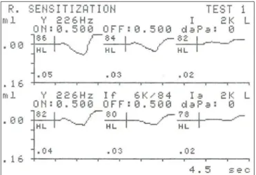
Figure 1 (below) depicts one example of acoustic reflex sensitization. Initially, the reflex threshold at 2kHz was obtained at 84 dB. When the threshold was assessed again, now with the introduction of a facilitating 6 kHz tone in the intensity at which the acoustic reflex had been identified (84 dB), the threshold drops to 80 dB.
Deutsch (1973)7 studied auditory sensitization in
adult patients with normal hearing and found reduced acoustic reflex thresholds in most subjects after stimula-tion by white noise. The author makes reference to the theory that considers the potential of enhancing VIII cranial nerve potential sensitivity by means of stimulation, and mentions the possibility of stimulation leading to fatigue and consequent recruitment (reduction on the acoustic
reflex threshold). Sesterhen, Breuninger (1976)8 also found
reduced acoustic reflex thresholds (30 dB) after the intro-duction of an 8 kHz tone concurrently to the second as-sessment of the acoustic reflex thresholds on 25 adult ears.
On a later study (1977)9, the same authors found reduced
acoustic reflex thresholds (20-30 dB) in normal ears and in ears with hearing loss after introducing concurrent 6 or 8 kHz facilitating tones of the same intensity as that of the tone used to elicit the acoustic reflex. Blood, Greenberg
(1981)10 also identified reduced acoustic reflex thresholds
when simultaneously introducing a facilitating tone in a single ear and in both ears.
Stelmachowicz, Gorga (1983)11 concluded that the
acoustic reflex threshold reduction does not depend on the frequency of the facilitating stimulus. They were able to observe threshold reductions at various frequencies (500 Hz, 1, 2, 4 and 6 kHz), suggesting that the cochlea tonoto-pic pattern does not interfere in the process. The detected independence from frequency indicates that acoustic reflex facilitation is not primarily mediated by afferent mecha-nisms. The authors propose that facilitation takes place at the efferent portion of the acoustic reflex arc.
Jeck et al. (1983)12 have also observed reduced
acoustic reflex thresholds of 10 to 12 dB in adults for fre-quencies of 500Hz, 1 and 2 kHz using a 6 kHz simultaneous facilitating tone. The authors suggested that sensitization may improve the signal-noise ratio in complex hearing scenarios by attenuating the lower frequencies.
Differently from other authors, Kumar and Barman
(2002)13 used broad band noise to find increased acoustic
reflex thresholds. They introduced contralateral white noise simultaneously to the acoustic reflex threshold assessment. Thresholds were increased by 4 dBNA at 1 kHz and by 3.6 dBNA at 2 kHz. Such effect was imputed to the auditory
efferent pathway activation by broad band noise.
Carvallo, Soares (2004)14 looked into acoustic reflex
sensitization in young women without audiologic com-plaints and with tonal hearing thresholds within normal ranges. The frequencies analyzed were of 500 Hz, 1, 2 and 4 kHz respectively, while the facilitating tone was of 6 kHz, introduced simultaneously and ipsilaterally. Acoustic reflex thresholds decreased in 6.7 to 13 dBNA on right ears and in 14.3 to 17.2 dBNA on left ears. The observed difference between the values found for right and left ears was not significant for any of the frequencies.
Studies analyzing the activation of the auditory efferent pathway have contributed to enhance the under-standing of acoustic reflex reduction in adults. However, its occurrence in neonates still remains doubtful, as it has not been described in papers studying such population. Functional studies of the efferent pathway in neonates using other principles such as the analysis of suppressed
otoacoustic emissions (Durante, Carvallo; 200215; During;
200416) have shown that the efferent pathway is present at
birth. Therefore it is reasonable to assume that acoustic re-flex threshold reduction may also be found in neonates.
OBJECTIVE
This paper aims at investigating acoustic reflex threshold variations resulting from simultaneous ipsilateral application of a 6 kHz facilitating stimulus in full term birth neonates, without indicated risk for hearing loss and with transient-evoked otoacoustic emissions.
MATERIALS AND METHOD
Sample
The sample comprised 40 full term babies, at proper weight for their ages, without prenatal, perinatal, or post-natal complication, and without indicated risk for hearing loss. Twenty babies of each gender were chosen for the study. Tests were carried out within the neonatal period.
The neonates participating in the study –- all un-der Informed Consent signed by their parents (research approved by the Ethics Committee of the USP University Hospital permit # 174/01) – fit the following inclusion criteria:
1) Presence of TOAE on both ears during hearing screening, according to the criteria set by Brass, Kemp
(1994)17, and Letourneau et al. (2000)18.
2) Ears with normal tympanometric curves, types A or D (double peak for neonates) and presence of acoustic reflex on at least two frequencies, so that the threshold comparison without and with 6 kHz tone stimulation could be done.
Materials
• “ILO 292 / ECHOPORT PLUS Otodynamics Analyser” – for the acquisition of OAEs using the ‘Quick-screener’ software, indicated for neonatal hearing screen-ing. This software consists of a standard non-linear mode composed of four stimuli, three of which are equal to one another while the remaining one is reversed and three times greater in terms of amplitude. Response analysis time was of 12ms. Stimuli were of the ‘click’ type, had a duration of 80us and intensity of 80 dB at peak NPS
equivalent (Otodynamics, 1992)19.
• Portable computer with a Pentium III processor, color monitor, with ILO V5 292 Echoport Plus Otodynam-ics Analyser installed.
• Grason Stadler TympStar Middle Ear Analizer re-lease 2 immittancemeter – microprocessed and equipped with three tone frequencies probe: 226, 678 and 1000 Hz. Tympanometric measures were carried out automatically by the device at 50 decaPascals per second (daPa/s). Results were plotted in a graph and printed out. The analysis of ipsilateral acoustic reflexes was done with stimuli calibrated in dBHL, played on a loudspeaker as-signed exclusively to the ipsilateral mode. The signal was digitally multiplexed, thus allowing the probe tone (226 Hz) to be separated from that of the stimulus avoiding overlapping waves and consequent generation of artifact. The equipment used to analyze ipsilateral acoustic reflex delivers a maximum output of 110 dBHL. It was calibrated for São Paulo city altitude, and all measures were taken from the standpoint of electrical installation so as to meet the manufacturer’s technical specifications.
(Grason-Sta-dler, 2001)20
Procedures
The procedures were carried out at the audiology department of the University Hospital of the University of São Paulo, in a quiet environment. The prenatal and neonatal health data and family information in regards to hearing and language skills were obtained by means of interviews with the mothers. Additional data on the pre-natal and peripre-natal periods was collected from the hospital admission charts.
Immittance assessment was done starting by the right ear for one half of the subjects in the sample and by the left ear for the other half. Immittance screening comprised studies on tympanometric curve and acoustic reflex at 100 dBHL. The frequency of the tone produced by the immittance probe was of 226 Hz. Neonates who did not present acoustic reflex in the screening mode at 100 dBHL were sent to analysis in diagnostic mode.
After immittance screening, subjects underwent analysis of ipsilateral acoustic reflexes through the device special mode, where sensitization would be carried out further. The frequencies of the analyzed thresholds were 1, 2 and 4 KHz, in 2 dB incremental steps. Tone length was 0.5 s, with on time of 0.5 s and off time of 0.25 s. Sensitization analysis was carried out separately for each of the frequencies mentioned previously after the first in-vestigation of acoustic reflex threshold was completed.
The facilitating stimulus used was an ipsilateral 6 kHz 0.5 second long tone (length equal to that of the reflex-eliciting tone). For each frequency, the intensity of the facilitating stimulus was kept equal to that in which the acoustic reflex threshold had been obtained. The search for the new threshold was done in incremental steps of 2 dB, with simultaneous exposure to the facilitating stimulus (6kHz).
One must bear in mind that the population analyzed by this study presents its inherent difficulties to being tested. Subjects were tested while asleep, and all proce-dures were carried out rapidly, as they could wake up at any moment. Many published papers report reductions in the acoustic reflex threshold by 10-12 dB (Jeck et al., 1983) and by 6-17 dB (Carvallo, Soares, 2004), to name a few. Thus, the second threshold analysis in this study was done at a frequency 10 dB below the first threshold obtained.
RESULTS
Tympanometry (screening mode)
In terms of tympanometric curves, for all 40 ne-onates, their two ears double peak curves (D) were present in 60% of the cases, while type A curves were found in the remaining 40%. From the two proportions equality test, it was found that there is no significant difference between curve types (p>0.05), as shown in Table 1.
Acoustic Reflex and Auditory Sensitization Analysis
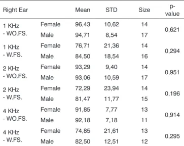
In regards to facilitating stimulus, the mean values for the acoustic reflex thresholds without (WO.FS) and with facilitating stimulus (W.FS) for males and females on each frequency for their right and left ears can be seen on tables 2 and 3 respectively. There was no significant difference between males and females, as in both Tables the p values were greater than 5%.
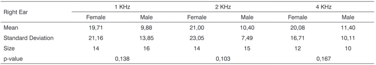
Tables 4 and 5 show the mean values for the differ-ence between thresholds obtained in the two scenarios of the study, i.e., without facilitating stimulus (W.FS) and with facilitating stimulus (W.FS). Although reduction is slightly higher among females in both ears, the difference is not statistically significant (p>0.05).
Tympanometric Curve RE LE Qty % Qty % Double Peak 24 60% 25 60% A 16 40% 16 40% Total 40 100% 40 100% p-value 0,074 0,074
Table 1. Percentage of tympanometric curves present.
Right Ear Mean STD Size value p-1 KHz
- WO.FS.
Female 96,43 10,62 14
0,621 Male 94,71 8,54 17
1 KHz - W.FS.
Female 76,71 21,36 14
0,294 Male 84,50 18,54 16
2 KHz - WO.FS.
Female 93,29 9,40 14
0,951 Male 93,06 10,59 17
2 KHz - W.FS.
Female 72,29 23,94 14
0,196 Male 81,47 11,77 15
4 KHz - WO.FS.
Female 91,85 7,77 13
0,914 Male 92,18 7,18 11
4 KHz - W.FS.
Female 74,85 21,61 13
0,295 Male 82,50 12,51 12
Table 2. Acoustic reflex thresholds without and with facilitating stimulus – RE.
Left Ear Mean STD Size value p-1 KHz
- WO.FS.
Female 92,67 9,00 15
0,643 Male 94,25 9,77 16
1 KHz - W.FS.
Female 78,63 20,12 16
0,159 Male 87,13 12,18 16
2 KHz - WO.FS.
Female 92,38 8,89 16
0,600 Male 94,13 9,76 16
2 KHz - W.FS.
Female 75,87 21,15 15
0,155 Male 85,20 12,78 15
4 KHz - WO.FS.
Female 91,67 8,86 12
0,219 Male 95,47 6,82 15
4 KHz - W.FS.
Female 75,82 19,05 11
0,112 Male 86,67 11,77 12
Right Ear 1 KHz 2 KHz 4 KHz Female Male Female Male Female Male Mean 19,71 9,88 21,00 10,40 20,08 11,40 Standard Deviation 21,16 13,85 23,05 7,49 16,71 10,11 Size 14 16 14 15 12 10 p-value 0,138 0,103 0,167
Table 4. Mean difference without and with facilitating stimulus - RE.
Table 5. Mean difference without and with facilitating stimulus - LE.
Left Ear 1 KHz 2 KHz 4 KHz Female Male Female Male Female Male Mean 15,07 7,13 15,73 8,40 15,27 8,33 Standard Deviation
16,95 7,27 16,65 7,49 13,84 11,81 Size 15 16 15 15 11 12 p-value 0,097 0,131 0,209
DISCUSSION
Tympanometry screening underscored a distri-bution of results between two different configurations. Without analyzing them separately by gender, 60% of the right ear tympanometric curves presented double peak (type D) configuration, while 40% were type A. Both are deemed normal for the neonatal population. The same was found for left ears. Another paper (Vincent, Gerber,
1987)4 studying infants identified double peak
tympano-metric curves, which did not prevent the obtainment of acoustic reflexes.
It is important to stress that all subjects included in this study – both males and females – presented acoustic reflex thresholds in at least one of their ears when analyzed in the device off of the screening mode. The goal here was to verify sensitization by comparing thresholds without and with the 6 kHz stimulus. That was possible only because all subjects presented acoustic reflexes.
The mean values found for acoustic reflex without facilitating stimulus ranged between 91 and 96 dBHL. Close
values were also found by Carvallo, Albernaz (1997)3 in
their research on ipsilateral acoustic reflex in a population similar to that of this study. They used frequencies of 1 kHz and 2 kHz, and found values between 96.3 dBHL at
1 kHz and 95.2 dBHL at 2 kHz. McMillan et al. (1985)5
also studied acoustic reflexes in infants at 500 Hz, 1 and 2 kHz, finding mean threshold values between 80 and 85 dBHL.
The presentation of a facilitating stimuli brought significant reduction to acoustic reflex thresholds at all
frequencies, for both ears of subjects from both genders,
as seen in the literature10,12,14 of adult populations.
The facts observed in this study agree with
vari-ous other papers10-12 insofar as frequency independence
is concerned, as sensitization occurred in the neonatal population regardless of the frequency utilized. Further studies are required to determine whether or not the same would happen for other facilitating stimuli.
From what has been explained one might say that, apart from the conventional investigation of the stape-dian reflex, it is also possible to assess the acoustic reflex threshold reduction (sensitization) in neonates. As authors agree that this occurrence is related to the auditory
effer-ent pathway11, it may be used to assess this same pathway
in neonates.
Additionally, the investigation on auditory sensitiza-tion in populasensitiza-tions with dysfuncsensitiza-tional efferent pathways and in individuals with altered auditory processing may be an important diagnostic tool. It will increase the cer-tainty around the location in the auditory system where sensitization or acoustic reflex threshold reduction oc-curs, thus contributing to further scientific investigation on hearing.
CONCLUSION
REFERENCES
1. Colletti V, Fiorino FG, Verlato MD, Carner M. Acoustic reflex in fre-quency selectivity: brain stem auditory evoked response and speech discrimination. In: Katz K, Stecher NA & Henderson D. Eds. Central Auditory Processing: A Transdisciplinary View. St Louis Mosby Year Book; 1992.
2. Carvallo RMM. O efeito do reflexo estapediano no controle da pas-sagem da informação sonora. In: Schochat E. (Org.) Processamento Auditivo. São Paulo: Editora Lovise; 1996. 57-73.
3. Carvallo RMM, Mangabeira Albernaz PL. Reflexos Acústicos em Lactentes. Acta AWHO 1997; 16 (3): 103-8.
4. Vincent VL, Gerber SE. Early development of the acoustic reflex au-diology 1987; 26: 356-62.
5. Mcmillan PM, Marchant CD, Shurin PA. Ipsilateral acoustic reflexes in infants. Ann Otol Rhinol Laryngol 1985; 94: 145-8.
6. Borg E. On the neuronal organization of the acoustic middle ear reflex. A physiological and anatomical study brain research. 1973; 49: 101-23.
7. Deutsch LJ. Auditory sensitization in the human stapedius reflex. J Aud Res 1973; 13: 301-4.
8. Sesterhenn G, Breuninger H. The acoustic reflex at low sensation levels. Audiology 1976; 15: 523-33.
9. Sesterhenn G, Breuninger H. Determination of hearing threshold for single frequencies from the acoustic reflex. Audiology 1977; 16: 201-14.
10. Blood IM, Greenberg HJ. Low-level acoustic reflex thresholds. Au-diology 1981; 20: 244-50.
11. Stelmachowicz PG, Gorga MP. Investigation of the frequency specifi-city of acoustic reflex facilitation. Audiology 1983; 22: 128-35. 12. Jeck LT, Ruth RA, Schoeny ZG. High-frequency sensitization of the
acoustic reflex. Ear Hear 1983; 4(2): 96-101.
13. Kumar A, Barman A. Effect of efferent-induced changes on acoustical reflex. Int J Audiol 2002; 41: 144-7.
14. Carvallo RMM, Soares JC. Efeito do estímulo facilitador no limiar de reflexo acústico. Rev Bras Otorrinolaringol 2004; 70(2): 200-6. 15. Durante AS, Carvallo RMM. Contralateral suppression of otoacustic
emissions in neonates. Int J Audiol 2002; 41: 211-5.
16. Durante AS. Supressão das emissões otoacústicas por transientes em neonatos com risco para alteração auditiva; 2004. 136f. Tese (Doutorado em Ciências) - Faculdade de Medicina, Universidade de São Paulo, São Paulo.
17. Brass D, Kemp DT. Quantitative assessment of methods for the detec-tion of otoacustic emission. Ear Hear 1994; 15: 378-89.
18. Letourneau KS, Mcdermott C, Vohr BR. Factors affecting maternal anxiety in a universal screen program. In: Preliminary Program and Registration Book. Vol. 1. American Academy of Audiology, Annual Convention, Chicago: American Academy of Audiology 2000; 110. 19. Otodynamics LTD Ipsilateral and Contralateral TEOAE Suppression
(for the ILO96 Research Level ILO92 & Research Level ILO 292). 20. Grason-Stadler GSI TympStar Version 2 Middle-Ear Analyzer Reference